Fibroadenoma of Ectopic Breast Tissue - A Case Report
Anuj Srinivasan*, Bose JC, Govindaraj K, Parthasarathy Ramalingam
Department of Surgical Oncology, Dr. G. Viswanathan Hospital, Trichy, India.
*Corresponding Author
Anuj Srinivasan,
Department of Surgical Oncology,
Dr. G. Viswanathan Hospital, Trichy, India.
E-mail: anujsrinivasan@gmail.com
Received: January 31, 2018; Accepted: February 27, 2018; Published: February 28, 2018
Citation: Anuj Srinivasan, Bose JC, Govindaraj K, Parthasarathy Ramalingam. Fibroadenoma of Ectopic Breast Tissue - A Case Report. Int J Surg Res. 2018;5(1):88-90.doi: dx.doi.org/10.19070/2379-156X-1800019
Copyright: Anuj Srinivasan© 2018. This is an open-access article distributed under the terms of the Creative Commons Attribution License, which permits unrestricted use, distribution and reproduction in any medium, provided the original author and source are credited.
Abstract
Fibroadenomas of the breast are highly common among females whereas fibroadenomas of ectopic breast tissue, which can occur anywhere along the early ‘milk lines’ of breast development are a rare occurrence. We report the case of a 38 year old lady who presented to us with a firm, right axillary swelling of more than a year. The FNAC of the swelling turned out to be axillary fibroadenoma for which an excision biopsy was done and the post operative histopathological diagnosis turned out to be the same. Ectopic Breast tissues can occur anywhere along the embryonic milk lines but has also been reported to be found in the face, vulva, anus and foot. It can occur as just supernumerary nipples or as polymastia or in other forms. Any abnormalities arising from ectopic breast tissue can be evaluated using mammogram, cytology and ultra sonogram that are also used for diagnosis of pathologies of normal breast. The treatment of asymptomatic benign lesions of ectopic breast tissue is mainly psychological assurance and repeated follow up whereas in symptomatic patients surgery is the best treatment option.
2.Introduction
3.Case Report
4.Discussion
5.Conclusion
6.References
Keywords
Axillary Fibroadenoma; Ectopic Breast Tissue; Fine Needle Aspiration Cytology, Fibroadenoma.
Introduction
Ectopic breast tissue (EBT) can occur anywhere along the embryonic milk lines [1]. They are persistent ectodermal cells that fail to disappear along the stages of breast development. The prevalence of EBT is has been observed to be between 0.2-6% [2].
Fibroadenoma of ectopic breast tissue are quite rare unlike fibroadenoma of normal breasts. Ectopic breast tissue is also associated with abnormalities of urogenital, cardiac and central nervous system [3].
We report a 38 year old lady who presented with a right axillary swelling for more than a year that was gradually progressing in size and associated with pain. When the FNAC was reported as axillary fibroadenoma, we evaluated the patient pertaining to the axillary swelling and also for any other abnormalities associated with the ectopic breast tissue.
Case Report
A 38 year old lady presented to us on the second day of her ongoing menstrual period with a swelling in the right axilla for more than 1 year. The swelling gradually increased in size and was associated with occasional pain. She came for consultation when there was an increase in the intensity of the pain, especially during menstruation.
On examination, a firm non-tender swelling measuring 5x4 cm and freely mobile was palpable in the right axilla in the subcutaneous plane. The swelling was 1 cm posterior to the anterior axillary line. The skin over the swelling appeared normal with no discolouration and was pinchable. The swelling was away from the lateral border of the right breast. Since the plane of the swelling was subcutaneous, it could be differentiated from a swelling of the axillary tail of Spence which lies deeper to the subcutaneous tissues. The right breast was found to be normal. No swelling was palpable in the left breast and the left axilla. There were no other palpable swellings anywhere else in the body or any generalized lymphadenopathy.
The patient was advised an Ultrasound (USG) guided Fine Needle Aspiration Cytology (FNAC) of the swelling and a Mammogram of both breasts. The FNAC reported benign ductal epithelial cells forming tight cohesive clusters with few cells showing apocrine change with no evidence of malignancy. The cells showed moderate cytoplasm, oval bare nuclei with indistinct nucleoli in a proteinaceous background which was suggestive of fibroadenoma of the right axilla.
The USG with Mammogram revealed a 5*4 cm, high density, well defined, lobulated swelling in the right axilla suggestive of a benign lesion. Both the breasts and the left axilla were normal with no evidence of microcalcifications or swellings in either breast.
An USG of the abdomen was done to rule out any urogenital malformations associated with ectopic breast tissue and was found to be normal. An echocardiogram, ECG and chest x-ray were also taken and were found to be normal.
The patient was then taken up for excision biopsy of the right axillary swelling after relevant investigations. A transverse skin crease incision was made over the right axilla and deepened in layers. A firm, nodular and whitish swelling on gross appearance was seen on the subcutaneous plane and was excised in toto. Excised swelling was then sent for histopathological examination which confirmed the diagnosis of fibroadenoma in ectopic breast tissue in the right axilla (Figure 1& 2).
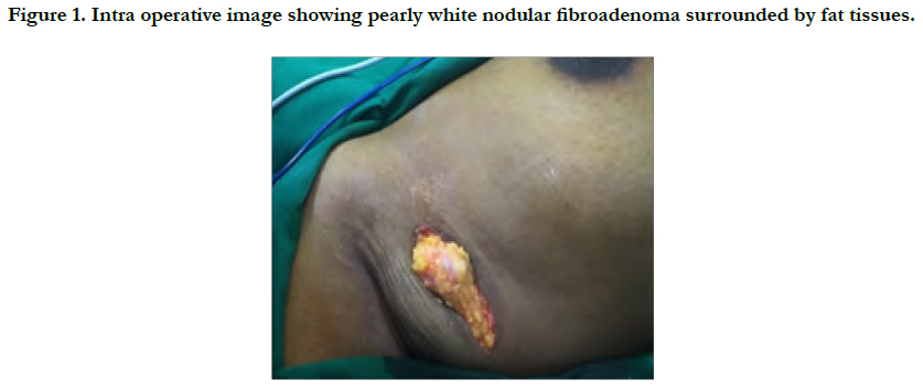
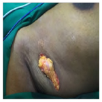
Figure 1. Intra operative image showing pearly white nodular fibroadenoma surrounded by fat tissues.
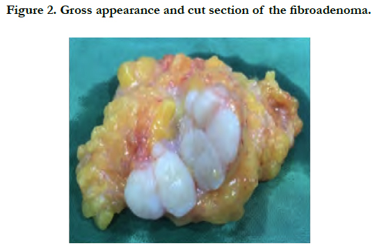
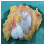
Figure 2. Gross appearance and cut section of the fibroadenoma.
Discussion
The development of breast begins early in the 4th week of gestation and is completed in the postpartum period. In the 4th week, ectodermal thickening known as milk ridges appear on the anterior part of the embryo, extending anywhere between the axilla to the groin in a convex curvilinear fashion. These milk lines proliferate downwards and branch out to eventually form the lactiferous ducts and the lobules. The breast continues to develop in the pectoral region due to the action of various hormones, mainly oestrogen and other growth factors. The breast tissue that forms along the milk ridge in the other regions excluding the pectoral area undergoes regression and disappears. Failure of the tissue to regress results in the formation of ectopic breast tissue [4, 5].
Ectopic Breast tissues can occur anywhere along the embryonic milk lines but has also been reported to be found in the face, vulva, anus and foot. It can occur as just supernumerary nipples or as polymastia or in other forms [6]. In 1915, Kajava introduced a classification for supernumerary breast tissue (Table 1) [6, 7].
Similar to eutopic breasts, ectopic breast tissues are also subject to influence by various hormones. There might be increase in the size or pain in ectopic breast tissues during pregnancy and lactation as well. Various diseases can arise in ectopic breast tissue similar to normal breasts like fibroadenomas, fibroadenosis, papillomas and malignancy [8]. This explains the increase in the intensity of pain during menstruation for the reported patient.
Any abnormalities arising from ectopic breast tissue can be evaluated using mammogram, cytology and ultra sonogram that are also used for diagnosis of pathologies of normal breast. As in our case, the FNAC clinched the diagnosis of axillary fibroadenoma. The treatment of asymptomatic benign lesions of ectopic breast tissue is mainly psychological assurance and repeated follow up whereas in symptomatic patients surgery is the best treatment option as it provides both pain and psychological relief for the patients [9, 10]. A malignancy of the ectopic breast tissue is reported to be treated with a wide local excision and regional lymph node dissection [11]. The confirmation of presence of ectopic breast tissue should also warrant further investigative process of the patient to rule out any urogenital malformations or malignancies, cardiac abnormalities and central nervous system disorders.
Conclusion
Ectopic breast tissue is a rare entity occurring along the embryonic milk lines. They often present as fibroadenoma and sometimes are also hormone dependent tumors. The differential diagnosis of axillary swellings is wide and ectopic breast tissue lesions should also be included due to its rising incidence.
References
- Velanovich V. Ectopic breast tissue, supernumerary breasts, and supernumerary nipples. South Med J. 1995 Sep;88(9):903-6. PubMed PMID: 7660204.
- Shuster M, Julie YT, Smith GP. Breast carcinoma arising in ectopic breast tissue presenting as an enlarging axillary nodule. Indian J Dermatol Venereol Leprol. 2015 Jul-Aug;81(4):422-4. doi: 10.4103/0378-6323.158655. PubMed PMID: 26087096.
- Goyal S, Bawa R, Sangwan S, Singh P. Fibroadenoma of axillary ectopic breast tissue: A rare clinical entity. Clinical Cancer Investigation Journal. 2014 May 1;3(3):242.
- De Filippis EM, Arleo EK. The ABCs of accessory breast tissue: basic information every radiologist should know. AJR Am J Roentgenol. 2014 May;202(5):1157-62. doi: 10.2214/AJR.13.10930. PubMed PMID: 24758674.
- Gabriel A, Maxwell G, et al. Breast Embryology: Overview, The Integument, The Embryologic Breast. MedScape[Internet][Updated 8 May 2015].
- Shatzel J, Blum A, Khoury T, Milligan J, Skitzki JJ. Gynecomastia-Like Hyperplasia of Axillary Ectopic Breast Tissue in a Young Female. Case Rep Pathol. 2013;2013:634248. doi: 10.1155/2013/634248. Epub 2013 Aug 1. PubMed PMID: 23984148.
- De Cholnoky T. Supernumerary breast. Archives of Surgery. 1939 Dec 1;39(6):926-41.
- Lakkawar NJ, Maran G, Srinivasan S, Rangaswamy T. Accessory breast tissue in the axilla in a puerperal woman-case study. Acta Med Medianae. 2010 Jan 1;49:45-8.
- Lesavoy MA, Gomez-Garcia A, Nejdl R, Yospur G, Syiau TJ, Chang P. Axillary breast tissue: clinical presentation and surgical treatment. Ann Plast Surg. 1995 Oct;35(4):356-60. PubMed PMID: 8585676.
- Goyal S, Puri T, Gupta R, Julka PK, Rath GK. Accessory breast tissue in axilla masquerading as breast cancer recurrence. J Cancer Res Ther. 2008 Apr-Jun;4(2):95-6. PubMed PMID: 18688128.
- Khatib Y, Kashikar A, Patel RD, Shrestha M. Varied presentations of ectopic breast-polymastia, fibroadenoma, and carcinoma arising from ectopic breast tissue. Clinical Cancer Investigation Journal. 2015 Jul 1;4(4):539.


