Craniometric Indices of Nigeria Skulls
Orish CN*, Ibeachu PC
Department of Anatomy, Faculty of Basic Medical Sciences, University of Port - Harcourt, Rivers State, Nigeria.
*Corresponding Author
Chinna Orish,
Department of Anatomy, Faculty of Basic Medical Sciences,
University of Port - Harcourt, Rivers State,
Nigeria.
E-mail: chinnaorish@yahoo.com
Received: December 01, 2015; Accepted: December 30, 2015; Published: January 04, 2016
Citation: Orish CN*, Ibeachu PC (2016) Craniometric indices of Nigeria skulls. Int J Anat Appl Physiol. 2(1), 6-13. doi: dx.doi.org/10.19070/2572-7451-160001
Copyright: Orish CN© 2016. This is an open-access article distributed under the terms of the Creative Commons Attribution License, which permits unrestricted use, distribution and reproduction in any medium, provided the original author and source are credited.
Abstract
Introduction: Craniometric indices show the percentage relationship between different dimensions. It is an important parameter for classification of race and sex of individuals of unknown identity. This study was undertaken to determine the craniometric indices of gnathic, palatal, orbital, cranial and nasal indices of Nigerian skulls.
Materials and Methods: One hundred adult dry skulls, (78 males, and 22 females) free from damage and deformities from eleven Departments of Anatomy in Nigerian Universities were used. Automatic digital and spreading callipers were used for the measurement. Data was analyzed with Graph Pad Prism 5.03. The mean, coefficient of variation, correlation, linear regression, percentiles, sexual dimorphism ratio were calculated.
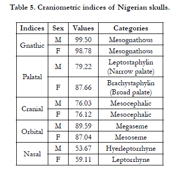
Results: The male/female ratios for the mean measurements were greater than unity. The mean value of all cranial parameters showed high level of sexual dimorphism. The male gnathic, palatal, orbital, cranial and nasal indices were found to be 99.50, 79.22, 89.59, 76.03, 53.67 respectively while female gnathic, palatal, orbital, cranial and nasal indices were 98.78, 87.66, 87.04, 76.12, 59.11 respectively. Male basion-nasion versus basion-prosthion length had positive correlation and the fit line sloped upward. There was no correlation between female basion-nasion versus basion-prosthion and the fit line was straight. Percentiles of indices showed a progressive increase from 10th - 90th.
Conclusion: The findings from this study will be handy tools in anatomical modelling, in addition to providing information for both cosmetic surgery and medico-legal guide in forensic science.
2.Introduction
3.Materials and Methods
3.1.Basion - Prosthion, Basion - Nasion
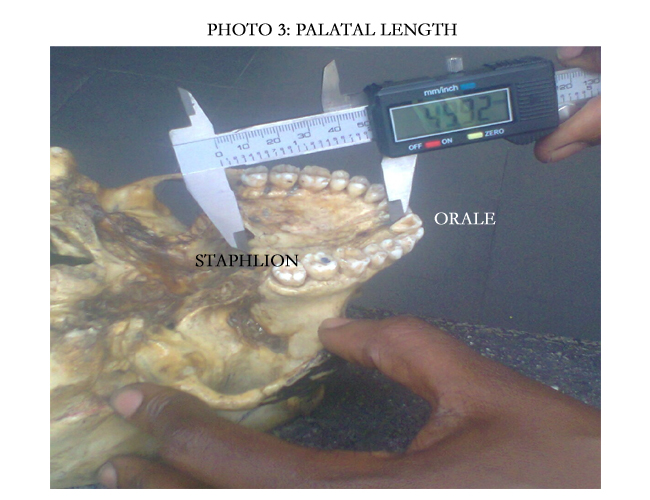
3.2.Palatal Length (Orale –Staphylion)
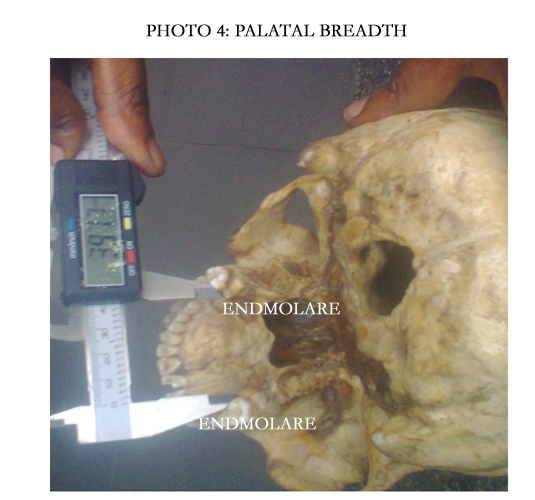
3.3.Palatal Breadth
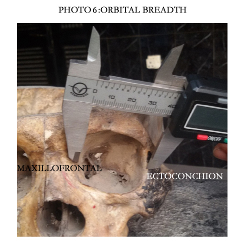
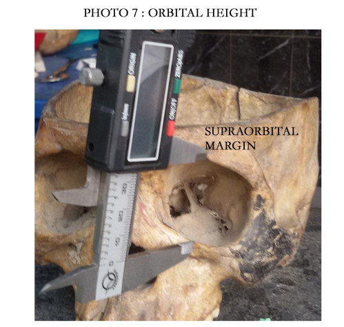
3.4.Orbital length and breadth
3.5.Maximum Cranial Length (Glabella - Opisthocranion)
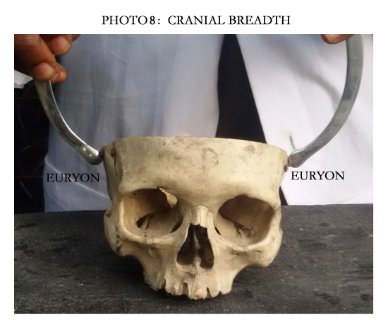
3.6.Maximum Cranial Breadth
3.7.Nasal Breadth
4.Discussion
5.Conclusion
6.References
KeyWords
Gnathic Index; Palatal Index; Skull; Anthropometry; Forensic Medicine.
Introduction
Craniometry is the scientific measurement of skull. It plays an important role in forensic practise. Skull is the skeleton of the head and consists of cranium and facial skeleton. The cranium is made up of occipital, temporal, parietal, frontal, sphenoid and ethmoid bones while the facial skeleton comprises of maxillae, palatine, nasal, zygomatic, inferior nasal conchae, lacrimal, mandible and vomer bones. Measurement of cranial bones play an important role in analysis of skeletal variation in determining population history and classification.
Indices show the relationship between different dimensions (length and breadth) which can also be expressed as ratios or percentages of comparison of two measurements. The general formula of index is the ratio of numerator (smaller measurement) to denominator (larger measurement) multiplied by hundred [22]. They are vital tools to forensic experts with respect to identification and classification of races and sexes. Indices can demonstrate the degree of disproportions in various parts of human body caused by hormonal and other disorders, congenital anomalies or trauma [12]. Also variations between and within population have been attributed to a complex interaction between genetic and environmental factors [17].
Gnathic index is the ratio of basion – prosthion to basion – nasion multiplied by hundred. Gnathic index has been classified into three namely, orthognathous which is 98 and below, mesognathous which ranges between 98-103, prognathous which is above 103. Palatal index is ratio of palatal breadth to palatal length multiplied by hundred and is also classified into three: leptostphylin or narrow palate which is less than 79.9, mesostphylin or medium palate which ranges from 80-84.9 and brachystphylin or broad palate which is 80 and above.
Orbital index is ratio of greatest height of the orbital cavity to its greatest breadth multiplied by hundred [22]. Three classes of orbit index have been described: Megaseme (large) with orbital index of 89 and above usually seen in yellow races [5]. Mesoseme (intermediate): The orbital index range between 89 and 83. This type is seen in the white races [24]. Microseme (small): Orbital index 83 or less. This type is characteristic of the black races where the orbital opening is rectangular [5]. Nasal index is ratio of the greatest width of the nasal aperture to the length or height multiplied by hundred [22]. There are three main nasal types; leptorrhine, (narrow) which ranges from 54.9 to 69.9. Mesorrhine (medium) which ranges from 70.0 to 84.9 and platyrrhine with a range from 85.0 to 99.9.
Cranial index is ratio of the maximum breadth of the bare skull to its maximum length multiplied by hundred [22]. Cranial index is classified into, four main types namely dolicocephalic which is less than 74.9, mesocephalic with cranial index between 75 to 79.9, brachycephalic with cranial index between 80 to 84.9 hyperbrachycephalic with cranial index from 85 to 89.9 [22].
Several researches have worked on various craniometric indices [35, 6]. Such measurements are also useful in the analysis and classification of fossil remains as well as study of living population [4]. Information on craniometric indices especially palatal and gnathic indices is sparse in Nigerian population. The present study is aimed at adding some fund of knowledge to address the information gap in craniometric indices of Nigerian skulls.
Materials and Methods
A total of 100 adult dry skulls (78 males and 22 females), free from damage and deformity, fully ossified collected from Departments of Anatomy in Nigerian Universities were used for this study. A digital caliper with a precision of 0.01mm (Mitutoyo®), spreading calliper, marker was used to measure the following length parameters basion-prosthion, basion-nasion, palatal length and breadth, orbital length and breadth, nasal height and breadth, cranial length and breadth.
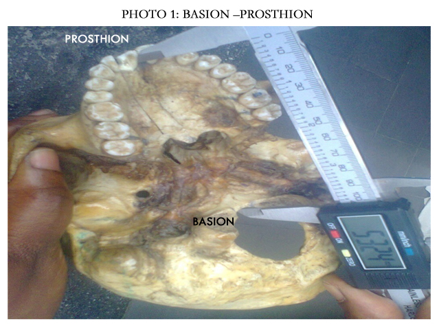
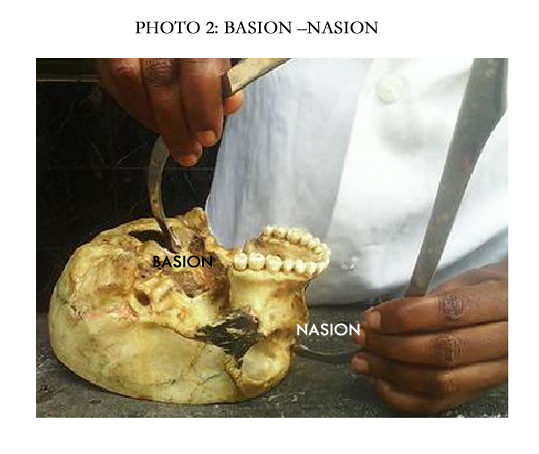
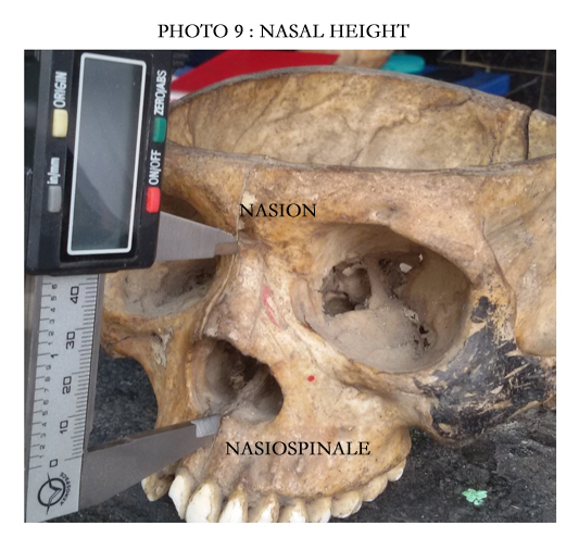
It measures the straight distance between basion – prosthion. Basion, the most anterior point of the great foramen magnum in the sagittal plane [3]. Prosthion, the lowest, most anterior point on the alveolar portion of the premaxilla in the median plane, between the upper central incisors [3]. Nasion: This is the midpoint of the sutures of the frontal and nasal bones [3].
It measures the straight distance between orale and staphylion. The skull was held with the norma basalis facing upward [3]. Staphylion: This is a single point on the posterior hard palate where the palatal suture is crossed by a line drawn tangent to the curves of the posterior margin of the palatal bones [3]. Orale: This is the most anterior point on the hard palate where a line drawn lingual to the central incisors intersects the palatal suture [3].
It measures the straight distance between the middle of the inner margin of the alveolar on the second molar i.e. endmolar to endmolar. Endmolar: This is the most medial point on the inner margin (lingual surface) of the socket of the second upper molar. It is used to measure palatal width. The skull was held in the norma basilis facing upward [3].
This measures the straight distance between maxillofrontale-ectoconchion. Maxillofrontale is the point where anterior lacrimal crest meet the frontomaxillary suture. Ectoconchion is the point where a line running parallel to the the upper orbital border cuts lateral orbital margin. The skull was held in the norma frontalis facing upward[3].
The length of the orbit was measured from supraorbital margin to the optical canal ie the maximum distance between the upper and lower margin of the orbital cavity taken perpendicular to the orbital breadth.
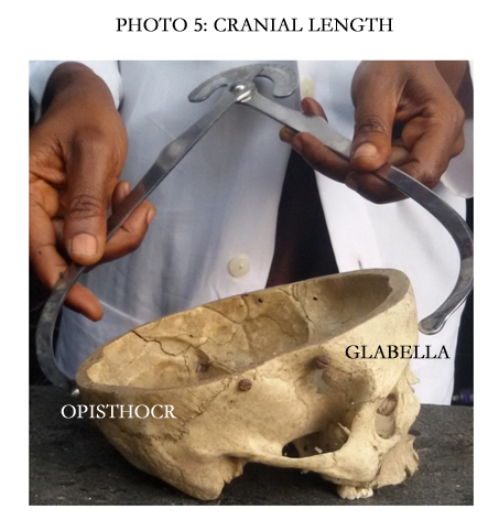
The straight distance from the glabella to the opisthocranion. Glabella is the most prominent point between the two superaorbital ridges above the fronto-nasal suture in the mid-sagittal plane. Opisthocranion is the most posterior point from the glabella in the mid-sagittal plane, excluding the inion.
The straight distance between the two euryon points. Euryon is the most lateral point on the skull, which can only be determined by measuring the maximum cranial breadth, as it is variable.
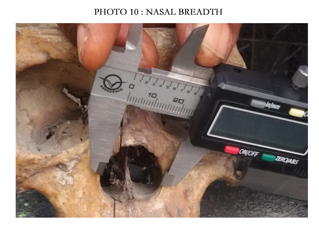
The maximum breadth between the two lateral margins of the nasal apertures.Nasal Height (nasion to nasospinale): The straight distance from the nasion to nasospinale. Nasospinale is the point where a line touching the lower margin of the nasal aperture crosses the mid-sagittal plane.
Data was anaylzed with Graph Pad Prism 5.03. The mean, standard deviation, correlation, linear regression, percentiles, sexual dimorphism ratio and cranial indices were calculated. Student‘s T-test was used to compare male-female and right-left measurements.
Gnathic index = (Basion - prosthion × 100 %) / Basion-nasion
Palatal index = (palatal breadth × 100 %) / Palatal length
Nasal index = (nasal breadth × 100 %) / Nasal height
Orbital index = (orbital height × 100 %) / Orbital width(breadth)
Cranial index = (cranial breadth × 100 %) / cranial length
Effect of sex on the following cranial parameters namely basion–prosthion, basion-nasion, palatal length and breadth, orbital length and breadth, nasal height and breadth, cranial length and breadth are shown on Table 1. Male parameters were significantly higher than female parameters except basion–prosthion, basionnasion.
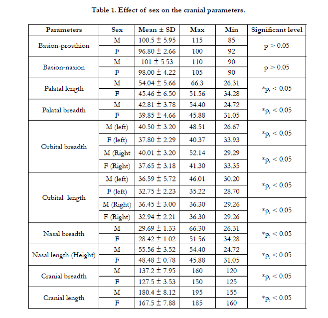

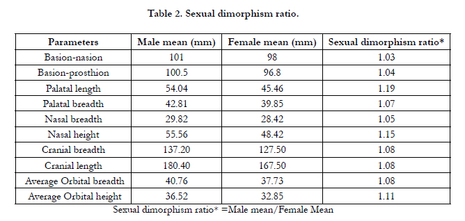
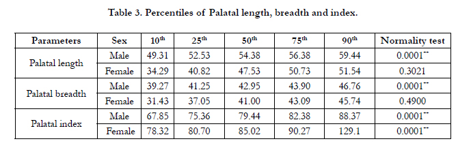
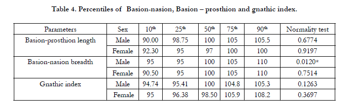
Table 2 shows sexual dimorphism ratio of the basion–prosthion, basion-nasion, palatal length and breadth, orbital length and breadth, nasal height and breadth, cranial length and breadth. The male/female ratios for the mean measurements were greater than unity in all. The percentiles of palatal length, breadth and index and basion-nasion versus Basion–prosthion and gnathic index are shown on Tables 3 and 4. There was progressive increase in these parameters from 10th to 90th percentiles. Table 5 shows cranial indices. The palatal, nasal and cranial indices were higher in female while gnathic and orbital indices were higher in male.
The incidence and classification of male and female gnathic indices are shown in Figure 1. Mesognathous and orthognathous showed higher incidence in males and females respectively. Figure 2 shows incidence and classification of male palatal and female palatal indices with leptostphylin and brachystphylin having the higher incidence respectively.
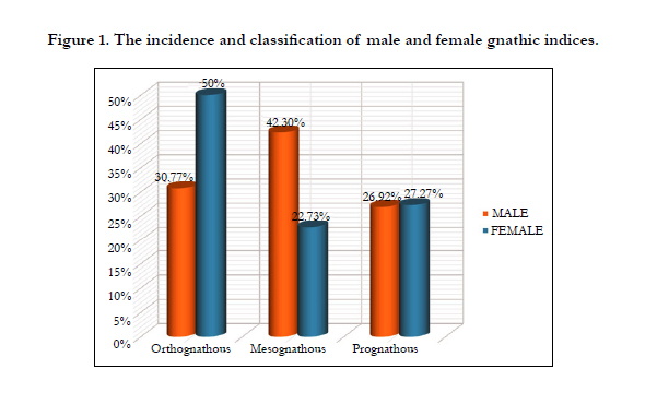
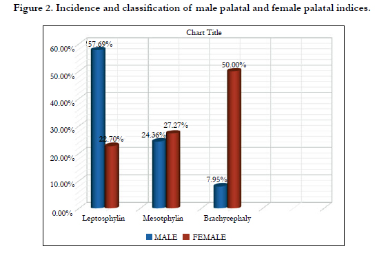
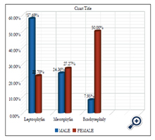
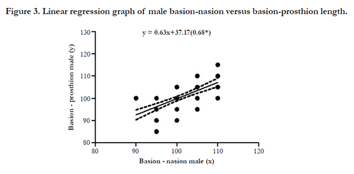
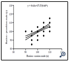
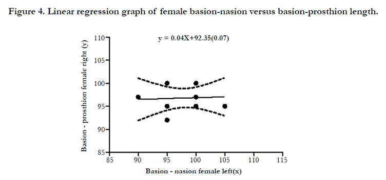
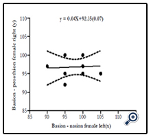
Figure 3 shows a scatter plot of the linear relationship between male basion-nasion versus basion- prosthion length. There was positive correlation between the left and right hence the fit line sloped upward. The scatter plot of the linear relationship between female basion-nasion versus basion-prosthion length (Figure 4). There was no correlation between them hence the fit line was straight (Figure 4).
Discussion
This study has shown that the cranial, parameters are higher in male than females. Our findings show that the basion-prosthion were 100.5 ± 0.67 and 96.80 ± 0.84mm, basion-nasion 101 ± 5.53 and 98.00 ± 4.22 for male and female respectively. Kajanjola 1966
reported basion-prosthion as 9.85 ± 0.07cm and 9.45 ± 0.76cm and basion nasion as 9.98 ± 0.43cm and 9.66 ± 0.47cm for male and female respectively. Deshmukh and Devershi (2006) reported Basion-Prosthion length 90 ± 4.21 and 89 ± 5.08 and basion nasion length as 99 ± 4.19 and 96 ± 4.61 for male and female respectively and was found to be significant by univariate analysis with p < 0.05. Steyna and Yasar (1998) found basion-prosthion length to be 95.4 ± 5.39 and 90.0 ± 5.03 and basion-nasion as 102.4 ± 4.40 and 96.2 ± 4.10mm for male and female respectively. Kraniotia et al 2008 in their work tilted Craniometric analysis of the modern Cretan population found Basion-prosthion length to be 93.11 ± 5.05 and 88.76 ± 5.70 and Basion-nasion length to be 102.01 ± 3.85 and 96.25 ± 6.54mm for male and female respectively. Rooppakhun et al (2011) showed that the mean of Basion-prosthion to be 96.1 ± 5.4 and 92.9 ± 5.5mm for male and female respectively. Ahmed et al 2011 in Northern Sudanese using crania (69 males, 41 females) found Basion-prosthion to be 98.39 ± 4.89 and 93.12 ± 4.98 and Basion-nasion 100.62 ± 4.09 and 94.41 ± 3.38mm for male and female respectively.
Deshmukh and Devershi (2006) reported palatal length of 45 ± 3.08 and 44 ± 3.37, palatal breadth 35 ± 1.50 and 34 ± 2.73mm for male and female respectively and was found to be significant (p < 0.05) by univariate analysis. Rooppakhun (2011) study on advanced medical imaging and reverse engineering technologies in craniometric studies using 104 skulls (63 males and 41 females) reported palatal length of 42.6 ± 4.2 and 42.6 ± 4.4mm and palatal breadth of 39.1 ± 3.1mm and 37.6 ± 2.4mm for male and female respectively. The above findings differ from our own research which reported higher values for male and female to be 54.04 ± 0.64 and 45.46 ± 2.06mm respectively.
Dave et al, 2013 reported incidence of palatal index of 63% as narrow (leptostaphylin), 24% had intermediate (mesostaphylin), and 13% had wide (brachystaphylin). This agrees well with male palatal index of the present study which showed that majority of the skulls 57.69% had narrow( leptostaphylin), 24.36% had intermediate (mesostphylin), 17.95% had wide (brachystaphylin). It contradicts however with female palatal index which reported majority of the skulls to be 50% brachystaphylin, 30% mesostaphylin and 20% leptostaphylin.
In this study orbital breadth and height have higher values which can be attributed to racial differences. In a study by Gosavi et al., (2014) from central India, the mean height of the orbit was observed as 32.31 ± 2.52mm. Kaur et al., 2012 recorded orbital height it as 32.05 ± 2.0mm in North Indian skulls. The orbital width was observed as 36.5 ± 1.92 and 36.41 ± 1.78mm on right and left sides respectively by Narasinga and Pramila 2015 whereas it was reported to be 39.25 ± 2.3mm by Kaur et al., (2001). Ebeye and Otikpo 2013 in Nigerian subjects observed orbital height as 32.46mm and mean orbital width as 41.43mm. Weaver et al., 2010 in their computer tomography scan based study in Caucasian population observed orbital height as 32.09 ± 2.2mm and orbital width as 37.01 ± 2.0mm. Also Ebeye and Otikpo 2013 in their study observed orbital index as 78.15 which is different from our reported orbital indices of 89.59 and 87.04 for male and female respectively. The orbital index which determines the shape of the face differs in different population groups. This means that the orbit with larger width than height will have smaller orbital indices while those with larger orbital index will have narrow faces. Kaur et al., 2012 observed orbital index as 81.65 while Narasinga and Pramila 2015 recorded right and left orbital indices as 86.13 and 90.69 respectively.
The Indian population according to Kaur et al 2012 that reported an orbital index of 81.65 belongs to the microseme category. Kaplanoglu et al 2013 in Turkey reported the average orbital index in females to be 85 in males 84.6 belonging to mesoseme category. The orbital indices for male and female from the present study are 89.59 and 87.04 suggesting that Nigerian skulls can be classified as megaseme and mesoseme for male and female respectively. This is in agreement with earlier study by Ukoha et al 2011 in Nigerian skulls with orbital index of 89.21. In Malawians, the skulls have been categorized as megaseme with the orbital indices in males and females as 94.35 and 96.03 respectively [37]. In an old study conducted by Casidy, the black race was categorized as microseme [27].
Kranioti and Iscan 2008 reported nasal breadth and height to be higher in male higher than female which is similar to our findings. In another skull study, Vidya et al 2012 reported nasal breadth as 2.36 ± 0.26 and 2.23 ± 0.24cm, nasal height 4.79 ± 0.57 and 4.54 ± 0.35cm for males and females respectively and the differences were found to be significant (p = 0.026). Nasal indices of 49.38 and 49.24 for male and female respectively have been reported by Vidya et al 2012, whereas the nasal indices from the present study are 58.6 and 53.4 for male and female respectively.
Kranioti et al 2008 reported cranial length as 181 ± 6.63 and 172.89 ± 6.48mm in males and females respectively. These data tend to agree with the present study that has cranial length of 180.4 ± 8.12, 167 .5 ± 7.28mm for male and female respectively. Vidya et al 2012 also reported cranial length of 16.81 ± 1.61, 16.67 ± 1.73 and breadth of 13.29 ± 1.93 and 13.28 ± 1.45mm and cranial indices of 78.40 and 79.13 for male and female respectively. Strouhal 1992 reported that the cranial index in ancient Egyptians skulls ranged from 71.80-76.10 while the present recorded cranial indices of 76.03 and 76.12 for male and female respectively.
In a study of 62 skulls, Seema and Gandhi 2011 reported that the cranial indices for male were 72.54 and female 72.06. Adejuwon et al 2011 reported cranial indices of 72.97 and 71.72 for males and females respectively. Jaysingh et al 1979 in their study of 300 human skulls reported a cranial index of 74.35.
In a Tibetan skull study by Morant, the cranial index was 75.25 [25]. Chaturvedi et al 1963 reported a cranial index of 70.75. In Mongoloid race dolicocephaly is rare while brachycephaly is rare in Negroid race [30]. The present study also showed the rarity of brachycephaly. Human knowledge of paleontology and available data suggest that early man was generally dolicocephalic. Brachycephaly developed later as a result of repeated mutation and various other factors [30]. The present study shows that the majority of the skull are dolicocephalic which is in agreement with the work of Vishal and Pradeep 2012 study on in South India skulls. However the mean of the cranial indices from the present study suggest mesocephalic for both sexes.
Taken together there was a progressive increase in the 10th percentiles to 90th percentiles of palatal length, breadth and index, basion-nasion, basion-prosthion and gnathic index. To the best of our knowledge, this is the only study that has reported the percentiles of palatal length, breadth and index, basion-nasion, basion-prosthion and gnathic index.
In a bid to model the relationship between two anthropometric parameters, this study has employed linear regression and correlation tools as aforementioned using the equation y = ax + b. With this equation the value y can be predicted when x is known. Hitherto information is scanty on the mathematical models of these craniometric parameters [28]. A positive regression coefficient indicates a positive relationship between two variables and from the graph the fit line sloped upward as in male Basion-nasion v Basion-prosthion while a negative regression coefficient indicates a negative relationship between two variables and from the graph the fit line sloped downward as in male Basion-bregma v Nasion bregma. Ahmed et al, 2011 reported that sexual dimorphism ratio (male/female ratios) for the mean measurements were greater than unity, indicating that the male crania were larger in all linear dimensions than female crania. This correlates well with the present study which reported that sexual dimorphism ratio for the mean measurements were greater than unity too, indicating that the male crania were larger in all linear dimensions than female crania. Knowledge of gnathic and palatal, orbital, cranial and nasal indices of Nigerians will be of immense help to forensic expert with respect to classification of races and sexes and also to surgeons in clinical practise.
Conclusion
Generally, the hematological characteristics of cannabis smoker differed significantly from those of non-smokers. A decrease in mean value of TWBC of cannabis smokers is a pointer to fact that immune defense in the body of test subjects, which also is regarded as body defense against infection, may have become porous.
The health risk of smoking cannabis has been shown to be so enormous that one can not justify this substance abuse. Depleted levels of Hb and PCV were evident in studied test subjects which may necessitate diet supplementation as well as enlightenment in the short and long term as observed trend may deteriorate with time.
Acknowledgements
The authors are grateful to John Orlu of University Teaching Hospital Port Harcourt Nigeria for his assistance during analyses.
References
- Adejuwon SA, Salawu OT, Eke CC, Femi-Akinlosotu W, Odaibo AB (2011) A craniometric study of adult humans skulls from Southwestern Nigeria. Asian Journal of Medical Sciences 3(1): 23-25.
- Ahmed AA, Mohammed HA, Hassan MA (2011) Sex determination from cranial measurements in recent northern Sudanese. Khartoum Medical Journal 4(1): 539-547.
- Ales Hrdlicka (1947) Hrdlicka's Practical Anthropometry. Wistar Institute of Anatomy and Biology, Philadelphia.109-197.
- Alex FR, Steven B, Timothy GL (1996) Human Body Composition. (4th edtn), Human Kinetics Publishers. 167-172.
- Cassidy PJ (1913) “Megaseme” Webster dictionary. answers.com (homepage on the internet),
Retrieved from http://www.answers.com/topic/megaseme - Dave MR, Gupta S, Vyas KK, Joshi HG (2013) A Study of Palatal Indices and Bony Prominences and Grooves in the Hard Palate of Adult Human Skulls. NJIRM 4(1): 7-11.
- Deshmukh AG, Devershi DB (2006) Comparison of Cranial Sex Determination by Univariate and Multivariate Analysis. J Anat Soc India 55(2): 48-51.
- Gopinathan K, Dhall U, Chhabra S (1998) Sutural bones in North Indian population. J Anat Soc India 47(2): 91-96.
- Ebeye OA, Otikpo O (2013) Orbital index in Urhobos of Nigeria. IOSR J Dental Med Sci 8(2): 51-53.
- Chaturvedi RP, Harneja NK (1963) A cephalometric study of human skulls. Journal of Anatomical Society of India 12: 93-96.
- Kranioti EF, Işcan MY, Michalodimitrakis M (2008) Craniometric analysis of the modern Cretan population. Forensic Sci Int 180(2-3): 110.e1–110.e5.
- Farkas LG, Munro IR (1987) Anthropometric facial proportions in medicine. Charles C Thomas Publisher, USA.
- Gosavi S (2014) A study of orbital morphometry in Indian dry skulls. Asian Journal of Biomedical and Pharmaceutical Sciences 4(29): 23-25.
- Kaur J, Yadav S, Singh Z (2012) Orbital dimensions - A direct measurement study using dry skulls. J Acad Indus Res 1(6): 293-295.
- Jaysingh P, Arora AK, Gupta CD, Dua S, Pandey DN (1979) Craniometric study of skulls of Uttar Pradesh. J Anat Soc India 28(3): 127-131.
- Kajanoja P (1966) Sex determination of Finnish crania by discriminant function analysis. Am J Phys Anthropol 24(1): 29-33.
- Kasai K, Richards LC, Brown T (1993) Comparative study of craniofacial morphology in Japanese and Australian aboriginal populations. Hum Biol 65(5): 821-834.
- Klepinger LL (2006) Fundamentals of Forensic Anthropology. John Wiley & Sons, USA.
- Krishan K (2008) Estimation of stature from cephalo-facial anthropometry in north Indian population. Forensic Sci Int 181(1-3): 52e1-52e6.
- Krogman WM, Iscan MY (1986) The Human Skeleton in Forensic Medicine. Charles C Thomas Publisher, USA.
- Lobo SW, Chandrashekar TS, Kumar S (2005) Cephalic Index of Gurung Community of Nepal - an Anthropometric study. Kathmandu Univ Med J 3(3): 263- 265.
- Martin R, Saller K (1957) Lehrbuch der Anthropologie. Gustav Fischer Verlag, Stuttgart.
- Steyn M, Işcan MY (1998) Sexual dimorphism in the crania and mandibles of South Africa whites. Forensic Sci Int 98(1-2): 9-16.
- Mcgraw Hill dictionary of scientific and technical terms “mesoconch” Mcgraw hill company Inc, answers. Com (homepage on the internet) 2003, Retrieved from http://www.answers.com/topic/mesoconch.
- Morant GM (1923) A first study of Tibetian skull. Biometrika 14(3-4): 193- 260.
- Rao NB, Padmini PM (2015) A Study of Orbital Index in dry Skulls of North Coastal Andhra Pradesh. International Journal of Basic and Applied Medical Sciences 5(2): 1-3.
- Novita M (2006) Facial, upper facial, and orbital index in Batak, Klaten, and Flores students of Jember University. Dent J (Maj Ked Gigi) 39(3): 116-119.
- Rooppakhun S, Chantarapanich N, Sitthiseripratip K (2011) Advanced Medical Imaging and Reverse Engineering Technologies in Craniometric Study. Forensic Medicine - From Old Problems to New Challenges 307-326.
- Shah GV, Jadhav HR (2004) The Study of Cephalic Index in Students of Gujarat. J Anat Soc India 53(1): 25-26.
- Sharma RN (2005) Criteria of racial classification. Physical Anthropology. Surjeet Publication. 226.
- Mahajan SA, Gandhi D (2011) Cephalometric study of adult human skulls of north indian origin. International Journal of Basic and Applied Medical Sciences 1(1): 81-83.
- Strouhal E (1973) Temporal and Spacial Analysis of Some Craniometric Features in Ancient Egyptians and Nubians. In Population Biology of the Ancient Egyptians. Academic Press, London. 121-142.
- Ukoha U, Egwu OA, Okafor IJ, Ogugua PC, Onwudinjo O, et al. (2011) Orbital dimensions of adult male Nigerians: a direct measurement study using dry skulls. Int J Biol Med Res 2(3): 688-690.
- Salve VM, Londhe PS (2012) A craniometric study of adult human skulls from Andhra Pradesh. National Journal of Integrated Research in Medicine 3(1): 63-66.
- Vidya CS, Prashantha B, Gangadhar MR (2012) Anthropometric Predictors for Sexual Dimorphism of Skulls of South Indian Origin. International Journal of Scientific and Research Publications 2(10): 1-3.
- Weaver AA, Loftis KL, Tan JC, Duma SM, Stitzel JD (2010) CT scan based three-dimensional measurement of orbit and eye anthropometry. Invest Ophthalmol Vis Sci 51(10): 4892-4897.
- Weiss RA, Haik BG, Saint-Louis LA, Ellsworth RM (1987) Advanced diagnostic imaging techniques in ophthalmology. Adv Ophthalmic Plastic Reconstr Surg 6: 207-263.


