Alterations in Hematological and Biochemical Parameters Affecting the Reproductive Performance in Female Camels (Camelus Dromedaries)
Zaher H2,3, El-Zahar H1, Al Sharifi S4, Shety T1*
1 Department of Animal Medicine, Faculty of Veterinary Medicine, Zagazig University, Egypt.
2 Department of Theriogenology, Faculty of Veterinary Medicine, Zagazig University, Egypt.
3 Abu Dhabi Control Authority, Animal Wealth Sector, Veterinary Laboratories Division, A.R.E., UAE.
4 Consultant at Abu Dhabi Food Control Authority, Abu Dhabi A.R.E., UAE.
*Corresponding Author
Tarek Shety
Department of Animal Medicine,
Faculty of Veterinary Medicine,
Zagazig University, Egypt.
Tel: 002 01007063608
Fax: 002 0552283683
E-mail: tarekshety@gmail.com
Received: February 04, 2017; Accepted: February 21, 2017; Published: February 23, 2017
Citation: Zaher H, El-Zahar H, Al Sharifi S, Shety T (2017) Alterations in Hematological and Biochemical Parameters Affecting the Reproductive Performance in Female Camels (Camelus Dromedaries). Int J Vet Health Sci Res. 5(1), 155-160. DOI : dx.doi.org/10.19070/2332-2748-1700032
Copyright: Shety T© 2017. This is an open-access article distributed under the terms of the Creative Commons Attribution License, which permits unrestricted use, distribution and reproduction in any medium, provided the original author and source are credited.
Abstract
The aim of the present study was to investigate the effect of alterations in hematological and biochemical parameters especially liver, renal enzymes and some element concentration on the reproductive performance of female camels. Blood samples were collected from 51 adult female camels. 39 adult female camels (diseased) suffered from anorexia, decreased body gain, some camels were emaciated and with varying degrees of reproductive abnormalities starting from repeat breeder, anestrous and infertility and 12 control camels that showed no evidence of clinical disease during clinical examination and samples collection. The results of hematological, biochemical and ultrasonographic findings were recorded. The hematological results showed significant reduction in erythrocyte count (RBCs), hemoglobin (Hgb), mean corpuscular volume (MCV) and mean corpuscular hemoglobin concentration (MCHC) with significant increase in packed cell volume (PCV%). The leucocyte count (WBCs) and neutrophils percentage were significantly increased in diseased camels compared to controls while, lymphocyte percentage was significantly decreased in diseased camels. The serum activity of aspartate aminotransferase (AST), lactate dehydrogenase (LDH), creatine kinas (CK) and γ-glutamyl transferase (γGT) was significantly increased in diseased camels. The concentration of selected elements in serum indicated significant reduction in copper, calcium and phosphorous concentration in diseased camels. Progesterone concentration was significantly decreased in diseased camels. Other tested parameters did not differ significantly in diseased and control camels. In conclusion, the hepatorenal dysfunction parallel with inanition might affect on the reproductive performance especially ovulation.
2.Introduction
3.Materials and Methods
3.1. Camels and Management
3.2. Blood Samples and Handling
3.3. Blood Hematology Analyses
3.4. Serum Biochemical Analyses
3.5. Ultrasonography
3.6. Statistical Analysis
4.Results
5.Discussion
6.Conclusion
7.References
Keywords
Camel; Infertility; Ultrasound; Liver; Enzymes; Copper.
Introduction
Camels play a very important role in the economy and social life of bedouins and pastoralists in different localities in the world. The breeding capacity is of major importance to camel owners and industry, the decrease in reproductive performance affects negatively on camel industry. Regular monitoring of camel herd for breeding problems and/or infertility is considered a major part of herd health management [1].
Generally, camels have low reproductive performance which seems to be a characteristic feature for camel under natural pastoral conditions [2]. There are different factors affecting on the reproductive performance of camels including short breeding season, age at first calving, low libido of male and late postpartum estrous [3]. Reproductive performance of camels kept under pastoral management was studied in northern Kenya by Kaufmann [1].
The change in the hematological and biochemical profiles of the blood is an important indicator of physiological and metabolic status of animals. The progesterone is metabolized in the liver and thus the disturbances in liver function might results in hormonal disturbances. Meanwhile, the progesterone concentration reaches its peak after ovulation and falls immediately before parturition [4].
To our knowledge, no research work was published focusing on the effect of liver, renal enzymes and serum elements concentration on the reproductive performance of female camels. Therefore the objectives of the present study were to investigate the effect of alterations in some hematological and biochemical profile parameters on the reproductive performance of female camels with special reference to liver, renal enzymes and some element concentration.
The samples were collected from camels reared in a private farm for camel breeding and management, the laboratory analyses were performed at Veterinary Laboratories Division, Animal Wealth Sector, Abu Dhabi Food Control Authority (adfca), Animal Wealth Sector, United Arab of Emirates. Fifty-one female camels (Camelus dromedarius) aged from 7 to 10 years with average body weight of (425 ± 18kg) were used in the present study. All camels were subjected to routine health check and divided into control camels (12 adult females) that were considered healthy on the basis of physical examination, with good body condition, free from common infectious diseases as proven by the veterinary authorities and with normal breeding capability. Diseased camels (39 adult females) suffered from anorexia, decrease body gain, indigestion and lowered reproductive performance expressed as delayed ovulation, anestrous and repeat breeder.
The camels were housed in an open yards during day and night with shelter all over the year. The animals fed a properly formulated concentrated ration that contain 65% total digestible nutrients and 14% crude protein according to their body weight requirements, alfa alfa hay was introduced all the time. Fresh water was presented twice daily.
Blood samples were collected from the jugular veins in an EDTA vacutainer for obtaining whole blood samples and on plain vacutainer for obtaining serum samples. Serum was harvested after centrifugation of the plain vacutainer at 3000 rpm for 15 minutes, and then stored at -20°C until analysis.
Hematological analyses were performed using Sysmex XT 2000i hematology analyzer (VLD-DPM-CBC-06, Mundelein, IL 60060, USA), for determination of RBCs, PCV%, Hgb concentration, WBCs, differential leucocytic count including neutrophils, lymphocytes, monocytes, eosinophils and basophils percentages. The hematological indices including MCV, MCH and MCHC were also measured during analysis.
Blood chemistry profile was performed using Beckman Coulter analyzers (VLD-DPM-CBC-02, Mundelein, IL 60060, USA). Serum samples were used to determine total protein, albumin, creatinine, BUN, glucose, calcium (Ca), phosphorous (P), copper (Cu), Iron (Fe), sodium (Na), potassium (K) and chloride (Cl). The activity of serum enzymes including γGT, AST, ALT, CK and LDH were also measured. Progesterone concentration was determined in serum samples using Electrochemiluminescent immunoassay kits (Roche Diagnostics, Indianapolis, Indiana, USA), with a measuring range of 0.030 to 60.00 ng/ml.
The genital tract was examined ultrasonographically using portable ultrasound scanner with 7.5 MHz transrectal transducer (Prosound 2, Model UST-660-7.5, Aloka, Japan). Ovaries were carefully examined for structure, size and consistency.
Data were statistically analyzed using SPSS Statistics® 17.0 (Version 17.0 released 2008, SPSS Inc., Chicago) and the results were expressed as mean values ± standard error (SE). Normal distribution test were performed using Shapiro Wilks W Test. The data were normally distributed. One way ANOVA test were performed to compare between the control group and the disease group. The level of significance was set at < 0.05.
Results
The results of the hematological and biochemical profiles parameters of female camels affected with varying degrees of infertility are listed in Tables (1 and 2); the results are expressed as mean ± SE. The control camels used in the present study were clinically healthy, the feeding status, clinical examination results were normal, there was no reproductive disorders. The diseased camels showed signs of anorexia, decrease food intake, varying degrees of emaciation, decrease body gain and different forms of reproductive impairments including repeat breeder, anestrous and delayed ovulation.
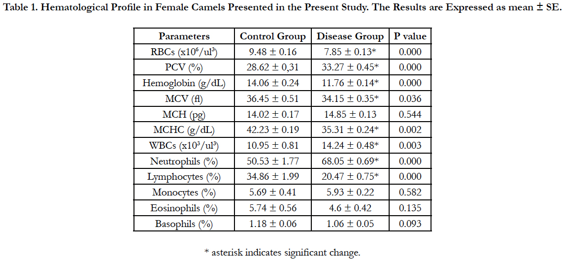

Table 1. Hematological Profile in Female Camels Presented in the Present Study. The Results are Expressed as mean ± SE.
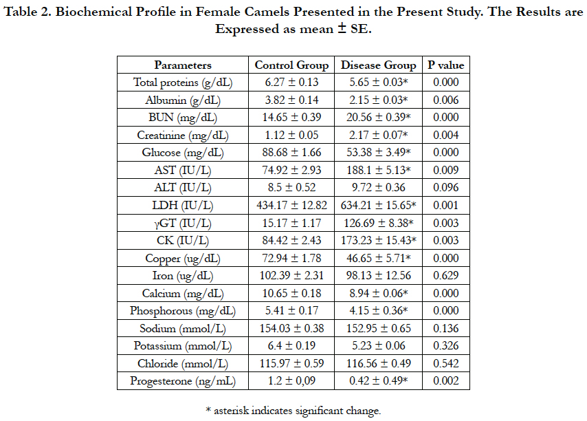
Table 2. Biochemical Profile in Female Camels Presented in the Present Study. The Results are Expressed as mean ± SE.
There was significant (P ≤ 0.05) reduction in the total erythrocyte count, hemoglobin, MCV and MCHC concentration and significant (P ≤ 0.05) increase in packed cell volume in diseased camels compared to clinically healthy. In addition, there was significant (P ≤ 0.05) increase in leucocyte count and neutrophils percentage while the lymphocyte percentage was significantly decreased.
The serum liver enzymes including ALT, AST, LDH and γGT were determined in serum samples. There was significant (P ≤ 0.05) increase in AST, LDH and γGT in diseased camels compared to clinically healthy, while ALT did not change. In addition, the concentration of CK showed significant (P ≤ 0.05) increase while progesterone concentration was significantly decreased in diseased camels compared to clinically healthy.
The protein status was measured by the concentration of renal enzymes (BUN and creatinine) and the concentration of total protein and albumin in serum. There was significant (P ≤ 0.05) increase in BUN and creatinine concentration and significant (P ≤ 0.05) decrease in total protein and albumin concentration in diseased camels compared to clinically healthy. The glucose concentration was significantly (P ≤ 0.05) decreased in diseased camels compared to clinically healthy.
The concentration of elements and electrolytes was determined. There was significant (P ≤ 0.05) reduction in the concentration of copper, calcium and phosphorous in diseased camels compared to clinically healthy. On the other hand, the concentration of iron, sodium, potassium and chloride did not change.
The results of ultrasonography on the ovaries of healthy camels showed the presence of normal ovarian follicle with a mean diameter of 13 mm and corpus luteum with a mean diameter of 16 mm (Figure 1 and Figure 2) respectively while, no structure was observed on the ovaries of diseased camels (Figure 3). No ovarian cyst or persistent corpus luteum. The uterine image was normal.
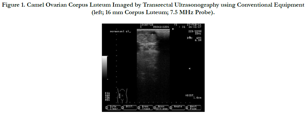
Figure 1. Camel Ovarian Corpus Luteum Imaged by Transrectal Ultrasonography using Conventional Equipment (left; 16 mm Corpus Luteum; 7.5 MHz Probe).
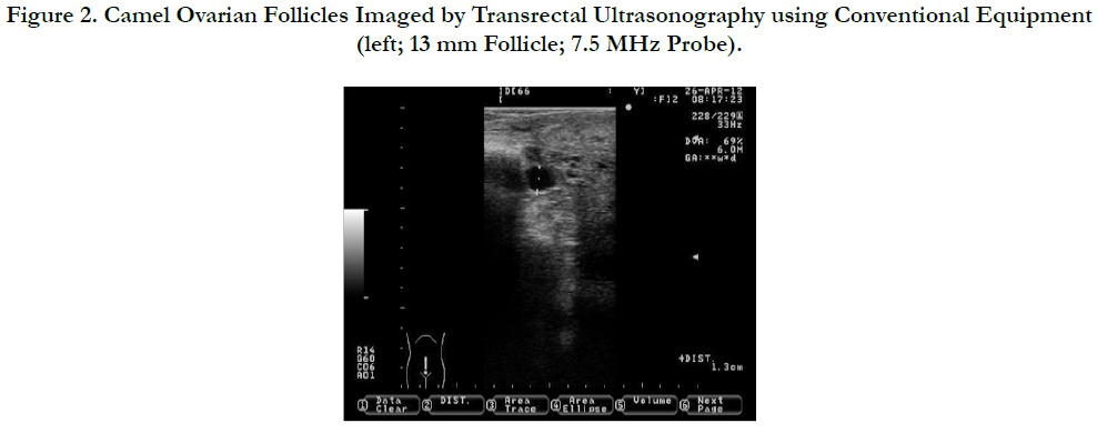
Figure 2. Camel Ovarian Follicles Imaged by Transrectal Ultrasonography using Conventional Equipment (left; 13 mm Follicle; 7.5 MHz Probe).
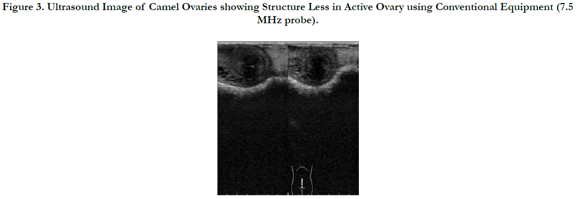
Figure 3. Ultrasound Image of Camel Ovaries showing Structure Less in Active Ovary using Conventional Equipment (7.5 MHz probe).
Discussion
In the present study, the diseased camels were presented with signs of anorexia, decreased body gain, some camels were emaciated and with varying degrees of reproductive abnormalities starting from repeat breeder, anestrous and infertility. Blood hematological and biochemical profiles is an important indicator of the physiological processes in animal's body. The values of the hematological and biochemical results obtained for healthy camels in the present study (Table 1) were in the normal ranges and are in agreement with previous studies by Mohamed and Hussein [5] and Alhadrami [6].
The hematological indices were performed. There were significant decrease in RBCs, Hgb, MCV and MCHC concentration while there was significant increase in PCV% in diseased camels compared to clinically healthy. This indicates a microcytic hypochromic type of anemia attributed to bad management and poor nourishment. The WBCs and neutrophils % were significantly increased and lymphocytes was significantly decreased in diseased camels compared to clinically healthy camels which might also be attributed to poor nutrition and environmental stresses [7].
The present study provides a new insight into the role of the liver and liver enzymes on fertility. It was observed that, the concentration of AST, γGT and GLDH were significantly increased in camels with signs of infertility compared to their values in healthy camels. Moreover, the progesterone concentration was significantly decreased in diseased camels compared to clinically healthy camels. The serum concentration of progesterone represents a balance between the production by the corpus luteum and the metabolism in the liver [8]. The altered liver enzymes are suggestive of liver dysfunction and altered metabolism which is reflected by the changes in the progesterone metabolism and expression [9], this result is supported in parallel with the ovarian inactivity on ovarian ultrasonogrphy.
The concentration of glucose is used to evaluate carbohydrate metabolism [10]. In the present study the glucose concentration is decreased in camels with signs of infertility compared to clinically healthy camels, this might be attributed to under nutrition and negative energy balance. This result are in agreement with previous studies in cattle where the glucose availability and insulin may decrease the LH expression and/or limit the ovarian response to gonadotropins [11].
Additionally, the creatine kinase concentration in the present study was significantly increased in diseased camels compared to clinically healthy; this might be attributed to malnutrition with the active breakdown of muscle protein to provide energy resulting in increased level of CK, this was in agreement with studies by Lehnert, et al., [12].
The blood urea nitrogen concentration is used for monitoring the protein status in animals. In the present study, it was observed that, BUN and creatinine concentration is increased in camels showed signs of infertility compared to clinically healthy camels. The increased level of BUN and creatinine might be attributed to renal disease which can interfere with the excretion of urea [13]. In addition, the high dietary protein intake causing increased BUN concentration which is associated with altered uterine environment and infertility including decreased conception and pregnancy rates [14].
The serum total protein and albumin were significantly decreased in camels with signs of impaired reproductive performance compared to healthy controls, this result are agreed to previous study by Pariza, et al., [15] on anestrous Zebu cows. The decreased serum total protein level resulted in deficiency of specific amino acids needed for the synthesis of gonadal hormones and thus results in inactive ovaries due to hormonal disturbances [16].
Elements such as Ca, P and Cu have an important role in digestive, physiological process in addition they are involved in the growth and reproduction for both male and female camels. In dairy cattle, they constitute part of the body fluids and enter in the structure of enzymes and hormones [17]. The deficiency and/or the imbalance in the major elements concentration can results in metabolic disturbances and infertility in most animal species [18].
Copper is an essential trace element that is necessary for the normal hemoglobin synthesis. In addition, it enters in the structure of some enzymes including ceruloplasmin and superoxide dismutase. In the present study copper concentration in diseased camels are significantly decreased compared to that of healthy camels, this supports the fact that the copper deficiency symptoms including suppressed estrous, impaired ovulation, decreased conception and infertility in cattle [19-21].
The disturbances in phosphorous level are mostly associated with reduction in the appetite, decreased body gain, altered estrus, decreased ovarian activity and impaired reproduction [17]. In addition, reduced serum calcium level might affect on the uterine motility and involution [22]. In the present study, there was significant decrease in phosphorous level in camels with infertility compared to the healthy camels.
The concentration of electrolytes during the routine analysis revealed that sodium, potassium and chloride were not changed in disease and healthy camels. Generally, camel serum is always showing a wide range of normal concentration of sodium and potassium [23].
Conclusion
We concluded that liver dysfunction originated either from liver disease or from malnutrition may result in delay ovulation due to impairment of progesterone hormone metabolism. On the other hand, copper is considered an important trace element supports the reproductive performance of camels and its deficiencies might results in infertility. However, renal enzymes also give an idea about the nutritional status and the turnover of wastes from the body. Therefore, regular hematological and biochemical profiling is very important for early detection of serious conditions and improving reproductive performance.
References
- Kaufmann BA (2005) Reproductive performance of camels (Camelus dromedarius) under pastoral management and its influence on herd development. Livestock Product Sci. 92(1): 17-29.
- Eknah MM (2000) Reproduction in Old World camels. Anim reprod sci. 60-61: 583-592.
- Skidmore JA (2005) Reproduction in dromedary camels: an update. Anim Reprod. 2(3): 161-171.
- Skidmore JA, Billah M, Allen WR (1996) The ovarian follicular wave pattern and induction of ovulation in the mated and non-mated one-humped camel (Camelus dromedarius). J reprod fertil. 106(2): 185-192.
- Mohamed HA, Hussein AN (1999) Studies on normal haematological and serum biochemical values of the 'Hijin' racing camels (Camelus dromedarius) in Kuwait. Vet res commun. 23(4): 241-248.
- Alhadrami GA (1997) Comparative haematology in the camel calf and adult racing camel (Camelus dromedaries). J Camel Pract Res. 4(1): 13.
- Partani AK, Rai AK, Kumaer AK, Kataria AK, Swarnkar CP (1995) Haematological and biochemical changes in camels naturally infected with gastrointestinal nematodes. J Camel Practice Res. 2: 33-36.
- Diaz FJ, Anderson LE, Wu YL, Rabot A, Tsai SJ, et al., (2002) Regulation of progesterone and prostaglandin F2alpha production in the CL. Mol cell endocrinol. 191(1): 65-80.
- Wiltbank MC, Souza AH, Carvalho PD, Cunha AP, Baez GM, et al., (2014) Physiological and practical effects of progesterone on reproduction in dairy cattle. Animal. 8(l): 70-81.
- Washington IM, Van Hoosier G (2012) Clinical Biochemistry and Hematology. The Laboratory Rabbit, Guinea Pig, Hamster, and Other Rodents. Boston: Academic Press. 57-116.
- Butler WR, Smith RD (1989) Interrelationships between energy balance and postpartum reproductive function in dairy cattle. J dairy sci. 72(3): 767-783.
- Lehnert SA, Byrne KA, Reverter A, Hudson NJ, Wang YH, et al., (2006) Gene expression profiling of bovine skeletal muscle in response to and during recovery from chronic and severe undernutrition. J anim sci. 84(12): 3239-3250.
- Ward JR, Henricks DM, Jenkins TC, Bridges WC, et al., (1992) Serum hormone and metabolite concentrations in fasted young bulls and steers. Domestic animal endocrinology. 9:97-103.
- Butler WR, Calaman JJ, Beam SW (1996) Plasma and milk urea nitrogen in relation to pregnancy rate in lactating dairy cattle. J anim sci. 74(4): 858-865.
- Pariza K, Alam J, Islam M, MM Hossain (2013) Investigation of hematological and biochemical profiles of anestrous zebu cows. Bangl J Vet Med. 11(1): 57-60.
- Arosh AJ, Kathiresan D, Devanathan TG, Rajasundaram RC, Rajasekaran J, et al., (2012) Blood biochemical profile in normal cyclical and anoestrus cows. Indian J Anim Sci. 68.
- Spain JN, Lucy MC, Hardin DK (2007) Effects of Nutrition on Reproduction in Dairy Cattle. Current Therapy in Large Animal Theriogenology. 2nd (Edn), Saint Louis: W.B. Saunders. 442-450.
- Faye B, Ratovonanahary M, Chacornac JP, Soubre P (1995) Metabolic profiles and risks of diseases in camels in temperature conditions. Comp biochem physiol A Physiol. 112(1): 67-73.
- Hidiroglou M (1979) Trace element deficiencies and fertility in ruminants: a review. J dairy sci. 62(8): 1195-1206.
- Corah LR, Ives S (1991) The effects of essential trace minerals on reproduction in beef cattle. Vet clin North Am Food anim pract. 7(1): 41-57.
- Bedwal RS, Bahuguna A (1994) Zinc, copper and selenium in reproduction. Experientia. 50(7): 626-640.
- Hurley WL, Doane RM (1989) Recent developments in the roles of vitamins and minerals in reproduction. J dairy sci. 72(3): 784-804.
- Salib AA, Saleh SY, Ragheb MF (1984) Foetal and maternal serum electrolytes during different stages of foetal growths in the camel. Acta veterinaria. 34..


