The Efficacy of Milbemycin Oxime in the Treatment of Naturally Acquired Infestations of Sarcoptes Scabiei on Dogs
Qianqian Xu1,2, Shijin Guo1,2, Jichang Li3, Yubo Wang4, Zhiqiang Shen1,2*, Yanping Wang1,2, Ying Zhang1,2,Zhimei Zhang1,2, Shijun Fu1,2, Li Ma1,2, Limei Yang1,2, Jianjun Wang1,2, Duanhui Ma1,2
1*Shandong Binzhou Animal Science and Veterinary Medicine Academy Binzhou, China.
2 Shandong Lvdu Ante Veterinary Drug Industry Co., Ltd, Binzhou, China.
3 College of Veterinary Medicine, Northeast Agricultural University Harbin, China.
4 Department of Thoracic Surgery Binzhou Medical College Hospital Binzhou, China
*Corresponding Author
Zhiqiang Shen,
Veterinary health care products department,
Shandong Binzhou Animal Science and Veterinary Medicine Academy,
Huanghe Second Road, Bincheng District,
Binzhou, P. R. China.
Tel: +86 15169990229 ; Fax: +86 0543-3403012
E-mail: qianqian19830125@163.com
Article Type: Review Article
Received: August 13, 2013; Accepted: August 30, 2013; Published: August 31, 2013
Citation: Shen Z, et al. (2013). The Efficacy of Milbemycin Oxime in the Treatment of Naturally Acquired Infestations of Sarcoptes Scabieion Dogs, Int J Vet Health Sci Res, 1(1), 1-5. doi: dx.doi.org/10.19070/2332-2748-130001
Copyright: Shen Z© 2013. This is an open-access article distributed under the terms of the Creative Commons Attribution License, which permits unrestricted use, distribution and reproduction in any medium, provided the original author and source are credited.
Abstract
Milbemycin oxime tablets were evaluated for efficacy against sarcoptic mange mites in naturally infested dogs.
Sixty-five dogs were allocated to two groups and were housed individually. Fifty of the dogs were treated orally with milbemycin oxime at the proposed dose. The other fifteen were treated orally with vehicle. Study day 0 was defined as the first day of treatment administration. Dogs were treated on days 0, 7 and 14, and efficacy was assessed by counting viable mites recovered from skin scrapings. To enumerate Sarcoptes scabiei mites, skin scrapings were taken on each of Days-1, 14, 28, 42 and 56. Clinical signs of mange and the extent of sarcoptic lesions were evaluated on each dog when scrapings were made. Evaluation of the efficacy of the treatment was based on the absence of mites supported by the absence of clinical signs associated with canine sarcoptic mange. Treatment with milbemycin oxime at weekly intervals resulted in a rapid reduction of mites and improved clinical signs. Percentage reductions in geometric mean mite counts for milbemycin oxime, compared with vehicle, on days 14, 28, 42, and 56 were 90, 96, 100, and 100%, respectively. The overall cure rates at Day 56, based on zero mite counts and resolution of clinical signs were 100% of dogs. Weekly treatment with maximum dose (2.0g/kg) rate of milbemycin oxime for continually three weeks, was effective against naturally acquired infestations of sarcoptes scabiei in dogs, reducing mites counts by 100%.
2.Introductiont
3.Materials and Methods
3.1 Test materials
3.2 Animals
3.3 Experimental design
3.4 Assessment
3.5 Data analysis
4.Results
4.1 Efficacy against sarcoptic mange
4.2 Clinical signs
5.Discussion and Conclusion
6.Sources of Funding
7.Limitations of this study
8.Acknowledgement
9.References
Key words
Milbemycin Oxime; Sarcoptes Scabiei; Dog; Scabies
Introduction
Canine sarcoptic mange caused by the burrowing epidermal mite,Sarcoptes Scabiei var. canis, is highly contagious,intensely pruritic and one of the most uncomfortable skin diseases that a dog can contract (Doering and Jensen, 1973). Mite infection occurs by direct contact with an infested dog (or fox) or by contact with an infected dog’s bedding (Curtis, 2004). And Sarcoptes scabiei is parasite of considerable importance in canine veterinary practice. In many animal species, the prevalence of scabies is very high and often confers the death of the untreated animals (Kemp et al., 2004). The mite is also transmissible to humans.
Canine scabies is often a progressive disease that is refractory to most symptomatic therapies (Bond, 1998). Of the options available for treating S. scabiei infestation in dogs, many involve treating the entire body surface with an acaricidal dip or wash (Sosna and Medleau, 1992). Several treatments are usually recommended for complete control of the infestation (Sosna and Medleau, 1992). Lime-sulfur or amitraz dips are usually effective and topical treatment with fipronil was also reported to be useful (Curtis, 2004). Metaflumizone plus amitraz showed cure rates of 75% and 83% for the monthly and two-weekly regimens based on zero mite counts and/or resolution of clinical signs (Fourie et al, 2007). Systemic treatment is based on the macrocyclic lactones. Selamectin (Shanks et al., 2000; Six et al, 2000) and moxidection in combination with imidacloprid (Krieger et al., 2005) have been used as effective topical treatments. Off-label use of macrocyclic lactones such as ivermectin and moxidectin at doses of 0.2–0.5 mg/kg given orally or by injection at treatment intervals of 1 or 2 weeks have been reported to be effective (Paradis, 1998; Wagner and Wendleberger, 2000; Fthenakis et al, 2000 Curtis, 2004). Otherwise, some herbal products also showed active effects (Das, 1996; Tabassam et al, 2008).
Milbemycin oxime (MBO), 5-hudroxyimino derivatives of milbemycin A4 and A3 (A4:A3≥80:20) (Tsukamoto et al, 1991), is effective against endo- and ectoparasites (Bergvall, 1998; Holm, 2003; Bredal, 1998; White et al, 2001), and belongs to semisynthesis macrocylic lactone anthelminthics, was launched as a parasiticide for dogs. MBO is a parasite control drug effective in controlling or preventing several of the most common parasitic diseases of dogs and cats. It is usually used to prevent heartworms (Stewart et al, 1992; Tagawa et al, 1993; Stewart et al, 1992; Snyder and Wiseman, 2012; Snyder et al, 2011a; Snyder et al, 2011b; Blagburn et al, 2011) and control disease caused by hookworms (Blagburn et al, 1992; Humbert-Droz et al, 2004; Niamatali et al, 1992; Bowman et al, 1990; Wade et al, 1991) in dogs and cats and certain whipworms in dogs (Blagburn et al, 1992; Reichard et al, 2007).
The objective of the studies reported here was to evaluate the efficacy and safety of milbemycin oxime, applied weekly for treatment of naturally occurring infestations of S. scabiei on dogs.
The commercial formulation for dogs, containing 2mg milbemycin oxime per pellet. The dosages of milbemycin oxime was administered ranged from 2.4 to 1.8 mg/kg of bodyweight.
Sixty-five dogs (27males, 28 females; age range: 6months to 6 years; weight range: 8.3 to 11.2kg) with naturally acquired infestations of S.scabiei were used in these studies. The animals presented clinical signs of sarcoptic mange including, erythematous lesions, crusts, alopecia, hyperkeratosis and intense pruritis. The dogs were of various breeds, mainly mongrels were otherwise healthy at veterinary assessment on Day -7. Animals selected for the studies had not received any avermectin or milbemycin drugs nor had they been treated with an ectoparasiticide for at least 60 days prior to the first administration of treatment. Dogs were housed individually under strict quarantine measures in pens that were approximately 3.7 m ×1.7 m with concrete flooring. The indoor sleeping area had under floor heating and the outdoor run area was covered to prevent exposure to rain. No contact between dogs was possible. The dogs were acclimated to the study conditions for 7 days prior to treatment, and were observed for general health at least once daily for the duration of the study.
The animals were fed an appropriate maintenance ration of a commercial dry dog feed for the duration of the study. Water was available ad libitum.
The dogs were divided into two groups, group MBO contains fifty dogs, were treated with milbemycin oxime (2 mg/kg bodyweight) orally, and applied weekly Zhiqiang Shen, International Journal of Veterinary Health Science & Research 2013, 1:201 3 for continually three weeks, group vehicle consisting of fifteen dogs, were treated with vehicle.
Efficacy was based on the presence or absence of mites or mite eggs, supported by the clinical signs associated with canine sarcoptic mange. To assess the numbers of S.scabiei mites, skin scrapings (~4 cm2) were taken on Days-1, 14, 28, 42 and 56 from five different body areas those sites believed to be the most likely to yield mites, i.e. elbows, ear margins, and any papules or crusts from lesions with an active appearance. The overlying hair was clipped and the area was scraped until capillary bleeding occurred. Skin scraping sites were recorded and these sites and/or sites of new lesions were scraped at each subsequent examination. Each scraping was mounted in mineral oil and examined microscopically for live mites on the presence of live or dead mites and mite eggs. The numbers of S. scabiei mites (immatures and adults) and eggs counted in each scraping were recorded separately. If dogs had no clinical signs of sarcoptic mange (papules, crusts, erythematous rash or alopecia) and were negative for mites on two previous scrapings, subsequent scrapings were not performed unless clinical signs (suitable sites) reappeared.
Because of difficulties in obtaining mites for accurate counts, false negatives could have been recorded, which would have resulted in an overestimation of the success of the treatments. To counter this possibility, clinical signs were also used to support the success of treatment. The clinical signs and the extent of sarcoptic mange lesions on each dog were assessed on the days when scrapings were made. The following parameters were assessed for each dog and recorded on silhouette diagrams (left- and right-hand side): Body areas covered by papules, encrustations, hyperkeratosis.
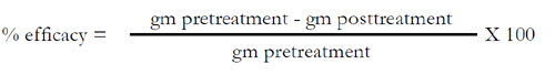
Data analysis followed method described by Fourie et al (Fourie et al, 2007). That is: The number of mites and eggs counted in the five skin scrapings were combined for each dog and served as the primary variable for treatment success. Percent efficacy, relative to pretreatment and based on geometric means (gm), was calculated from mite counts as follows:
For treatment efficacy, only dogs assessed at the posttreatment evaluation were used in the pretreatment calculation. The treatment success for each group was calculated from the proportion of dogs with no live mites or eggs and expressed as a percentage. The occurrence of clinical signs of mange was calculated as the percentage of dogs positive for each sign.
A semi-quantitative assessment of hair regrowth was made relative to pretreatment for each post-treatment evaluation. The skin surface of the dogs on which hair regrowth occurred, compared to the pretreatment observations, was graded as < 50%, 50–90% and >90% hair regrowth occurred.
An overall success rate was calculated and was defined as a dog that complied with all of the following conditions: No live mites or eggs; A complete resolution of the occurrence of papules and crusts as assessed on Days 28 and 56; Markedly improved hair regrowth in area of former alopecia.
There only signs of mild depression on the day of treatment, and a transient and slight intestinal hyperperistalsis were observed in some dogs. All dogs recovered completely by day 2 after treatment.
All of the dogs were positive for live mites or mite eggs prior to the first treatment (Day -1). Mite and egg counts as well as clinical evaluations showed that treatment with milbemycin oxime (2 mg/kg) following three 7-day treatment interval regiments resulted in a rapid reduction in the signs of sarcoptic mange (Tables 1, 2). In the group MBO, post-treatment number of dogs with mites or mite eggs in skin scrapings between 2 (Day 28) to 0 (D42) of 50 dogs, mean mite counts varied between 0.2 (Day 28) and 0 (Day 56), while in the group Vehicle post-treatment number of dogs with mites or mite eggs in skin scrapings between 15 (Day 0) to 13 (D56) of 15 dogs, mean mite counts varied between 7.0 (Day 0) and 6.5 (Day 56). The mean mite counts from skin scrapings performed on days 14, 28, 42, and 56 demonstrated that, compared with the vehicle, milbemycin oxime treatment produced reductions of 83.3%, 96%, 100% and 100%, respectively. Five dogs and two dogs were positive for live mites or mite eggs on day 14 and day 28, resulting in 83.3% and 96% values (Table 1). Success rates (proportion of mite free dogs) of milbemycin oxime and vehicle treatment at the end of the study were 100% and 13.3%.
Analysis of variance showed that on day 0 (prior to treatment), the mean values were not significantly different between treatment (P=0.545). analysis of variance performed after treatment showed that on each assessment day, the mean values for the milbemycin oxime treatment was significantly lower than that for the vehicle treatment (P=0.000, P=0.000, P=0.000, and P=0.000 for days 14, 28, 42, and 56; Table 1). Analysis of variance, using Savage Scores, confirmed these statistical significances. Fisher’s Exact Test demonstrated that significantly fewer milbemycin oxime-treated dogs had mites detected on skin scrapings than did the vehicle-treated dogs since day14 (P=0.000) (Table 1).
a. Treatment were administered on days 0, 7 and 14. Skin scrapings were performed on days -1, 14, 28, 42, and 56;
b. Statistical significance of the difference in the presence or absence of mites milbemycin oxime and vehicle treatments,using Fisher’s Exact Test( two-tailed);
c. Percent of dogs that were mite free;
d. Reduction calculated relative to the vehicle groups’ mean count;
e. Statistical significance of the difference of in mean mite or mite egg values between milbemycin oxime and vehicle.
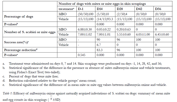

Table 1: Efficacy of milbemycin oxime against naturally acquired infestations of S. scabiei on dogs: summary of mean mite and egg counts in skin scrapings ( x‾ ±SD).
The presence of papules, encrustations, hyperkeratosis and hair regeneration was evaluated for all dogs by using clinical assessments made on days -1, 14, 28, 42 and 56 (Table 2). The prevalence of each sign assessed on each day showed marked trends in overall reductions in severity for milbemycin oxime-treated dogs incomparison with those receiving the vehicle only, as illustrated by the assessments on days -1, 14, 28, and 56 (Table 2). On day -1, papules were observed in at least 10% of animals; encrustations and hyperkeratosis were observed in at least 66% of both vehicle- and milbemycin oxime-treated dogs. As the studies progressed, clinical signs of sarcoptic mange and alopecia were markedly reduced following milbemycin oxime treatment. At the end of the study, papules, encrustations and hyperkeratosis has almost disappeared on Day 56, hair regrowth of all the dogs achieved 90%. However, there was no improvement in the vehicle-treated dogs on day 56, clinical signs were still evident in those dogs.
a. Measure of hair regrowth relative to Day-1 (reduction in the relative area affected by alopecia)
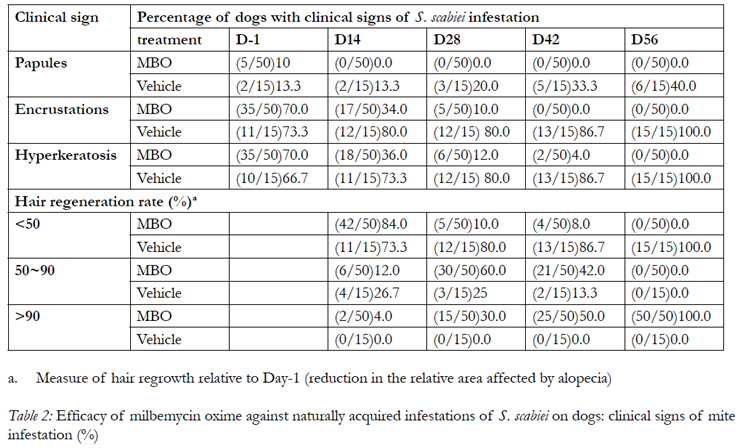
Table 2: Efficacy of milbemycin oxime against naturally acquired infestations of S. scabiei on dogs: clinical signs of mite infestation (%).
Discussion and Conclusion
The study was designed to demonstrate the efficacy of milbemycin oxime against infestations of S. scabiei on dogs under field conditions, on the basis of skin scriapings, no S.scabiei mites or eggs were recovered from milbemycin oxime–treated dogs after three doses, at 7-day interval, was highly effective in the treatment of naturally acquired infestations of S. scabiei on dogs. There was a 100% reduction in mean mite counts from deep skin scripings by 42 days after the first application. The treatments were well tolerated and safe. The quantitative mite count data were supported by the assessment of clinical signs associated with canine sarcoptic mange, and milbemycin oxime was shown to markedly reduce the prevalence of these signs compared with their prevalence in vehicle-treated dogs. The results of this and other studies (Paradis, 1998; Mueller et al, 1999) show that the agents used as positive controls are efficacious in the treatment of S. scabiei infestation in dogs when used according to their label recommendations, although repeated treatment may be necessary for control of the infestation. Milbemycin oxime also has been demonstrated to be highly effective against naturally acquired infestations of S. scabiei in dogs in other clinical studies following oral application (De Jaham and Henry, 1995; Miller et al, 1996).
Signs of toxicosis of avermectins are neurological and can include ataxia, abnormal behaviour, tremors, mydriasis, lethargy, weakness, recumbency, apparent blindness, hypersalivation/ptyalism, coma, and death (Lovell, 1990). Milbemycin oxime in the treatment for demodicosis can lead to lethargy and vomiting (Garfield and Reedy, 1992; Mueller and Bettenay, 1995; Paradis, 1999). During the study, there only signs of mild depression on the day of treatment, and a transient and slight intestinal hyperperistalsis were observed in some dogs. All dogs recovered completely by day 2 after treatment.
Milbemycin oxime has been shown to have a broad range of efficacy against endo- and ecto-parasites of dogs. The results of the study presented here indicate that weekly administration of milbemycin oxime is highly effective against natural infestations of S. scabiei in dogs.
This study showed that treatment with milbemycin oxime at the proposed minimum commercial dose rate applied at 7-day intervals rapidly reduced Sarcoptes mite infestations on dogs and resulted in a marked clinical improvement in the symptoms of mange. Milbemycin oxime pellet can potentially be used as a safe therapeutic agent for sarcoptic mange control in dogs.
Sources of Funding
This work was financially supported by the “Eleventh Five-Year” National Science and Technology Support Program( No. 2006BAD31B02).
Acknowledgement
The authors acknowledge Mr. Zhang Yaoteng( Northeast Agricultural University) for technical support. The veterinary drug, MBO, was kindly provided by Zhejiang Hisun Pharmaceutical Co. Ltd..
References
- Bergvall: Clinical efficacy of milbemycin oxime in the treatment of canine scabies: a study of 56 cases. Vet Dermatol 9: 231-233, 1998.
- Blagburn BL, DillonAR, Arther RG, et al: Comparative efficacy of four commercially available heartworm preventive products against the MP3 laboratory strain of Dirofilaria immitis. Vet Parasitol 176: 189–194, 2011.
- Blagburn BL, Hendrix CM, Lindsay DS, et al: Efficacy of milbemycin oxime against naturally acquired or experimentally induced Ancylostoma spp and Trichuris vulpis infections in dogs. Am J Vet Res 53: 513-516, 1992.
- Bond R: Diagnosis and treatment of canine scabies. In Pract 20: 308–315, 1998.
- Bowman DD, Johnson RC, Hepler DI: Effects of milbemycin oxime on adult hookworms in dogs with naturally acquired infections. Am J Vet Res 51: 487-490, 1990.
- Bredal W: Use of milbemycin oxime in the treatment of dogs with nasal mite (Pneumonyssoides caninum) infection. J Small Anim Pract 39: 126-130, 1998.
- Curtis CF: Current trends in the treatment of Sarcoptes, Cheyletiella and Otodectes mite infestations in cats and dogs. Vet Dermatol 15:108–114, 2004.
- Das SS: Effect of a herbal compound for treatment of sarcoptic mange infestations on dogs. Vet Parasitol 63: 03-306, 1996.
- De Jaham C, Henry CJ: Treatment of canine sarcoptic mange using milbemycin oxime. Can Vet J 36:42–43, 1995.
- Doering GG, Jensen HE( ed): Clinical Dermatology of Small Animals: A Stereoscopic Presentation. C.V. Mosby, Saint Louis, MO, 1973.
- Fourie LJ, Heine J, Horak IG: The efficacy of an imidacloprid/ moxidectin combination against naturally acquiredSarcoptes scabiei infestations on dogs. Aust Vet. J 84: 17-21, 2006.
- Fourie LJ, Kok DJ, Plessis A du, et al: Efficacy of a novel formulation of metaflumizone plus amitraz for the treatment of sarcoptic mange in dogs. Vet Parasitol 150: 275–281, 2007.
- Fthenakis GC, Papadopoulos E, Himonas C, et al: Efficacy of moxidectin against sarcoptic mange and effects on milk yield of ewes and growth of lambs. Vet Parasitol 87: 207–216, 2000.
- Garfield M, Reedy L: The use of oral milbemycin oxime (interceptor R) in the treatment of chronic generalized canine demodicosis. Vet Dermatol. 3: 231–235, 1992.
- Holm BR: Efficacy of milbemycin oxime in the treatment of canine generalized demodicosis: a retrospective study of 99 dogs (1995- 2000). Vet Dermatol 14: 189-195, 2003.
- Humbert-Droz E, Büscher G, Cavalleri D: Efficacy of milbemycin oxime against fourth-stage larvae and adults of Ancylostoma tubaeforme in experimentally infected cats. Vet Rec 154: 140-143, 2004.
- Kemp DJ, Walton SF, Harumal P, et al: The scourage of scabies. Biologist 49: 19–24, 2004.
- Krieger K, Heine J, Dumont P, et al: Efficacy and safety of imidacloprid 10% plus moxidectin 2.5% spot-on in the treatment of sarcoptic mange and otoacarosis in dogs: results of a European field study. Parasitol Res 97: S81–S88, 2005.
- Lovell RA, et al: Ivermectin and piperazine toxicoses in dogs and cats. Vet Clin N Am 20: 453–468, 1990.
- Miller WH, DeJaham C, Scott DW, et al: Treatment of canine scabies with milbemycin oxime. Can Vet J 37: 219–221, 1996.
- Mueller RS, Hastie K, Bettenay SV: Daily oral ivermectin for the treatment of generalised demodicosis in the dog- 23 cases. Aust Vet Pract29: 132–137, 1999.
- Niamatali S, Bhopale V, Schad GA: Efficacy of milbemycin oxime against experimentally induced Ancylostoma caninum and Uncinaria stenocephala infections in dogs. J Am Vet Med Assoc 201: 1385-1387, 1992.
- Paradis M: Ivermectin in small animal dermatology. Part II. Extralabel applications. Comp Cont Educ Pract Vet 20: 459–469, 1998.
- Paradis M: Newapproaches to the treatment of canine demodicosis. Vet Clin N Am Small 29: 1425–1436, 1999.
- Reichard MV, Wolf RF, Carey DW, et al: Efficacy of fenbendazole and milbemycin oxime for treating baboons (Papio cynocephalus anubis) infected with Trichuris trichiura. J Am Assoc Lab Anim Sci 46: 42-45, 2007.
- Shanks DJ, McTier TL, Behan S, et al: The efficacy of selamectin in the treatment of naturally aquired infestations of Sarcoptes scabiei on dogs. Vet Parasitol 91: 269–281, 2000.
- Six RH, Clemence RG, Thomas CA, et al: Efficacy and safety of selamectin against Sarcoptes scabiei on dogs and Otodectes cynotis on dogs and cats presented as veterinary patients, Vet Parasitol 91: 291–309, 2000.
- Snyder DE, Wisema S, Cruthers LR, et al: Ivermectin and Milbemycin Oxime in Experimental Adult Heartworm (Dirofilaria immitis) Infection of Dogs. J Vet Intern Med 25: 61–64, 2011a.
- Snyder DE, Wiseman S, Bowman DD, et al: Assessment of the effectiveness of a combination product of spinosad and milbemycin oxime on the prophylaxis of canine heartworm infection. Vet Parasitol, 180: 262–266, 2011b.
- Snyder DE, Wiseman S: Dose confirmation and non-interference evaluations of the oral efficacy of a combination of milbemycin oxime and spinosad against the dose limiting parasites, adult cat flea (Ctenocephalides felis) and hookworm (Ancylostoma caninum), in dogs. Vet Parasitol, 184: 284–290, 2012.
- Sosna CB, Medleau L: Treating parasitic skin conditions. Vet Med 573–586, 1992.
- Stewart VA, Hepler DI, Grieve RB: Efficacy of milbemycin oxime in chemoprophylaxis of dirofilariasis in cats. Am J Vet Res 53: 2274- 2277, 1992.
- Tabassam SM, Iqbal Z, Jabbar A, et al: Efficacy of crude neem seed kernel extracts against natural infestation of Sarcoptes scabiei var. ovis, J Ethnopharmacol 115: 284–287, 2008.
- Tagawa M, Okano S, Hayashi Y, et al: Prophylactic effect of milbemycin oxime against Dirofilaria immitis infection in dogs: optimum dose and administration time. J Vet Med Sci 55: 693-694, 1993.
- Tsukamoto Y, Sato K, Mio S, Sugai S, et al: Synthesis of 5-keto- 5-oxime derivatives of milbemycins and their activities against microfilafiae. Agric Biol Chem 55: 2615-2621, 1991.
- Wade CG, Mercer SH, Hepler DI, et al: Effect of milbemycin oxime against Ancylostoma caninum in dogs with naturally acquired infection. Am J Vet Res 52: 951-953, 1991.
- Wagner R, Wendleberger U: Field efficacy of moxidectin in dogs and rabbits naturally infested with Sarcoptes spp., Demodex spp. and Psoroptes spp. mites. Vet Parasitol 93: 149–158, 2000.
- White SD, Rosychuk RA, Fieseler KV, et al: Clinicopathologic findings, sensitivity to house dust mites and efficacy of milbemycin oxime treatment of dogs with Cheyletiella sp. Infestation. Vet Dermatol 12: 13-18, 2001.
- William H, Miller Jr, Caroline de, Jaham. Treatment of canine scabies with milbemycin oxime. Can Vet J 37: 219-221, 1996.


