Obtaining Mesenchymal Stem Cells From Adipose Tissue Of Murin Origin: Experimental Study
Altomare R1, Cannella V3*, Abruzzo A1, Palumbo VD1,2,4,6, Damiano G2,4, Spinelli G2,4, Ficarella S4, Cicero L3, Cassata G3, Di Bella S3, Di Marco P3,Purpari G3, Gioviale MC4, Damiani F1,4, Sinagra E1, Pisano C4, Marino A5, LO Monte G5, Tomasello G2,4,6, Guercio A3, Lo Monte AI2,4
1 Phd School in Surgical Biotechnology and Regenerative Medicine. School of Medicine – School of Biotechnology, University of Palermo, Italy.
2 DICHIRONS Department, School of Medicine. University of Palermo. Italy.
3 Istituto Zooprofilattico Sperimentale della Sicilia A.Mirri, Palermo, Italy
4 AUOP “P. Giaccone”, Universitary Hospital, Palermo, Italy.
5 School of Biotechnology, University of Palermo, Italy.
6 Euromediterranean Institute of Science and Technology (IEMEST), Palermo, Italy.
*Corresponding Author
Vincenza Cannella,
Executive-cell culture lab, Diagnostic Virologic Area,
Istituto Zooprofilattico Sperimentale (IZS) della Sicilia A.Mirri,
via Gino Marinuzzi 3,90129, Palermo, Italy.
E-mail: vincenza.cannella@izssicilia.it
Recieved: September 26, 2014; Accepted: November 05, 2014; Published: November 06, 2014
Citation: Cannella V, et al., (2014) Obtaining Mesenchymal Stem Cells from Adipose Tissue of Murin Origin: Experimental Study. Int J Stem Cell Res Transplant. 2(5), 86-90. doi: dx.doi.org/10.19070/2328-3548-1400014
Copyright: Cannella V© 2014. This is an open-access article distributed under the terms of the Creative Commons Attribution License, which permits unrestricted use, distribution and reproduction in any medium, provided the original author and source are credited.
Abstract
The aim of this study was to isolate and characterize rat Adipose Derived Mesenchymal Stem Cells (AD-MSCs) in order to evaluate their proliferative potential and their ability to differentiate in different cell types. AD-MSCs and Derived Mesenchymal Stem Cells (BM-MSCs) have the same characteristics in terms of plasticity. The advantage of adipose tissue is that it is an easier accessible source and it offers a large amount of MSCs by less invasive surgical tecniques. MSCs were obtained from subcutaneous adipose tissue of Wistar rats. First of all microbiological controls were made to exclude the presence of bacteria or fungi in the tissue. Adipose tissue was mechanically and enzimatically fragmented and stomal cell fraction was seeded in adherent culture flasks in DMEM 20% FBS. After 48h the medium was replaced. Cells were characterized by evaluating:
1) their ability to adhere to the plastic;
2) the clonogenic potential by Colony Forming Unit (CFU) assay;
3) their ability to differentiate in 3 mesodermal lineages (adipocytes, osteocytes and chondrocytes).
AD-MSCs are able to differentiate in adipocytes, osteocytes and chondrocytes as confirmed by Oil Red’O staining, von Kossa staining and histological analysis respectively. This first characterization is essential for the second part of our study in which we are planning to use AD-MSCs in vivo to restore renal function after an induced ischemic damage in experimental animals.
2.Introduction
3.Materials and Methods
3.1.Isolation of MSCs from Adipose Tissue
3.2.CFU Assay
3.3.Differentiation of AD-MSCs (Osteogenesis, Chondrogenesis And Adipogenesis)
3.3.1.Osteogenesis
3.3.2.Chondrogenesis
3.3.3.Adipogenesis
4.Results and Discussion
4.1.Isolation and Culture of Mesenchymal Stem Cells from Adipose Tissue
4.2.Microbiological Controls
4.3.MSCs Characterization
4.3.1.CFU Assay
4.4.Differentiation of MSCs
4.4.1.Osteogenesis
4.4.2.Condrogenesis
4.4.3.Adipogenesis
5.References
Keywords
Adipose tissue, Mesenchymal Stem Cells, Regenerative Medicine.
Introduction
Stem cells are primitive not specialized cells, able to regenerate themselves and to differentiate into specific cell types [1]. According to their differentiative ability, stem cells are classified in totipotent (isolated by the first phases of the embryo development until the eight-cell stage), able to give rise to all cell types of the organism, including extra-embryonic tissues [1,2]; pluripotent, isolated from the inner cell mass of the blastocyst and able to differentiate into cell types derived from the three germ layers (endoderm, mesoderm and ectoderm) but not in extra-embryonic tissues [2]; multipotent (identified in the fetus and in the adult tissues), capable of generating a limited number of cell types, restricted to a single germ layer; unipotent, able to generate a single cell type [3]. According to the stage of development of the organism from which they are obtained, stem cells can also be classified in embryonic and adult stem cells [1].
Embryonic stem cells (ESC), pluripotent, have a high grade of plasticity and self-renewal, with an important therapeutic potential, but may form tumors when injected and may determine host immune rejection . Moreover, their use opens a wide spectrum of ethical concerns [4]. Adult stem cells (ASC), multipotent, have a more limited differentiation and proliferative potential compared to ESC but do not present ethical problems. ASCs are naturally present in each organism during the whole life and their role is repairing tissues after injuries and replace old cells [1,2].
Among ASCs, mesenchymal stem cells (MSCs), multipotent, represent a promising tool in clinical applications for their differentiative potential towards mesoderm-derived lineage (liver, kidney, muscle, epithelial, neuronal and cardiac cells) [5].
The International Society for Cell Therapy (ISCT) proposed a set of standard criteria to identify MSCs:
(a) plastic adherent ability;
(b) expression of CD73, CD90 and CD105 and lack the expression of CD14, CD19, CD31,CD34,CD45 and HLA-DR surface molecules;
(c) mesodermal differentiation capability into osteoblasts, chondrocytes and adipocytes and
(d) immunomodulatory functions.
Considering this assumption it is possible to distinguish mesenchymal stem cells from other cells thanks to their fibroblastoid morphology and their ability to grow adherent to plastic in culture.
These cells can be easily detected through the assessment of their immunophenotype and the absence of positivity for all hematopoietic markers (CD34,CD45,CD11b,CD33,CD117 and HLADR). Investigators often use immunomagnetic beads or flow citometry to select this subpopulation of cells [6].
In clinical practice, the main source of MSCs is the adult bone marrow [7]. However, cultures of mesenchymal cells derived from bone marrow is associated to several problems: it is a weak accessible source and it involves the use of invasive techniques. The percentage of MSCs in the bone marrow is very low (0.001- 0.01%) and it decreases with ageing. For these reasons it is important to find alternative sources of MSCs [8], such as adipose tissue, peripheral blood or foetal tissues [5,9].
Adipose tissue is well distributed in the body and cells isolated form this source (Adipose Derived Mesenchymal Stem Cells, AD-MSCs) have the same characteristics as Bone Marrow derived MSCs [10] as they are able to grow on plastic surface, they show fibroblastoid morphology, they are able to generate Colony Forming Units (CFU), they express typical MSCs markers and they are able to differentiate into adipocites, chondrocites and osteoblasts [11,12]. MSCs can be characterized by the expression of specific surface markers, like CD105, CD73 and CD90, or by the expression of typical stem cell genes which are OCT-4 (Octamer- Binding Transcription Factor 4), MNANOG and SOX2 (SRY boxcontaining factor 2) [13].
AD-MSCs are a suitable choice for regenerative medicine because they can be used in authologous manner and they can be easily isolated in human and animals from subcutaneous adipose tissue to be harvested in vitro for different culture passages [14,15].
Selection of appropriate animal models for preclinical evaluations is crucial for optimization and validation of therapeutic protocols in regenerative medicine. For this reason, in vitro studies are necessary to future in vivo MSC applications. For this study we chose Rat model, which is an established small animal model for a large number of in vitro studies.
Materials and Methods
Subcutaneous fat was obtained from 20 Wistar breed male rats whose average weight was 350g. All animals were sedated with an intramuscular injection of midazolam and anesthesia was maintained with isoflurane and oxygen gas mixture administered with a mask. For adipose tissue procurement, a small incision of about 2 cm was performed at the root of animal’s thigh. Through blunt dissection, the fat was separated from the muscular layer and finally, excised. The skin was closed with single interrupted stitches.
Adipose tissue samples were carried to the laboratory into a sterile box containing HBSS (Hanks balanced salt solution; Gibco) supplemented with 5% antibiotics.
Following 1 hour of tissue recovery, samples were processed under sterile conditions. Before starting the cell isolation procedure, sterility controls on the adipose tissue were performed by using specific bacterial and fungi media such as Blood-Agar, Plate Count Agar (PCA) e Agar Sabouraud to be sure that no contamination events occurred during the sampling.
First of all adipose tissue sample was washed extensively (about 3 times) with HBSS enriched of 2% a penicillin-streptomycin in order to remove debris and blood. The piece was then weighed and placed in 90 mm petri plates with 15-20 ml of penicillin-streptomycin enriched saline solution to be mechanically fragmented into small pieces and subjected to further washes with the saline solution.
Fragmented sample was then placed in a flask with a solution of 0.2% collagenase type IA in PBS (20 ml of collagenase per gram of adipose tissue to digest) supplemented with 1% antibiotics (penicillin, streptomycin and amphotericin) . Enzymatic digestion was performed in thermostatic bath at 37°C under stirring for 2-3 hours until the complete disruption of the extracellular matrix and the release of the cellular fraction.
Standard cell culture medium consisting of Dulbecco’s Modified Eagle Medium (DMEM) supplemented with 10% FBS and 1% penicillin-streptomycin was then added for neutralizing the activity of the enzyme.
The digested tissue was filtered with a sterile gauze, placed in 50ml tubes and centrifuged at 2700g for 10 minutes at room temperature.
The supernatantwas therefore aspirated and the cell pellet obtained was resuspended in 10 ml of complete medium (Dulbecco low glucose + 20% SFB + 1% antibiotic-antimycotic), centrifuged 2 times at 1400g for 10 minutes at room temperature to remove any residual enzymatic solution.
After spinning, cell suspension was resuspended in 10 ml of complete medium, filtered through a cell strainer (pore size 70 μm) and centrifuged. Sterility controls were performed again as described above to be sure that no contamination events occurred during the isolation procedure.
Cell suspension obtained was added with 3ml of DMEM and cells were counted in a Burker Camera by using Trypan Blue. Cells were then plated in 25 cm2 culture flasks in DMEM-Low Glucose medium supplemented with glutamine, foetal bovine serum (FBS) and 1% penicillin-streptomycin. Cultures were maintained in an incubator with an humidified atmosphere containing 5% CO2. The CO2 incubator has a constant temperature of 37°C, ideal for human and animal cells, and a relative humidity of 100 % to prevent evaporation of culture medium.
Medium was changed after 48h incubation in order to remove the non-adherent fraction, consisting of hematopoietic cells, debris and other cell types not able to adhere to the plastic.
When cells reached 80% confluence, subcultures were performed by using trypsine-EDTA enzyme to remove adherent cells from the flask.
The identity of AD-MSCs was verified by their ability to attach to the plastic surface of culture flasks, form CFU (colony-forming units) and differentiate into cells of mesodermal lineages: chondrocytes, adipocytes and osteocytes. The presence of transcription factors indicative of self-renewal and undifferentiation was also investigated.
The colony forming efficiency on plastic was assayed by plating isolated cells at three different seeding densities (150, 60 and 30 cells/cm2) in 6-well plates in DMEM Low Glucose with 5% FBS. Cells were incubated for 2 weeks in an incubator with humidified air and 5% CO2. The colonies were stained with Giemsa solution and scored.
Cells derived from subcutaneous and visceral fat and expanded in the DMEM Low Glucose medium with 20% FBS were used for differentiation studies. The MSCs were cultured in appropriate differentiation media to obtain the 3 mesodermal lineages (osteogenic, chondrogenic and adipogenic). All studies were carried out with the same number of controls.
Cells were plated at 4500 cells/cm2 on 6-well plates and treated with NH OsteoDiff Medium (Mylteny Biotec) for 3 weeks, with a medium change once every 3 days. von Kossa staining was used to detect calcified extracellular matrix deposits.
Micromass cultures of cells (1×10⁶ cells/ml) were incubated in 15 ml test tubes with NH Chondro Diff Medium (Mylteny Biotec) for 30 days, with a medium change once every 3 days. The micromasses were fixed and stained with hematoxylin-eosin
Cells were plated at 7500 cells/cm2 on 6-well plates, and treated with Complete MesenCult Adipogenic Medium (Stemcell Technologies) for 21 days with a medium change once every 3 days. Oil Red O staining was used to examine lipid droplet formation.
Results and Discussion
MSCs were successfully isolated from adipose tissue; they reached the semiconfluence (80%) in 25 cm2 flasks in 5-6 days, maintaining the typical fibroblastoid appearance. We were able to obtain a significant amount of cells (3x10⁶) in 5-6 days from a single adipose tissue sample.Subcultures were made until it was possible to observe the maintenance of the characteristic phenotype and appreciable levels of vitality in culture. For some samples, it was possible to observe cell growth up to the tenth passage, the other samples were observed until the fifth subculture.
The microbiological controls, performed on the samples of adipose tissue and on cells performed after the extraction protocol, were negative for contamination by bacteria and fungi. It has therefore confirmed that the tissue procurement and handling of cell cultures were performed under sterile conditions.
The clonogenic assay for testing the efficiency of the cells in culture was performed on all samples at cell passages 1, 3, 5. Results obtained demonstrated that MSCs maintain clonogenic ability (figure 1).
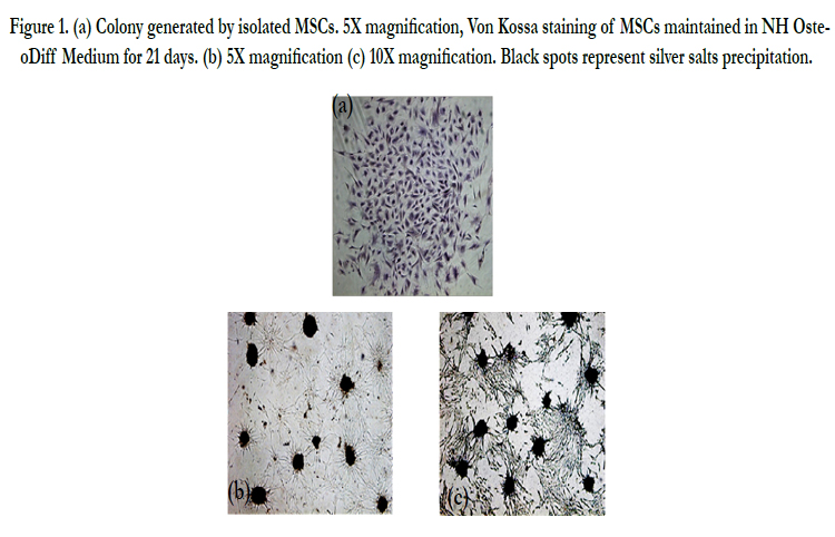
Figure 1. (a) Colony generated by isolated MSCs. 5X magnification, Von Kossa staining of MSCs maintained in NH OsteoDiff Medium for 21 days. (b) 5X magnification (c) 10X magnification. Black spots represent silver salts precipitation.
MSCs isolated show differentiation capacity towards the three mesodermal cell lines osteocitic, adipocytic, condrociti.
MSCs cultured in OsteoDiff Medium showed cuboidal phenotype, continue to actively proliferate and form cell aggregates. After "von Kossa" staining on cells induced to differentiate, it was notable the deposition of calcified extracellular matrix, displayed in black, derived from the precipitation of silver salts (figure 2). In control cells, coltured with maintenance medium, the phenotype did not change.
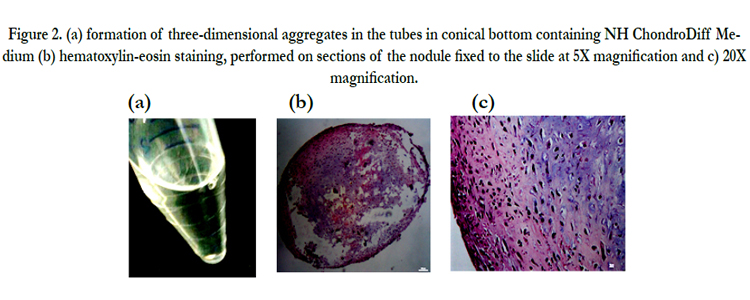
Figure 2. (a) formation of three-dimensional aggregates in the tubes in conical bottom containing NH ChondroDiff Medium (b) hematoxylin-eosin staining, performed on sections of the nodule fixed to the slide at 5X magnification and c) 20X magnification.
The formation of three-dimensional aggregates was observed in the tubes in conical bottom containing NH ChondroDiff Medium,(figure 2a) but not in control tubes with the same concentration of cells in maintenance medium.
The hematoxylin-eosin staining, performed on sections of thenodule fixed to the slide, showed that cells acquired a structural organization similar to that of cartilage (figure 2b).
After 4-5 days, MSCs cultured in Adipogenic Medium with MesenCult, formed numerous lipid accumulation in the cytoplasm (figure 3a). The increase in size of the lipid droplets was verified in subsequent observations. At day 21 after induction of differentiation Oil Red O and hematoxylin staining was performed to highlight the cell nuclei in blue and fat deposits in red (figure 3b).No differentiation was observed in control cells (figure 3c).
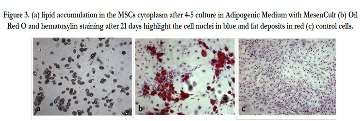
Figure 3. (a) lipid accumulation in the MSCs cytoplasm after 4-5 culture in Adipogenic Medium with MesenCult (b) Oil Red O and hematoxylin staining after 21 days highlight the cell nuclei in blue and fat deposits in red (c) control cells.
Mesenchymal stem cells isolated from adipose tissue show high proliferative potential in culture and ability to differentiate into different cell types. Adipose tissue is almost ubiquitously distributed, the levy provides minimally invasive techniques and is a source of stem cells phenotypically comparable to bone marrow MSCs, until now reputed the best source of choice for the supply of autologous stem cells.
In this work, we studied the characteristics of MSCs isolated from subcutaneous adipose tissue of Wistar rats. Cells were characterized by assessing the adherence to the plastic material, the fibroblast- like morphology, the clonogenic potential and the differentiation potential in mesodermal cell lines. MSCs isolated retain their typical fibroblastoid morphology and constant proliferation for at least 5 passages in basal culture medium.
The aim of this study, approved and granted by Italian Board of Health, was to isolate and characterize rat Adipose Derived Mesenchymal Stem Cells (AD-MSCs) in order to evaluate their proliferative potential and their ability to differentiate into different cell types [16]. This first characterization is essential for the second part of our study in which we are planning to use AD-MSCs in vivo to restore renal function after an induced ischemic damage in experimental animals [17]. Acute renal failure is a medical emergency; in Italy about 5 millions people are affected and there are 10.000 new cases every year. This disease is mostly caused by a damage of endothelial cells of the kidney which inhibits reperfusion and restoring of organ function. After an ischemic offense, Endothelial Progenitor Cells (EPCs) move from Bone Marrow (BM) to the damaged site [18]. However, subjects affected by chronic renal failure, have lost the regenerative ability of their kidney progenitor cells and of their BM which become aplastic [19]. For this reason it is important to find alternative sources of cells able to restore the damaged site and we are going to investigate the role of AD-MSCs.
As the future goal of the study is to use these cells in vivo, we wanted to test if it is possible to get an adequate amount of cells, with characteristics of stem cell, to avoid any mutations or alterations gene that may occur to proceed subcultures. Since the retrieval does not require the sacrifice of the animal, it can be concluded that the subcutaneous adipose tissue is preferable especially for the later stages of the in vivo study in which each animal could receive autologous MSCs. The possibility of using these cells in the field of cell therapy and regenerative medicine is reinforced by the fact that a significant number of cells can be obtained and that their use does not involve any kind of ethical or legal. The three-dimensional growth of these cells on scaffolds could be the challenge for the future to rebuild organs and tissues in the laboratory. [20-29].
References
- Zipori D (2005) The stem state: plasticity is essential, whereas self-renewal and hierarchy are optional. Stem Cells 23(6):719-726.
- Mitalipov S ,Wolf D (2009) Totipotency, Pluripotency and Nuclear Reprogramming. Adv Biochem Eng Biotechnol 114: 185–199.
- Rao MS (2004) Stem sense: a proposal for the classification of stem cells. Stem Cells Dev 13(5):452-455.
- Leeb C, Jurga M, McGuckin C, Forraz N, Thallinger C et al. (2011) New perspectives in stem cell research: beyond embryonic stem cells. Cell Prolif 44 (Suppl. 1), 9–14.
- Ma T (2010) Mesenchymal stem cells: From bench to beside. World Journal of Stem Cells 2(2):13-17.
- Dominici M, Le Blank K, Mueller, Slaper-Cortenbach I, Marini F et al (2006) Minimal criteria for defining multipotent mesenchymal stromal cells. The International Society for Cellular Therapy position statement. Cytotherapy 8(4):315-317
- Trounson A, Thakar RG, Lomax G, Gibbons D (2011) Clinical trials for stem cell therapies. BMC Medicine 9:52.
- Bellavia M, Altomare R, Cacciabaudo F, Santoro A, Allegra A et al (2013) Towards an ideal source of mesenchymal stem cell isolation for possible therapeutic application in regenerative medicine. Biomed Pap Med Fac Univ Palacky Olomouc Czech Repub 158(3):356-60
- Allegra A Altomare R, Curcio P, Santoro A, Lo Monte AI (2013) Gene expression of stem cells at different stages of ontological human development. Eur J Obstet Gynecol Reprod Biol.170(2):381-6.
- Hauner H (2004) The new concept of adipose tissue function. Physiol Behav 83(4):653-8.
- Rastegar F (2010) Mesenchymal stem cells: Molecular characteristics and clinical applications. World Journal of Stem Cells 2(4):67-80.
- Choi YH, Burdick MD, Strieter RM (2010) Human circulating fibrocytes have the capacity to differentiate osteoblast and chondrocytes. Int J Biochem Cell Biol 42:662-671.
- Gaiba S, Pereira de França L, Pereira de França J, Masako Ferreira L (2012) Characterization of human adipose-derived stem cells. Acta Cir Bras 27(7):471-476.
- MacArthur BD, Ma’aya A and Lemischka IR (2009) Systems biology of stem cell fate and cellular reprogramming. Nat Rev Mol Cell Biol 10(10):672–681.
- Beahm EK, Walton RL, Patrick CW Jr (2003) Progress in adipose tissue construct development. Clin Plast Surg 30(4):547-58.
- Konno M (2013) Adipose-derived mesenchymal stem cells and regenerative medicine. Development, Growth & Differentiation 55(3)309–318.
- Guercio A, Di Bella S, Casella S, Di Marco P, Russo C, et al. (2013) Canine mesenchymal stem cells (MSCs): characterization in relation to donor age and adipose tissue-harvesting site. Cell Biol Int 37(8):789-98
- Ye Y, Wang B, Jiang X, Hu W, Feng J et al. (2011) Proliferative capacity of stem/progenitor-like cells in the kidney may associate with the outcome of patients with acute tubular necrosis. Human Pathology 42(8):1132-41.
- Alejandro R (2009) Endotelial progenitor cells restore renal function in chronic experimental renovascular disease. Circulation 119:547-547.
- Lam AQ, Freedman BS, Bonventre JV (2014) Directed differentiation of pluripotent stem cells to kidney cells. Semin Nephrol 34(4):445-461.
- Lo Monte AI, Licciardi M, Bellavia M, Damiano G, Palumbo VD, et al. (2012) Biocompatibility and biodegradability of electrospun phea-pla scaffolds: Our preliminary experience in a murine animal model. Dig J Nanomater Biostruct 7(2):841-851.
- Jahani H, Jalilian FA, Kaviani S, Soleimani M, Abassi N, et al. (2014) Controlled surface morphology and hydrophilicity of polycaprolactone towards selective differentiation of mesenchymal stem cells to neural like cells. J Biomed Mater Res A [Epub ahead of print].
- Kim SJ, Park MH, Moon HJ, Park JH, Ko DY, et al. (2014) Polypeptide thermogels as a three dimensional culture scaffold for hepatogenic differentiation of human tonsil-derived mesenchymal stem cells. ACS Appl Mater Interfaces 6(19):17034-43.
- Kedong S, Wenfang L, Yanxia Z, Hong W, Ze Y, et al. (2014) Dynamic fabrication of tissue-engineered bone substitutes based on derived cancellous bone scaffold in a spinner flask bioreactor system. Appl Biochem Biotechnol 174(4):1331-43.
- Shim JB, Ankeny RF, Kim H, Nerem RM, Khang G (2014) Study of a three-dimensional PLGA sponge containing natural polymers co-cultured with endothelial and mesenchymal stem cells as a tissue engineering scaffold. Biomed Mater 9(4):045015.
- Rettinger CL, Fourcaudot AB, Hong SJ, Mustoe TA, Hale RG, et al. (2014) In vitro characterization of scaffold-free three-dimensional mesenchymal stem cell aggregates. Cell Tissue Res 358(2):395-405.
- Karam JP, Muscari C, Sindji L, Bastiat G, Bonafè F, et al. (2014) Pharmacologically active microcarriers associated with thermosensitive hydrogel as a growth factor releasing biomimetic 3D scaffold for cardiac tissue-engineering. J Control Release 192:82-94.
- Palumbo VD, Bruno A, Tomasello G, Damiano G, Lo Monte AI (2014) Bioengineered vascular scaffolds: the state of the art. Int J Artif Organs 37(7):503-12.
- Abruzzo A, Fiorica C, Palumbo VD, Altomare R, Damiano G, et al. (2014) Using polymeric scaffolds for vascular tissue engineering. Int J Polym Sci ,Article ID 689390, 9 pages.


