Solitary Cutaneous Myxoma in a Child: Rare Entity
Barbach Y1*, Mazti A2, Baybay H1, Chaouche M1, Cherif AD1, Hamas N2, Mernissi FZ1
1 Dermatology Department, University Hospital Hassan II, Fez, Morocco.
2 Anatomic Pathology Department, University Hospital Hassan II, Fez, Morocco.
*Corresponding Author
Younes Barbach,
Dermatology Department, University Hospital Hassan II, Morocco.
Tel: +212671797158
Fax: +212535613729
E-mail: dr.younes2011@gmail.com
Received: October 07, 2018; Accepted: November 26, 2018; Published: November 28, 2018
Citation: Barbach Y, Mazti A, Baybay H, Chaouche M, Cherif AD, Hamas N, et al., Solitary Cutaneous Myxoma in a Child: Rare Entity. Int J Pediat Health Care Adv. 2018;5(5):92-94. doi: dx.doi.org/10.19070/2572-7354-1800026
Copyright: Barbach Y© 2018. This is an open-access article distributed under the terms of the Creative Commons Attribution License, which permits unrestricted use, distribution and reproduction in any medium, provided the original author and source are credited.
Abstract
Solitary myxomas without other manifestations of Carney’s complex usually arise within the skeletal or cardiac muscles, and less commonly in the intestine, pelvis and subcutis, and very rarely in the skin. Solitary cutaneous myxomas (SCM), more descriptively called solitary superficial angiomyxomas, are uncommon tumours having characteristic histological findings. They occur most commonly on the trunk, leg, head and neck, and more rarely at acral sites of adults. They are characterized histologically by a well-defined, hypocellular, myxoid tumour with many vascular components in the dermis or subcutaneous fat. We report a case of solitary skin myxoma sitting on the scalp of a young child.
2.Abbreviations
3.Introduction
4.Observation
5.Discussion
6.Conclusion
7.References
Keywords
Cutaneous; Myxoma; Carney Complex.
Keywords
SCM: Solitary Cutaneous Myxomas.
Introduction
Solitary cutaneous myxoma (SCM), sometimes referred to as superficial angiomyxoma because of its vascular component, is relatively rare. They usually occur as an asymptomatic nodule with slow growth on the head, neck, and trunk usually in adults [1, 2]. Rarely, they locate at the acral level [2]. SCM should be distinguished from the complex of myxomas (cardiac, cutaneous and breast), speckled pigmentation (cutaneous and mucocutaneous) and endocrine hyperactivity (Cushing's syndrome, sexual precocity and acromegaly) described by Carney et al., [3].
We report a case of solitary cutaneous myxoma sitting on the scalp of a young child.
Observation
An 11-year-old child, with no notable pathological history, presented with swelling in the scalp for 3 years, with a very progressive increase in size. Dermatological examination showed the presence of a 1.5 cm nodule, pale pink in color, bilobed smooth surface, fixed in relation to the deep plane, painless and non-pruriginous. The diagnoses evoked were a keloid scar or a pilomatricoma. The histological study showed tumor proliferation in the superficial dermis made of fusiform stellate cells arranged on a loose and myxoid base with a fine vascularization. An immunohistochemical study made of PS100 came back negative. The diagnosis of benign cutaneous myxoma was retained. The child underwent surgical resection of the nodule, with a recoil of 10 months without recurrence.
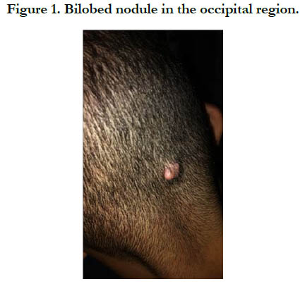
Figure 1. Bilobed nodule in the occipital region.
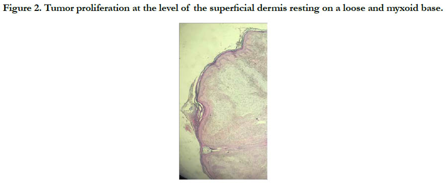
Figure 2. Tumor proliferation at the level of the superficial dermis resting on a loose and myxoid base.
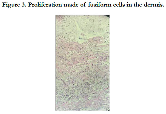
Figure 3. Proliferation made of fusiform cells in the dermis.
Discussion
SCM has been described as a "hairy flesh colored nodule" or as a "soft, lobulated nodule"[1]. Over the years, only a few cases have been reported of cutaneous solitary or multiple cutaneous myxomas without systemic features [1, 2]. Multiple cutaneous myxomas have generally been attributed to the Carney complex[4], NAME [5], or LAMB [6] syndromes. The cutaneous myxomas that accompany the Carney complex usually affect young patients, and are usually less than 1 cm in size. They are multiple and have a widespread distribution with a predilection for eyelids, ears and nipples. Usually, they are described as asymptomatic, sessile, subcutaneous papules or nodules with a non-ulcerated surface; rarely, they are rough and papillomatous. These cutaneous myxomas announce the presence of a potentially serious cardiac tumor, and after the excision, tend to the local recurrence, in spite of their benign nature. Solitary myxomas, without other manifestations of Carney Complex, are generally localized in the skeleton or cardiac muscle, less frequently in the intestines, the pelvis, subcutaneously [3], and very rarely in the skin. Dutz et al., [7] reported 15 myxomas from subcutaneous tissue in children, but not in the dermis. Other reports in the literature [8, 9] describe small lesions classified as cutaneous focal mucinosis, which are not considered to be true myxomas. They are characterized by local accumulations of mucin in the dermis with spindle-shaped cells, but with less vascularization than myxomas [2].
Solitary cutaneous myxomas are located in the dermis or subcutaneous tissue. They are generally well-limited, without peripheral capsule when they are localized at the level of the dermis and sometimes encapsulated when they are located at the subcutaneous fat level. The stroma is very hypocellular, myxoid, and contains mucopolysaccharides. Within this stroma, fusiform and star-shaped cells with oval nuclei and poorly defined cytoplasm are present [10]. Other lesions include myxoid deposits such as myxoid neurofibroma, neurotheleomeoma and fibromyxoid tumor (ossifying and non-ossifying) which must be differentiated from cutaneous myxoma through histological examination with immunohistochemical study. We describe a case of solitary cutaneous myxoma on the scalp of an 11-year-old child with no other features of the Carney complex, no evidence of cardiac myxoma, and no family history of this autosomal dominant disorder. No evidence of local recurrence is noted 10 months after surgical removal of the tumor.
Conclusion
Although it is necessary for the clinician and the pathologist not o misunderstand the association between cutaneous myxoid tumors and cardiac myxomas given the seriousness of this table considered as a potentially fatal risk, we would like to emphasize the occurrence of sporadic cases of cutaneous myxomas as an ntity in itself, and to distinguish it from other cutaneous myxoid neoplasms.
References
- Choi HJ, Kim YJ, Yim JH, Kim MY, Kim HO, Park YM. Unusual presentation of solitary cutaneous myxoma. J Eur Acad Dermatol Venereol. 2007 Mar;21(3):403-4. PubMed PMID: 17309474.
- Alaiti S, Nelson FP, Ryoo JW. Solitary cutaneous myxoma. J Am Acad Dermatol. 2000 Aug;43(2 Pt 2):377-9. PubMed PMID: 10901728.
- Armstrong DKB, Irvine AD, Handley M, Walsh MY, HADDENj DR, Bingham EA. Carney complex: Report of a kindred with predominantly cutaneous manifestations. Br J Dermatol. 1997 Apr;136(4):578-82. PubMed PMID: 9155962.
- Carney JA, Gordon H, Carpenter PC, Shenoy BV, Go VL. The complex of myxomas, spotty pigmentation, and endocrine overactivity. Medicine (Baltimore). 1985 Jul;64(4):270-83. PubMed PMID: 4010501.
- Atherton DJ, Pitcher DW, Wells RS, MacDonald DM. A syndrome of various cutaneous pigmented lesions, myxoid neurofibromata and atrial myxoma: the NAME syndrome. Br J Dermatol. 1980 Oct;103(4):421-9. PubMed PMID: 7437308.
- Rhodes AR, Silverman RA, Harrist TJ, Perez-Atayde AR. Mucocutaneous lentigines, cardiomucocutaneous myxomas, and multiple blue nevi: the ‘LAMB’ syndrome. J Am Acad Dermatol. 1984 Jan;10(1):72-82. PubMed PMID: 6693605.
- Dutz W, Stout AP. The myxoma in childhood. Cancer. 1961 May- Jun;14:629-35. PubMed PMID: 13725297.
- Kuo KL, Lee LY, Kuo TT. Solitary cutaneous focal mucinosis: A clinicopathological study of 11 cases of soft fibroma-like cutaneous mucinous lesions. J Dermatol. 2017 Mar;44(3):335-338. doi: 10.1111/1346-8138.13523. PubMed PMID: 27450934.
- Gutte R, Garg G, Kharkar V, Khopkar U. Asymptomatic nodule over the shin. Cutaneous focal mucinosis (CFM). Indian J Dermatol Venereol Leprol. 2012 Jan-Feb;78(1):123. doi: 10.4103/0378-6323.90978. PubMed PMID: 22199090.
- Abarzúa-Araya A, Lallas A, Piana S, Longo C, Moscarella E, Argenziano G. Superficial angiomyxoma of the skin. Dermatol Pract Concept. 2016 Jul 31;6(3):47-9. doi: 10.5826/dpc.0603a09. PubMed PMID: 27648383.


