High Resolution TEM Characterisation of Hydrogen Peroxide Treated Tooth Structures
Mohan V1*, Young ND1, Kaiser M2, Bolten H2
1 Oral Healthcare, Philips Research Laboratories, Cambridge, United Kingdom.
2 Philips Innovation labs, Material Analysis, Eindhoven, The Netherlands.
*Corresponding Author
Veena Mohan,
Oral Healthcare, Philips Research Laboratories,
Cambridge, United Kingdom.
E-mail: veena.mohan@philips.com
Received: January 22, 2016; Accepted: February 18, 2016l; Published: February 19, 2016
Citation: Mohan V, Young ND, Kaiser M, Bolten H (2016) High Resolution TEM Characterisation of Hydrogen Peroxide Treated Tooth Structures. Int J Dentistry Oral Sci. S7:001, 1-7. DOI : dx.doi.org/10.19070/2377-8075-SI07001
Copyright: Mohan V© 2016. This is an open-access article distributed under the terms of the Creative Commons Attribution License, which permits unrestricted use, distribution and reproduction in any medium, provided the original author and source are credited.
Abstract
Objective: Previous studies reported that bleaching agents are capable of altering the outer enamel or exposed dentine surfaces. In this study, transmission electron microscopy (TEM) characterisation in combination with energy dispersive x-ray spectroscopy (EDS) elemental mapping was performed to investigate the effect of hydrogen peroxide (HP) on the sub-surface of tooth structures.
Materials and Methods: Bovine incisors (n=6) were assigned to three groups for treatments: 30% H2O2 pH 3, 30% H2O2 pH 7 and 1M sodium hydroxide (NaOH) for 16 hours. Samples were exposed to direct treatment with agents to allow easy access of the agents and uniform treatment across the entire sample sub-surfaces. Specimens were immersed in Karnovsky’s fixative for three days, embedded in epoxy and polished. Using focussed ion beam (FIB), samples were milled ~50-100 μm below the treated polished surface to feature the ultra-fine structures for TEM analysis.
Results: Enamel rods, inter-rods and crystalline structures within rods were discerned for sound and H2O2 treated enamel. No structural difference in the mineral dense peritubular region or fibrous protein rich inter-tubular dentine was observed after acidic or neutral H2O2 treatment. A complete loss in the structural integrity of enamel rod was observed after NaOH treatment for 16 hours without having any impact on dentine structure.
Conclusions: TEM analysis produced high quality sub-surface images of tooth structures and revealed no deleterious effect on the structural integrity of the sub-surface enamel or dentine after direct bleaching with hydrogen peroxide.
2.Materials and Methods
2.1 Specimen preparation
2.2 Chemical solutions for treatment
2.3 Dental sample preparation for TEM examination
2.4 Mechanical polishing
2.5 Focused Ion Beam milling
2.6 Microscopic and elemental analysis
3.Results
3.1 Effect of treatments on enamel
3.2 Effect of treatments on dentine
3.3 Energy Dispersive X-ray Spectroscopy (EDS) mapping
4.Discussion
5.Acknowledgements
6.References
Introduction
In recent years, aesthetic dental treatments such as tooth whitening has gained popular demand from patients and established as a rapidly growing field of dentistry. Consumers are more conscious of their image and believe in a brighter smile as a way of maintaining beauty and health. Bleaching of teeth has a long history since 1864 and the observation of tooth lightening with Carbamide peroxide in 1989 marked the beginning of using 10% CP in a mouth guard [1].
The fundamental bleaching procedure to bleach the discoloured teeth exists in the form of dentist-supervised night guard bleaching, in-office bleaching, and OTC bleaching [2]. Various forms of OTC bleaching products are available in market in the form of gels, paint on brush, mouth rinses, chewing gums, tooth pastes, strips etc. The choice of procedure and resulting efficacy of the whitening treatment depends on many factors such as origin or cause of tooth discoloration, type of discolouration, location and depth of stain residing tooth, affinity of the stain to enamel and dentine tissues, age and gender of patient, concentration and time of exposure to the bleaching agent.
Today many tooth whitening products are available in market with a range of active agents including hydrogen peroxide, carbamide peroxide, sodium perborate, chlorite and others. These products have been used alone or in combination with activators to increase the efficacy of whitening.
Hydrogen peroxide is the most commonly used bleaching agent of this era [3]. It decomposes and produces high oxidising potential free radicals, perhydroxyl anions, which affect pigment macromolecules and resin matrices [4]. Although enamel is a hard and dense tissue, its highly porous structure enables hydrogen peroxide to penetrate this layer by diffusion [5]. Jiang et al. noted that hydrogen peroxide attacks the mineral and organic matrix of the dentine layer, and hypothesize that the organic components are removed, and the mineral components collapse and form a layer to protect the underlying dentine layers from any further attack [6].
To date, most of the studies reported on mineral content, surface morphology, surface and sub-surface micro hardness and other mechanical properties associated with hydrogen peroxide treatment utilised techniques such as scanning electron microscopy, atomic force microscopy, confocal laser microscopy, attenuated total reflection-Fourier transform infrared spectroscopy and Raman spectroscopy [7-17]. The combination of using focussed ion beam transmission electron microscopy (FIB-TEM) for studying the mineralized dentine has been reported over the years [18, 19]. To our knowledge limited information is available on the use of high resolution (TEM) for examining the sub surface microstructure of enamel and dentine after hydrogen peroxide treatment.
Compared to US, tooth whitening in Europe is not very popular and this might be because of the lingering concerns about the safety associated with whitening procedures [2]. Several studies have been conducted to evaluate the safety of using tooth whitening products based on carbamide peroxide and hydrogen peroxide. It has been suggested that considering the dosage and application mode of tooth bleaching, the exposure to the material during the bleaching procedure is inadequate to cause acute systemic toxicity [20].
A review on the safety of using hydrogen peroxide based materials confirmed that there is no evidence of significant health risks associated with tooth whitening and any such potential adverse effects occur with the in-appropriate usage of whitening products for tooth whitening [21]. It was also reported that the peroxide containing products and solutions have no significant deleterious effect on tooth surface morphology, chemistry, surface and subsurface micro hardness or ultra-structure. In contrast, the studies that showed an effect of peroxide have limitations in their method of not considering the in vivo situations of saliva remineralisation or using products that have acidic pH [22]. Considering the disparities relating to the safety of hydrogen peroxide, a wellcontrolled fundamental study is essential to gain insights into the impact of peroxide on the ultra-fine structure of teeth.
The aim of this study is to investigate the effect of hydrogen on teeth down to nanometre level using high resolution transmission electron microscopy to evaluate the safety of performing peroxide based tooth whitening procedures and any associated risks. In a previous study reported by Eimar et al. (2012), sodium hydroxide was used as a deproteinizing agent for studying the mechanism of whitening agents on teeth. Therefore, the effect of hydrogen peroxide on the organic content of tooth was tested by comparing the samples treated with deproteinizing solution of sodium hydroxide. EDS was used to perform elemental analysis of enamel and dentine on controls and treated sample with agents.
Materials and Methods
The study used non carious BSE certified bovine fragments of dimensions 10 x 10 mm without any cracks or defects. The hydrated teeth samples were sectioned (low speed saw, Isomet) from the crown to the root at a distance of 1-1.5mm from the labial aspect of tooth. Each tooth was cut into two equal parts to serve samples for treatment and internal control. Samples were polished, etched with 10% citric acid for 30 seconds and cleaned in ultrasound bath. The untreated control samples for each treatment were stored in PBS at 37°C for 16h.
The following solutions were prepared for treating the samples with enamel and dentine.
- 30% hydrogen peroxide solution (HP) pH 3: sample was immersed in 30% w/w HP solution, (Sigma Aldrich, UK) for 16h at 37°C.
- 30% hydrogen peroxide solution pH 7: pH of the 30% HP solution was adjusted to 7 using 1M sodium hydroxide (NaOH) and the sample was immersed in the solution for 16h at 37°C.
- Deproteinizing solution: 1M NaOH solution (Sigma Aldrich, UK) was prepared and the sample was immersed in the solution for 16h at 37°C.
The GP subgroup, created in order to verify the efficacy of the physiotherapy in association with RADICA, has been evaluated with the above mentioned semiological parameters, measured at T0 and T1 and compared with the entire sample (Table 4).
For examination in TEM, damage-free electron transparent lamellae of 50-100 nm thickness were obtained from a two-step routine procedure.
Samples were embedded in an electron microscopy compatible epoxy (Struers Epofix Resin) and mechanically polished (using a StruersRotoPol 21 mill) until ~50-100 μm below the original polished surface to avoid surface effects and sample preparation artifacts. This was followed by a second embedding step to fill in small voids like tubules and a subsequent ~250nm polishing step. As the samples were exposed uniformly to treatments via direct bleaching method, the locations for the TEM samples could be chosen randomly from the polished tooth surfaces.
TEM cross-sections of samples were prepared in a FEI Nova 200 Nanolab Small Dual Beam (SDB), equipped with a FIB and a field emission scanning electron microscope (SEM) column. To avoid charging and prevent ion-milling damage, C and Pt based protection layers were deposited both ex- and in-situ. The FIB liftout method was used to retrieve TEM samples. Ga ions (from a Ga Liquid Metal Ion Source) were used to mill away excess material. Different energy milling steps ranging between 20nA and 100pA performed at 30 kV were used to obtain thin TEM-lamella. As a final cleaning step, a 5 kV milling step was performed. Instead of using a cryo-stage, current densities were reduced by a factor 4 to 6 and dwell times were shortened by a factor 18 to obtain FIB-artefact free samples. The preparation method was similar for enamel and dentine samples. As unfilled tubules would have led to re-deposition and curtaining artefacts during milling, extra care was taken during epoxy filling of tubules. Final TEM lamellae were created with dimensions of 15-20 μm width x 5 μm height and 50-100 nm thickness. The time between extraction from the
SDB and insertion into the TEM was kept below 3 minutes to minimize ambient influences.
TEM studies were performed using a JEOL ARM 200 CFEG (Cold FEG) TEM equipped with a windowless 100mm2 silicon drift detector; energy dispersive x-ray spectroscopy (SDD EDS) detector. Imaging was performed in bright field and high angle annular dark field-scanning TEM (HAADF-STEM) mode. TEM was operated at 80kV during all examinations to minimize electron beam damage.
In bright field TEM mode both elemental and diffraction contrast contributed to the image. Heavy material appeared dark and vice versa, whereas crystalline material like enamel yielded different grey values depending on the material’s crystal orientation.
The HAADF-detector detected electrons that are elastically scattered after interaction with the TEM-sample, yielding mass sensitive contrast; higher brightness in the image corresponded to the presence of (larger concentration of) heavier atoms.
Two-dimensional elemental mappings were acquired in HAADFSTEM mode in combination with the EDS detector using JEOL Analysis Station software. In scanning TEM, a small electron beam diameter (~0.1 nm) in combination with the EDS detector enabled elemental analysis on the (sub) nano meter scale. In the collected EDS spectral mapping the detected signal was plotted as a function of the (element characteristic) X-ray energy.
Results
Typical detailing of the ultra-structure of sound and 16 hours treated enamel samples were examined under transmission electron microscopy and are shown in Figure 1.
The structural analysis of sound enamel demonstrated rod and interrod structures (Figure 1a, i). The rods and interrods with numerous densely packed and well organised crystalline facets were clearly visible in the bright field image at higher magnification (Figure 1e). The HAADF image (Figure 1i) showed pores at the prism-junction as dark holes within the structure.
As observed in reference sample, the overview images of 30% HP (pH 3 and 7) treated enamel depicted the crystalline structure of rods and interrods (Figure 1b-c, 1f-g). In the TEM lamella, the rod structure could be differentiated clearly from the interrod structure with the difference in the orientation of crystals (Figure 1b). The interfaces between the rods displayed voids as the untreated sample. However, a wider region of rod sheath was observed for 30% HP treated samples (Figure 1b-c and 1j-k).
The characteristic crystalline structures within prisms were visible on enamel treated with NaOH (Figure 1d). However, a significant change in the overall structure of sample was observed with the absence of structured rods and well defined boundaries between rods and interrods. The crystals were merged and no regular spacing between the facets was visible. In the detailed HAADF image (Figure 1l), the characteristic voids representing the pores between the rods are absent. In Figure 1h, porous material was visible at many of the prisms borders compared to other samples (denoted by white arrow). Using EDS, this porous material contain fluoride.
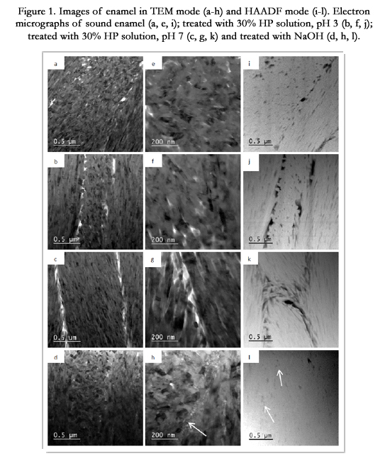
Figure 1. Images of enamel in TEM mode (a-h) and HAADF mode (i-l). Electron micrographs of sound enamel (a, e, i); treated with 30% HP solution, pH 3 (b, f, j); treated with 30% HP solution, pH 7 (c, g, k) and treated with NaOH (d, h, l).
The electron micrographs obtained from the dentine crosssections of control and treated tooth samples in bright field TEM and HAADF-mode are shown here.
The examination of micrographs sound dentine at higher magnifications on TEM and HAADF mode revealed a highly dense peritubular (PT) region surrounding the tubule. In TEM, the PT is characterised as a dark region (Figure 2a) whereas it appeared as a bright region in HAADF image (Figure 2i). The dark speckles appeared in TEM of PT were not visible as bright speckles in HAADF, indicated that the speckles were not correlated to denser material but to the presence of crystals. In HAADF, small voids which are intrinsic to the tooth material were visible in the PT region. The intertubular region displayed a very open fibre structure and often these regions contain far less or no material. Collagen fibres are found in the intertubular regions, characterised by the periodic banding in contrast in both modes (Figure 2e, i).
The electron micrograph of dentine treated with 30% hydrogen peroxide solutions for 16h showed the presence of PT region that was asymmetric in positioning around the tubule and inhomogeneous in thickness (Figure 2b-c, j-k). Differences and inhomogeneities in the thickness of the peritubular region were observed for control and treated samples and hence the thickness of PT region cannot be used as a measurement from the impact of treatment (Figure 2i-l). As observed in the control sample, collagen fibres were exposed, represented by the periodic banding (Figure 2f) and crystals were visible as small dark speckles in the denser material (Figure 2g).
The NaOH treated sample displayed the denser PT region and collagen fibres in the intertubular region (Figure 2d, h and l). The tubules were not filled with resin for this sample and this could be easily distinguished from other images. It appeared that the dental material was deposited into tubules during early stages of preparation. The dark dots in the denser material around the tubule are small crystals. HAADF image (Figure 2l) showed the denser material around the tubule. The image also showed small voids in the denser material of the sample similar to other samples.
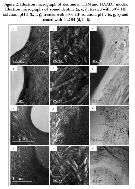
Figure 2. Electron micrograph of dentine in TEM and HAADF modes. Electron micrographs of sound dentine (a, e, i); treated with 30% HP solution, pH 3 (b, f, j); treated with 30% HP solution, pH 7 (c, g, k) and treated with NaOH (d, h, l).
The HAADF-STEM images shown in Figures 1 and 2 indicate the areas that were scanned during EDS acquisition.
A representative EDS spectral mapping acquired from untreated dentine is shown in Figure 3. Apart from the major constituent elements such as Ca and P, minor amounts of N (6 atom %), F (about 1 atom %) and Mg, Na (< 0.5 atom %) were detected in the regions analysed.
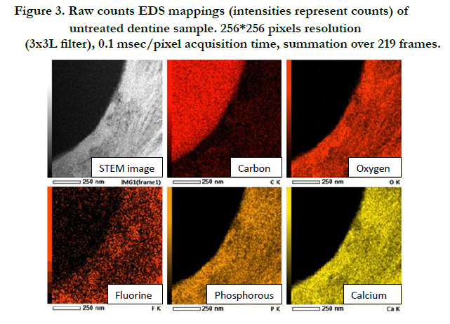
Figure 3. Raw counts EDS mappings (intensities represent counts) of untreated dentine sample. 256*256 pixels resolution (3x3L filter), 0.1 msec/pixel acquisition time, summation over 219 frames.
Figure 4 shows the two-dimensional EDS map of carbon in the enamel as a function of treatment. Although C was detected in between the rods or rod sheath of untreated teeth, the signal was not acquired in hydrogen peroxide and sodium hydroxide treated enamel samples. The C content can be linked to the presence of protein material. It was reported earlier that HP and NaOH attacks proteins, which are C-based material [34] and this could explain the absence of C in treated sample.
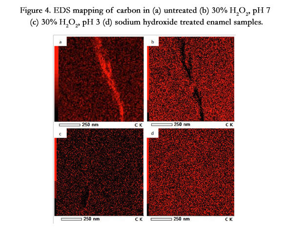
Figure 4. EDS mapping of carbon in (a) untreated (b) 30% H2O2, pH 7 (c) 30% H2O2, pH 3 (d) sodium hydroxide treated enamel samples.
The entire set of EDS mappings of enamel and dentine samples treated with HP and NaOH can be found in the supplementary material.
Discussion
Dentine is a complex hydrated biological composite structure for which only limited structure-property relationships are available [23]. An increase in the demand for tooth whitening led to the release of enormous number of products in the market. Therefore a better understanding of the ultrastructure of dentin after whitening treatments is essential to provide valuable insights into the safety of using whitening products.
In this study, we investigated the effect of hydrogen peroxide and sodium hydroxide on enamel and dentine using a direct bleaching method to explore any underlying deleterious effect from treatments. Acidic and neutral hydrogen peroxide solutions were tested to understand the effect of pH of whitening products and sodium hydroxide as an agent to analyse the role of proteins in the overall tooth structural integrity. FIB produced thin lamellae of mineralised teeth samples with minimal damage to examine the morphological changes after 30% HP treatment and deproteinization.
Bovine enamel and dentine were selected for this feasibility study as the crystalline orientations of bovine enamel matches to human, has more uniform composition and is more susceptible to acid attack due to variations in the hydroxyapatite lattice [24, 25]. Moreover, bovine enamel is more porous and demineralises faster than human enamel [26]. Therefore with bovine teeth, any structural changes that eventuate from hydrogen peroxide or sodium hydroxide treatments could be clearly and rapidly captured under high resolution microscopy. The use of bovine teeth also enables comparison with several other studies, which have reported their use in adhesion studies, caries-like lesions, micro hardness and lesion depth studies as an alternate substrate to human teeth [26-30].
TEM examination of 30% hydrogen peroxide treated sample revealed no difference in the structural integrity of enamel rods or fibrous structure of dentine at ~50-100μm depth. This is in agreement with many previous studies reported on the minimum or insignificant effect of acidic and alkaline 30% hydrogen peroxide solution [14, 31-34]. However, studies conducted by Sun et al. (2011) observed less deleterious effects of neutral 30% HP on enamel than acidic solution [10]. Moreover, several other studies have reported a decrease in the microhardness of dentin following 30% HP solution at pH 3 and reduced in vitro fracture resistance of dentin after prolonged use of leaching products [35, 36]. As reported by Chng et al., the surface alteration on intertubular dentine resulted from hydrogen peroxide treatment occurred predominantly from its oxidising property rather than the acidic pH [37]. The observation is similar to our study where no alteration in the ultra-structure of dentine was visualized with acidic or neutral hydrogen peroxide solutions.
The wide difference between the studies could be derived from the preferred in vitro method or technique used, type of tooth studied, source of peroxide, concentration of peroxide and time of exposure, effect of other non-active ingredients in formulation or presence of saliva as a potential remineralising agent etcetera [22]. In order to avoid the complexity derive from the presence of components in saliva, the immersion of tooth samples in artificial saliva before or after treatments was excluded from the protocol of this current study.
In contrast to HP treated teeth, enamel treated with sodium hydroxide revealed a complete loss of the defined structure of enamel rods. This might be due to the fact that deproteinsation of enamel resulted in the destruction of highly organized array of very fine hydroxyapatite crystals embedded in protein rich rod sheaths and within the rods themselves [38]. Proteins which constitute only a small fraction of enamel disperse through enamel and bind together the hydroxyapatite crystals within the rods and the rod sheath [39].
High resolution imaging of the dentine revealed the presence of highly dense peritubular regions (HAADF mode), needle like crystallite structures and characteristic periodic banding of protein matrix featuring the collagen fibril structures. TEM analysis of treated dentine did not show any evidence of the destruction of peritubular region or fibres within the intertubular region. The microstructure of enamel and dentine observed to be similar between tooth structures treated with acidic and neutral hydrogen peroxide. This might have been a result of the examination of samples ~100μm below the original exposed surface. Therefore the final TEM lamella might not have captured any superficial morphology change resulted from acidic peroxide.
With EDS mapping, the presence of Ca, P, O, F and traces of Na, Mg and Cl were detected in the enamel of control and treated samples. Carbon was only present in the rod sheath area of control enamel with the absence of nitrogen in the enamel of both control and treated samples. We postulate that the absence of carbon in treated samples could be due to the removal of extrinsic hydrocarbons such as stains. The presence of porous material was observed in sodium hydroxide treated sample at several prism borders unlike other samples and using EDS, this porous material appeared to contain F. EDS analysis of dentine specimens showed the presence of C and N, representing the existence of organic contents such as proteins. However, no effects of treatment were found in the EDS data of the dentine samples.
The safety of using tooth whitening products and risks associated with the treatment procedure is still unclear and controversial. Experts in this field recommended the involvement of dental professionals in bleaching treatments to reduce the risk that might occur during the treatment and for correct diagnosis of the problem which resulted in tooth discolouration [20]. We demonstrated (at least to first order) that the ingression of hydrogen peroxide whitening agents into the complex hard tissues of the teeth does not cause significant damage to the mineral and protein constituents. Although the sub-surfaces of the enamel and dentine are not affected by HP treatment, it is possible that some alteration were occurred on tooth surfaces, which might not be clinically significant. The normal structural morphology could be restored with the natural remineralisation of saliva and with the practice of following a suitable post treatment care under the supervision of a professional dentist. However, further studies using extracted human teeth in an ideal oral environment and subsequent clinical studies are required to confirm such suggestions and for accurately predicting the effect of whitening agents on teeth.
Acknowledgements
We would like to acknowledge Ria Vrolijks, Ivo Lemmens and Carlo Manders from Philips Innovation services for mechanical polishing and SEM studies on the tooth samples.
References
- Haywood VB, Heymann HO (1989) Nightguard vital bleaching. Quintessence Int 20(3): 173-176.
- Heymann HO (2005)Tooth whitening: facts and fallacies. Br Dent J 198(8): 514.
- Madhu K, Hegde S, Mathew S, Lata D, Bhandi SH (2013) Comparison of radicular peroxide leakage from four commonly used bleaching agents following Intracoronal bleaching in endodontically treated teeth - an In Vitro Study. J Int Oral Health 5(4): 49-55.
- Chen HP, Chang CH, Liu JK, Chuang SF, Yang JY (2008) Effect of fluoride containing bleaching agents on enamel surface properties. J Dent 36(9): 718-725.
- Parreiras SO, Vianna P, Kossatz S, Loguercio AD, Reis A (2014) Effects of light activated In-office bleaching on permeability, microhardness, and mineral content of enamel. Oper Dent 39(5): E225-230.
- Jiang T, Ma X, Wang Y, Zhu Z, Tong H, et al. (2007) Effects of hydrogen peroxide on human dentin structure. J Dent Res 86(11): 1040-1045.
- Attin T, Schmidlin PR, Wegehaupt F, Wiegand A (2009) Influence of study design on the impact of bleaching agents on dental enamel microhardness: A review. Dent Mater 25(2): 143-157.
- Berger SB, Cavalli V, Martin AA, Soares LE, Arruda MA, et al. (2010) Effects of combined use of light irradiation and 35% hydrogen peroxide for dental bleaching on human enamel mineral content. Photomed Laser Surg 28(4): 533-558.
- Li Q, Xu BT, Li R, Yu H, Wang YN (2010) Quantitative evaluation of colour regression and mineral content change of bleached teeth. J Dent 38(3): 253-260.
- Sun L, Liang S, Sa Y, Wang Z, Ma X (2011) Surface alteration of human tooth enamel subjected to acidic and neutral 30% hydrogen peroxide. J Dent 39(10): 686-692.
- Pashley DH, Tao L, Boyd L, King GE, Horner JA (1988) Scanning electron microscopy of the substructure of smear layers in human dentine. Arch Oral Biol 33(4): 265-270.
- Yoshiyama M, Masada J, Uchida A, Ishida H (1989) Scanning electron microscopic characterization of sensitive vs insensitive human radicular dentin. J Dent Res 68(11): 1498-1502.
- Lin CP, Douglas WH, Erlandsen SL (1993) Scanning electron microscopy of type I collagen at the dentin-enamel junction of human teeth. J Histochem Cytochem 41(3): 381-388.
- Park HJ, Kwon TY, Nam SH, Kim HJ, Kim KH, et al. (2004) Changes in bovine enamel after treatment with a 30% hydrogen peroxide bleaching agent. Dent Mater J 23(4): 517-521.
- Ge J, Cui FZ, Wang XM, Feng HL (2005) Property variations in the prism and the organic sheath within enamel by nanoindentation. Biomaterials 26(16): 3333-3339.
- Marzuki AF, Masudi SM (2008) Confocal laser scanning microscopy study of dentinal tubules in dental caries stained with alizarin red. Arch Orofacial Sci 3(1): 2-6.
- Contaldo M, Serpico R, Lucchese A (2014) In vivo imaging of enamel by reflectance confocal microscopy (RCM): non invasive analysis of dental surface. Odontology 102(2): 325-329.
- Porter AE, Nalla RK, Minor A, Jinschek JR, Kisielowski C, et al. (2005) A transmission electron microscopy study of mineralization in age-induced transparent dentin. Biomaterials 26(36): 7650-7660.
- Jantou V, Turmaine M, West GD, Horton MA, McComb DW (2009) Focused ion beam milling and ultramicrotomy of mineralised ivory dentine for analytical transmission electron microscopy. Micron 40(4): 495-501.
- Li Y (2011) Safety controversies in tooth bleaching. Dent Clin North Am 55(2): 255-263.
- Li Y, Greenwall L (2013) Safety issues of tooth whitening using peroxidebased materials. Br Dent J 215(1): 29-34.
- Joiner A (2007) Review of the effects of peroxide on enamel and dentine properties. J Dent 35(12): 889-896.
- Marshall GW (1993) Dentin: microstructure and characterisation. Quintessence Int 24(9): 606-617.
- Yassen GH, Platt JA, Hara AT (2011) Bovine teeth as substitute for human teeth in dental research: a review of literature. J Oral Sci 53(3): 273-282.
- Sydney-Zax M, Mayer I, Deutsch D (1991) Carbonate content in developing human and bovine enamel. J Dent Res 70(5): 913-916.
- Mellberg JR (1992) Hard-tissue substrates for evaluation of cariogenic and anti-cariogenic activity in situ. J Dent Res 71: 913-919.
- Schilkea R, Lisson JA, Bauss O, Geurtsen W (2000) Comparison of the number and diameter of dentinal tubules in human and bovine dentine by scanning electron microscopic investigation. Arch Oral Biol 45(5): 355-361.
- Pearce EI (1983) A microradiographic and chemical comparison of in vitro systems for the simulation of incipient caries in abraded bovine enamel. J Dent Res 62(9): 969-974.
- Arends J, Schuthof J, Jongebloed WG (1980) Lesion depth and microhardness indentations on artificial white spot lesions. Caries Res 14(4): 190-195.
- Reis AF, Giannini M, Kavaguchi A, Soares CJ, Line SR (2004) Comparison of microtensile bond strength to enamel and dentin of human, bovine, and porcine teeth. J Adhes Dent 6(2): 117-121.
- Ernst CP, Marroquin BB, Willershausen-Zonnchen B (1996) Effects of hydrogen peroxide-containing bleaching agents on the morphology of human enamel. Quintessence Int 27(1): 53-56.
- Eimar H, Siciliano R, Abdallah MN, Nader SA, Amin WM, et al. (2012) Hydrogen peroxide whitens teeth by oxidizing the organic structure. J Dent 40(Suppl 2): e25-e33.
- Xu B, Li Q, Wang Y (2011) Effects of pH values of hydrogen peroxide bleaching agents on enamel surface properties. Oper Dent 36(5): 554-562.
- Sulieman M, Addy M, Macdonald E, Rees JS (2004) A safety study in vitro for the effects of an in-office bleaching system on the integrity of enamel and dentine. J Dent 32(7): 581-590.
- Lewinstein I, Hirschfeld Z, Stabholz A, Rotstein I (1994) Effect of hydrogen peroxide and sodium perborate on the microhardness of human enamel and dentin. J Endod 20(2): 61-63.
- Tam LE, Kuo VY, Noroozi A (2007) Effect of prolonged direct and indirect peroxide bleaching on fracture toughness of human dentin. J Esthet Restor Dent 19(2): 100-109.
- Chng HK, Ramli HN, Yap AU, Lim CT (2005) Effect of hydrogen peroxide on intertubular dentine. J Dent 33(5): 363-369.
- Robinson C, Connell S, Brookes SJ, Kirkham J, Shore RC, et al. (2005) Surface chemistry of enamel apatite during maturation in relation to pH: implications for protein removal and crystal growth. Arch Oral Biol 50(2):267-270.
- Ravindranath RM, Moradian-Oldak J, Fincham AG (1999) Tyrosyl motif in amelogenins binds N-acetyl-D-glucosamine. J Biol Chem 274(4): 2464-2471.


