Effect of post-polymerization heat treatment on a denture base acrylic resin: histopathological analysis in rats
Meister LM1, Kovalik AC1, Pellissari CV2, Bail M1, Campagnoli EB1, Jorge JH2*, Campanha NH1
1 State University of Ponta Grossa, Department of Dentistry, Paraná, Brazil.
2 Department of Dental Materials and Prosthodontics, Araraquara Dental School, UNESP - Univ Estadual Paulista, Araraquara, SP, Brazil.
*Corresponding Author
Janaina Habib Jorge,
Department of Dental Materials and Prosthodontics,
Araraquara Dental School,
Univ. Estadual Paulista, UNESP, Postal address: Rua Humaità,
1680, Centro; Araraquara, SP, Brazil.
Tel: 55 (16) 3301-6550
E-mail: janainahj@foar.unesp.br
Article Type: Research Article
Received: March 24, 2015; Accepted: April 22, 2015;Published: April 24, 2015
Citation: Jorge JH, et al., (2015) Effect of Post-Polymerization Heat Treatment on a Denture Base Acrylic Resin: Histopathological Analysis in Rats. Int J Dentistry Oral Sci. S2:001, 1-7. dx.doi.org/10.19070/2377-8075-SI02001
Copyright: Jorge JH© 2015. This is an open-access article distributed under the terms of the Creative Commons Attribution License, which permits unrestricted use, distribution and reproduction in any medium, provided the original author and source are credited.
Abstract
Aims: This work examined the histological effects, on the rat palatal mucosa, of a denture base acrylic resin, submitted or not to a post-polymerization heat-treatment.
Methods: Fifteen adult female Wistar rats, with sixty days old, weighting 150 g – 250 g were divided in G1: animals being maintained under the same conditions as the experimental groups following described, but without the use acrylic palatal plates (control group); G2: use of heat-polymerized acrylic resin palatal plates made of Lucitone 550; G3: use of palatal plates identical to G2, but subjected to a post-polymerization treatment in a water bath at 55°C for 60 min. The plates covered all the palate and were fixed in the molar region with light-cured resin, thus being kept there for 14 days. After the sacrifice, the palate was removed, fixed in formaldehyde 10% and decalcified with EDTA. Sections were stained using haematoxylin and eosin. Images in duplicate were made from the central region of the cuts, to measure the thickness (μm) of the keratin layers (TKC), epithelium total (TET) and connective tissue (TCC). Statistical analyses were carried out by one-way ANOVA and Tukey post-tests (α=0.05).
Results: According to the results there was significant difference in the thickness of keratin between G2 and G3, with G1 having the intermediate value and similar to the other groups. There was a significant difference in the connective tissue with G3 <G1 <G2 (p <0.0001).
Conclusion: Regarding the total epithelium, group G3 presented a statistically significant difference with both G1 and with G2 (p <0.0001), which were similar to each other. When using the proposed heat treatment it was found to be effective, from the viewpoint of biocompatibility, for the acrylic resin denture base investigated.
2.Introduction
3.Materials and Methods
4.Results
5.Discussion
6.Conclusion
7.References
Keywords
Biocompatibility; Acrylic Resin; Wistar Rat; Palatal Mucosa; Palatal Appliances.
Introduction
The material of choice for oral rehabilitation of partially or totally edentulous patients from the 1930s to the present day has been acrylic resin [1]. Used as the basis for full and partially removable dentures, and artificial teeth, when used in the oral cavity important features need to be taken into consideration such as: color and translucency, chemical stability, sufficient hardness, good resistance and resilience to wear, it should be insoluble and impervious to oral fluids, and should be biocompatible. However, one of the major factors limiting the use of this material is their biocompatibility. Biocompatibility can be defined as the acceptance (or rejection) of artificial materials to perform with an appropriate host response when applied as intended. Thus, studies in order to test new materials and techniques to improve physical, chemical and biological properties have been performed [2-5].
Denture resins may be classified by polymerization mode and include those that are heat-polymerized (by microwave or conventionally), auto-polymerized, and visible light-polymerized [6]. Acrylic resins consist of polymethylmethacrylate, a mixture of a polymer powder containing benzoyl peroxide and a liquid monomer, typically methyl methacrylate. The polymerization of the material occurs with a rapid decomposition of benzoyl peroxide, after being activated, which releases a large amount of free radicals, polymerizing the monomer. The process of formation of polymers and the growth of chains continues at a considerable speed. Theoretically, these reactions should proceed with increased heat until all the monomer had been converted to polymer. However, with a decrease in the polymerization temperature, polymer formation also decreases and, consequently, an amount of residual monomer remains in the polymerized resin [6]. There are many reports suggesting that residual monomer in the denture base is related to mucosal irritation and sensitization of tissues[1, 6-12]. The leaching of formaldehyde, methacrylic acid, and benzoic acid from acrylic resin dental materials has also been detected [13-16].There have been several investigations into ways of improving the polymerization of these materials and their behavior in relation to treatments to decrease the cytotoxic effect of their products and by-products from the polymerization reaction [5, 16, 24-35].
Toxic substances and their effects on tissues have been observed through animal studies, clinical observations and in vitro cell cultures[17, 18]. A cytotoxicity assay which uses the cell culture method is considered to be a preliminary test for assessing the biocompatibility of a material; it is simple, reproducible, effective and controlled [19, 20]. There are several studies in the literature with initial tests which determine the cytotoxicity of acrylic resins [21-24]. Acrylic resins remain in direct contact with the tissues of the oral cavity, requiring in vivo tests in animals to observe the histological changes that may occur due to prolonged use of these materials. The reactions most frequently found in the mucosa are redness, erosion in the oral mucosa, and burning in the mucosa and tongue [7,8, 36]. However, little information can be found related to the biological behavior of these materials in secondary tests, i.e. made directly in animals [37-39]. Few in vivo studies using experimental apparatus adapted for the palate of animals were found in the literature [37, 41, 42]. Therefore, the purpose of the study was to examine the histological effects of post-polymerization treatment on an acrylic resin denture base, on palatal mucosa in a Wistar rat model. The null hypotheses investigated in the present study were that: 1. palatal plates of heat-polymerized acrylic resin would not result in histological changes in the palatine mucosa of rats; 2. post-polymerization heat treatment of palatal plates would not diminish histological changes in the palatine mucosa of rats, relative to the untreated group.
Material and Methods
The methodological sequence followed the study by Meister et al.[43] Fifteen sixty-day old adult female rats (Rattus Norvegicus Albinus Wistar), weighing 150 – 250g were used for this study. The animals were maintained in separate cages at 23°C with 56% relative humidity, alternating light/dark cycles of 12 h. They were fed with water ad libitum and a nutritionally complete powdered diet (Nuvilab CR-1, Nuvital, PR, Curitiba, Brazil). The animals were kept on a paste diet one week before the experiments and were allowed nothing orally except for free access to water for 12h before the procedures. This study was approved in accordance with the recommendations of the Ethics Committee on Animal Use (CEUA) of the State University of Ponta Grossa (File: 11/2010, Protocol: 12673/2010) and the guidelines on animal handling of the COBEA (Brazilian College of Animal Experimentation) were followed.
A preliminary impression of a Wistar rat palate of an animal that had the approximate age and weight to those used in the experiments was taken. Ten trays were then constructed with putty silicone on a master dental stone cast from this preliminary impression. To take the master impressions and to fit the appliances, it was necessary to anaesthetize all the animals. This was accomplished by the administration of a solution of ketamin (100 mg/ mL) and xylasine (100 mg/mL) that was made by diluting 3.75 mL of ketamin plus 0.5mL of xylasine in 5.75 mL of distilled water. Intraperitonial injection of 0.2mL/100 g of this solution [44] was used to sedate the rats. The time of work obtained by this technique was approximately 30 min.
After the sedation of the animals, molding, using an addition silicone material of light consistency, was performed (Futura AD, DFL Indústria e Comercio S.A., RJ, Rio de Janeiro, Brazil). For this, the animals were placed in a stabilizing plate, with the mouth kept open and secured by incisors with the aid of handles, to facilitate the molding of the palate.
Palatal appliances, customized for each animal, were made using Lucitone 550 (Dentsply Indústria e Comercio Ltda, Petrópolis, Brazil) heat polymerized acrylic resin. The appliances were constructed over the plate area, comprising the region between the molars, extending to the first palatal fold, covering the occlusal surface of the molars and extending to the buccal surface of the molars. The acrylic resin denture material was polymerized according to the manufacturer’s specifications (90 min at 73°C and 30 min at 100°C - short cycle) and the quantity used was 0.42 g of powder and 0.2mL of liquid. Immediately after specimen fabrication, the acrylic resin devices were stored in distilled water for 48 h at 37°C prior to insertion, in order to eliminate the residual monomer [45].
At this moment, the animals were randomly allocated into three test groups: G1: without the use of plates, being maintained under the same conditions as the experimental groups (control group); G2 using Lucitone 550 heat polymerized acrylic resin palatal plates; G3: using plates identical to G2, but subjected to a postpolymerization treatment in a water bath at 55°C for 60 min [34].
In order to promote a mechanical retention of molars and fixing of the palatal device, a groove in the molar coverage region was made using a spherical bur #6. Composite resin (Opallis, FGM, SC, Joinville, Brazil) was used to fill the molar region groove and the resin was photo-polymerized while the device was set in position. Upon completion of the procedure, the rats were placed at rest to recover from anesthesia. From this moment, the rats were placed on a paste diet to avoid food debris gathering under the denture system [46]. The animals, and both groups, remained with the plates in the palate for 14 days.
After 14 days, the animals of all the groups were killed by euthanasia, according to resolution number 714 of 20/6/02 of the Federal Council of Veterinary Medicine (FCVM). The animals were anesthetized in accordance with the methodology, and under deep anesthesia, intracardiac perfusion with potassium chloride 10% (1 ml/100 g) was applied through the left ventricle. The palates of the animals were dissected, fixed in 10% buffered formalin for 48 h and decalcified in 4.17% EDTA for approximately four months. The pieces went through a dehydration process, embedded in paraffin, so that the back part of the palate stayed down. The samples embedded in paraffin were cut with a microtome (Leica, Berlin, Germany) in sections of 5μm and then stained with haematoxylin and eosin (H&E). Slides were mounted and observed by using light microscopy (Nikon, model YS 100, Tokyo, Japan). Five ribbons were made with five sections of each animal, then, for analysis, two images, standardized in the central region of the cuts, near the nasopalatine plexus and the midline of the palatine raphe, at 200x magnification to check for changes histological were made. Overall thickness of the epithelium (TET), cellular compartment (TCC) and keratin layer (TKC) were calculated. For the measurement, the Image-Pro Plus (software version 4.5.0.29-Media Cybernetics, Inc, Bethesda, USA) program was used in groups G1 (control group), G2 (Lucitone without heat treatment) and G3 (Lucitone with heat treatment) with a grid with ten horizontal lines for each analyzed layer, obtaining ten measurements, and then the averages of the measurements were analyzed using Microsoft Excel. This resulted in two averages for each animal, to perform statistical analysis.
The averages of the two readings (μm) of each parameter (i.e. keratin thickness, epithelium thickness, and connective tissue thickness) were calculated and compared for each animal by ANOVA of one variation factor, which was the type of plate that the animal used (no plate, control - G1; palatal plate with no heat treatment - G2; palatal plate with heat treatment - G3). The averages of groups G1, G2 and G3 were compared by Tukey’s posttest, with α = 0.05.
Results
According to the results, there was significant difference in the thickness of keratin between G2 and G3, with G1 being the intermediate value and similar to the other groups (Figure 1). There was a significant difference in the connective tissue with G3 <G1 <G2 (p <0.0001) (Figure 2). Regarding the total epithelium, group G3 presented a statistically significant difference with both G1 and with G2 (p<0,0001), which were similar to each other(Figure 3).
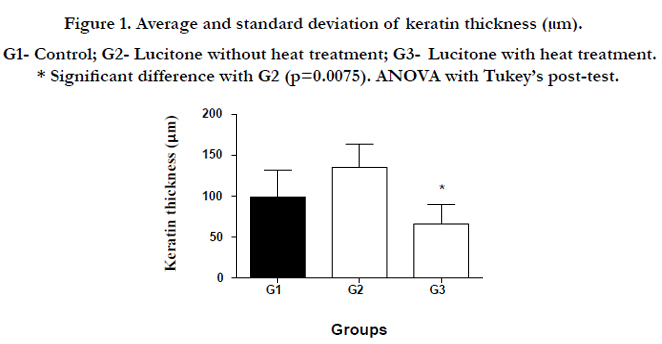
Figure 1. Average and standard deviation of keratin thickness (μm). G1- Control; G2- Lucitone without heat treatment; G3- Lucitone with heat treatment. * Significant difference with G2 (p=0.0075). ANOVA with Tukey’s post-test.
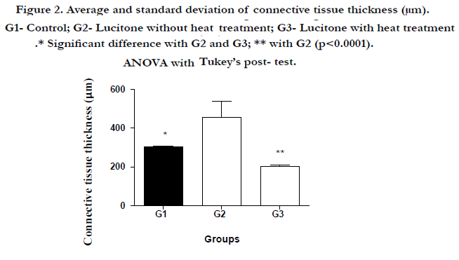
Figure 2. Average and standard deviation of connective tissue thickness (μm). G1- Control; G2- Lucitone without heat treatment; G3- Lucitone with heat treatment.* Significant difference with G2 and G3; ** with G2 (p<0.0001). ANOVA with Tukey’s post- test.
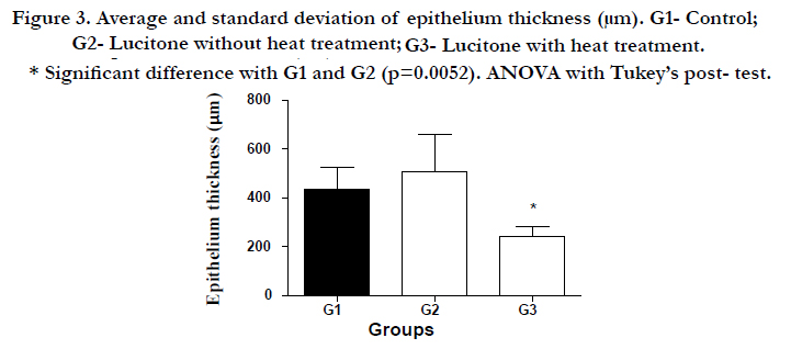
Figure 3. Average and standard deviation of epithelium thickness (μm). G1- Control; G2- Lucitone without heat treatment; G3- Lucitone with heat treatment. * Significant difference with G1 and G2 (p=0.0052). ANOVA with Tukey’s post- test.
Discussion
A number of studies have carried out in vitro tests to evaluate the biocompatibility of acrylic resins through the use of cell cultures, in which it was possible to observe the proliferation or inhibition of cell growth resulting from contact with cytotoxic substances [23, 27, 34, 47]. Whilst acknowledging the value of such studies, they are recommended mainly for the initial selection of materials and techniques and their results should not be directly extrapolated clinically [20-24, 48 ,49]. Animal model studies have been used to clarify the pathogenesis of human diseases, as well as evaluating new therapeutic targets, and they represent an efficient and accessible way to study disease processes in vivo, since ethical issues preclude the use of some invasive procedures in humans [50]. Among these, in vivo studies using the method of the subcutaneous implantation of materials in animals have been described [38, 39, 51, 52]. However, these studies were not likely to reflect the true clinical situation. Pathological conditions of oral mucosa under denture bases, such as the deformation of mucosa, decubital ulcers or denture stomatitis, are frequently found [53]. Little information is available regarding the biological behaviour of materials in secondary tests, performed directly on animals with the use of palatal devices [37, 40-42, 46, 54]. Mucous membrane irritation tests by means of removable partial palatal dentures or fixed-bridge type appliances in small animals were considered to be extremely difficult, expensive, and time consuming [55] mainly due to the fact that previously described intraoral devices were considered primitive and inadequate [42].
Due to the difficulties associated with palatal devices described in the literature, and in view of the importance of performing in vivo studies, in the present study with regard to the preliminary tests prior to the clinical tests, palatine plates, fixed in Wistar rats, made with Lucitone 550 acrylic resin according to the methodology described by Meister et al., [43] were used. The material selected for use in the present study was a heat-polymerized acrylic resin consisting of polymethyl methacrylate (PMMA) and ethylene glycol dimethacrylate, and it presents two possible polymerization cycles. One is called ‘short cycle’, where the dental flask is immersed in water at 73°C for 90 min and then the temperature is raised to 100°C and the flask is maintained for an additional 30 min period at this temperature. The other possible polymerization technique is called ‘long cycle’ and the flask is kept for 9 hours at 71°C, not submitting to a final polymerization in boiling cycle. It has been found that the ‘short cycle’ promoted a lower amount of residual monomer (0.08%) when using Lucitone 550 compared with the ‘long cycle’ (0.24%) [4] and also a lower level of toxicity in vitro [34]. Consequently, the ‘short cycle’ was selected for the present study. However, under the conditions of this study, using the same ‘short cycle’ described above, the use of palatal plates needed changes to some of the assessed parameters, e.g. group G2 showed higher levels of keratin thickness and connective tissue compared to G1, i.e. the control group, consisting of animals without plates. This increase in tissue thickness can be explained by the cytotoxic effect of the denture base resin used, already widely studied by in vitro tests, which has been proven to release residual monomers and other substances [1, 6-12, 24, 34] as confirmed in the present study. Our results are in agreement with Axelsson and Nyquist [25], who evaluated the levels and the effects of residual monomer on the mucosa due to the fact that they had observed a hyperkeratosis in various patients over a period of years. In 1963, Kapur and Shklar [56] also observed keratinization and a slight increase in connective tissue, suggesting that the tissue response might be a consequence of a chemical irritation.
However, the results for the groups G2 and G3 might also be a response to the continuous pressure exerted by the prosthesis on the oral mucosa due to occlusal forces exerted during mastication. More specific and more recent tests, such as the study of diabetes mellitus by Maruo et al. [54], used acrylic plates cemented in Wistar rats, to verify tissue reaction. The plates were cemented without pressure, with continuous and intermittent pressure, and it was found that there was a reduction in the total thickness of the epithelium and also that mechanical stimuli decreased susceptibility to cellular changes. Tsuruoka et al.[41] conducted a study on the physiological, histological and molecular levels to determine the effects of mechanical compression on prosthesis mucus supported on the palate of rats, They concluded that the effect of mechanical compression was greatest in cells of the periosteum and that these cells synthesize HSP70, which is responsible for bone remodelling and vascular endothelial growth factor (VEGF). VEGF is a protein that is produced by cells which stimulate angiogenesis and vasculogenesis. It is an integral part of the system that provides oxygen to tissues when blood circulation is insufficient and also if it is abundant it may contribute to diseases.
Analyzing data from the G3 group, values very close to normal (G1) were observed, meaning that the post-polymerization heat treatment that was performed to decrease the release of toxic compounds on the mucosa was probably efficient.
In the present study, a post-polymerization treatment in a water bath at 55°C for 60 min was performed, in accordance with Jorge et al. [34], a study which evaluated the cytotoxicity of several denture base acrylic
resins assessed in vitro by MTT and by 3Hthymidine. The null hypothesis formulated for this present study was not accepted and differences were observed between groups G2 and G3 when comparing thickness of connective tissue, keratin and epithelium. In all cases, the measurements of Group G3 always showed smaller thicknesses compared with G1 and G2. This was probably because there was a reduction in the irritation resulting from the use of plates compared to G2 due to the heat treatment that was tested. Regarding group G1, one can say that the pressure exerted on the overall thickness of epithelium was in agreement with Maruo et al. [54].
In carrying out post- polymerization treatments, heating the samples provides an increase in the mobility of monomer molecules, which are at rest in the polymeric mass, increasing the degree of conversion [57]. Vallittu et al. [28] demonstrated that the release of residual compounds is a process which depends on temperature; thus, if temperature is increased, greater diffusion occurs. It has been shown that the release mechanisms of monomers and other substances can be induced by treatments performed after polymerization, such as immersion in hot water or irradiation with microwave energy, thus significantly reducing the amount of monomer present in heat-polymerized acrylic resins and conventional auto-polymerized acrylic resins [2, 15, 58, 59]. Methods for reducing the monomer contents of polymerized acrylic resins have been described in the literature, to minimize the risk of adverse reactions for those who wear acrylic resin dentures.
Analyzing the image of the G1 group (Figure 4), the histological section displays orthokeratinized stratified squamous epithelium. The granular layer is very clear and the epithelial ridges are discrete. The lamina propria is with normal characteristics and there is the presence of vital bone tissue (HE 200X) There was a significant difference in the thickness of the epithelium compared with G2. Analyzing the image obtained from G2 (Figure 5), the histological section displays hyper- orthokeratinized stratified squamous epithelium and there was an increase in the thickness of the cell compartment and also the keratin layer in the epithelial tissue, showing that there was a difference when compared with the other groups. Note also the absence of inflammatory infiltrate in the connective tissue. The lamina propria is normal and without the presence of significant inflammatory infiltrate. Bone tissue is vital and with normal characteristics. (HE, 200X magnification). In the image from G3 (Figure. 6), there is a smaller increase in the thickness of the cell compartment and also in the keratin layer in the epithelial tissue. In this group, there was little change in tissue morphology. There was also the absence of inflammatory infiltrate in the connective tissue (HE, 200X magnification), confirming the hypothesis that the heat treatment decreases cytotoxicity.
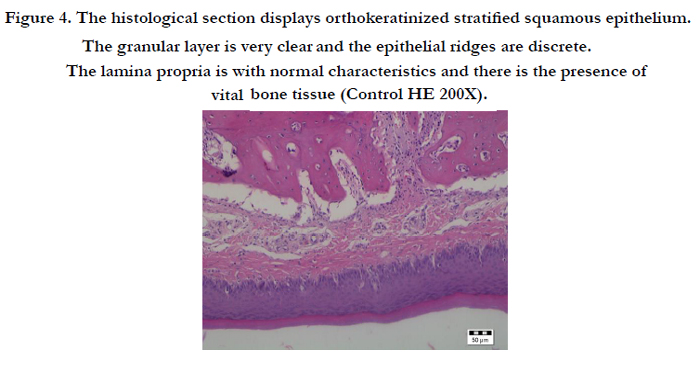
Figure 4. The histological section displays orthokeratinized stratified squamous epithelium. The granular layer is very clear and the epithelial ridges are discrete. The lamina propria is with normal characteristics and there is the presence of vital bone tissue (Control HE 200X).
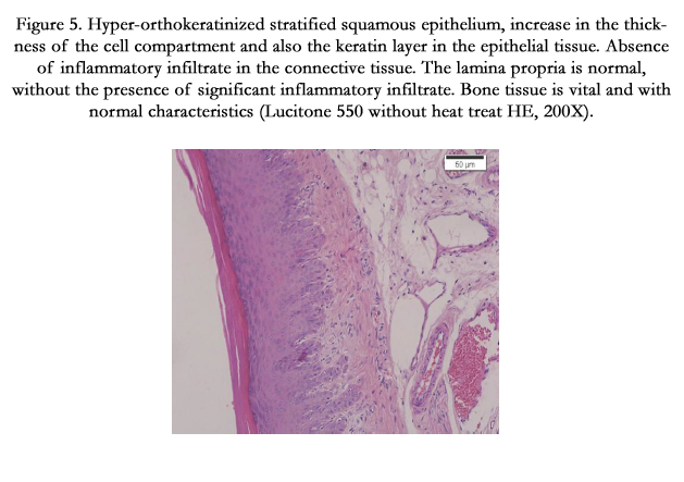
Figure 5. Hyper-orthokeratinized stratified squamous epithelium, increase in the thickness of the cell compartment and also the keratin layer in the epithelial tissue. Absence of inflammatory infiltrate in the connective tissue. The lamina propria is normal, without the presence of significant inflammatory infiltrate. Bone tissue is vital and with normal characteristics (Lucitone 550 without heat treat HE, 200X).
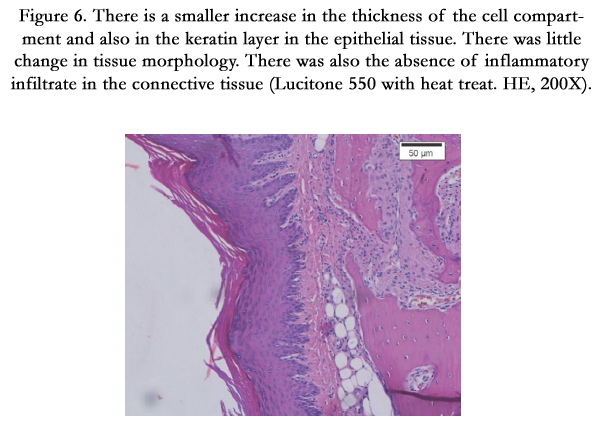
Figure 6. There is a smaller increase in the thickness of the cell compartment and also in the keratin layer in the epithelial tissue. There was little change in tissue morphology. There was also the absence of inflammatory infiltrate in the connective tissue (Lucitone 550 with heat treat. HE, 200X).
Conclusion
Thus, in accordance with the present results, the importance of adequate processing of material and histological analysis for comparison among treatments is evident. When using the proposed heat treatment it was found to be effective, from the viewpoint of biocompatibility, for the acrylic resin denture base investigated. Future studies are needed to detect, quantify and qualify the effects in the oral mucosa of substances released by acrylic resins from denture bases as well as to determine the cytotoxic effect of these substances.
References
- Craig RG (1997) Prosthetics applications of polymers. In: Restorative dental materials. (10th edn) St. Louis,Mosby. 500-551.
- Lamb DJ, Ellis B, Priestley D (1983) The effects of process variables on levels of residual monomer in autopolymerizing dental acrylic resin. J Dent 11(1): 80-88.
- De Clerck JP (1987) Microwave polymerization of acrylic resins used in dental prostheses. J Prosthet Dent 57(5): 650-659.
- Urban VM, Machado AL, Oliveira RV, Vergani CE, Pavarina AC, et al. (2007) Residual monomer of reline acrylic resins. Effect of water-bath and microwave post-polimerization treatments. Dent Mater. 23(3): 363-368.
- Novais PM, Giampaolo ET, Vergani CE, Machado AL, Pavarina AC, et al.(2009) The occurrence of porosity in reline acrylic resins. Effect of microwave desinfection. Gerodontology 26(1): 65-71.
- Anusavice KJ (1998) Resina para base de dentadura In: Phillips ateriais dentários. (10th edn) Rio de Janeiro: Guanabara Koogan. 140-160.
- Fisher AA (1954) Allergic sensitization of the skin and oral mucosa to acrylic denture materials. J Am Med Assoc 18(3): 238-242.
- Ali A, Reynolds AJ, Walker DM (1986) The burning mouth sensation related to the wearing of acrylic dentures: an investigation. Br Dent J 161: 444-447.
- Craig RG, O'brien W, Powers JM (1992) Plastics in prosthetics In: Dental materials: properties and manipulation. (5th edn) St. Louis: Mosby. 267- 292.
- Lygre H, Solheim E, Gjerdet NR (1995) Leaching from denture base materials in vitro. Acta Odontol Scand 53(2): 5-80.
- Vallittu PK, Ruyter IE, Buykuilmaz S (1998) Effect of polymerization temperature and time on the residual monomer content of denture base polymers. Eur J Oral Sci 106(1): 588-593.
- Kedjarune U, Charoenworaluk N, Koontongkaew S (1999) Release of methyl methacrylate from heat-cured and autopolymerized resins: cytotoxicity testing related to residual monomer. Aust Dent J 44(1): 25-30.
- Smith DC, Bains ME (1956) The detection and estimation of residual monomer in polymethyl methacrylate. J Dent Res 35(1): 16-24.
- Ruyter IE (1980) Release of formaldehyde from denture base polymers. Acta Odontol Scand 38(1): 17-27.
- Tsuchiya H, Hoshino Y, Kato H, Takagi N (1993) Flow injection analysis of formaldehyde leached from denture-base acrylic resins. J Dent 21(4): 240-243.
- Lefebvre CA, Schuster GS (1994) Biocompatibility of visible light-cured resin systems in prosthodontics. J Prosthet Dent 71(2): 178-185.
- Schweikl H, Schmalz G (1996) Toxicity parameters for cytotoxicity testing of dental materials in two different mammalian cell lines. Eur J Oral Sci 104(3): 292-299.
- de Andrade Lima Chaves C, Machado AL, Vergani CE, de Souza RF, Giampaolo ET (2012) Cytotoxicity of denture base and hard chairside reline materials: a systematic review. J Prosthet Dent 107(2): 114-127.
- Hensten-Pettersen A, Wictorin L (1981) The cytotoxicity effect of denture base polymers. Acta Odontol Scand 39(2): 101-106.
- Hensten-Pettersen A (1988) Comparison of the methods available for assessing cytotoxicity. Int Endod J 21(2): 89-99.
- Schuster GS, Lefebvre CA, Dirksen TR, Knoernschild KL, Caughman GB (1995) Relationships between denture base resin cytotoxicity and cell lipid metabolism. Int J Prosthodont 8(6): 580-586.
- Sheridan PJ, Koka S, Ewoldsen NO, Lefebvre CA, Lavin MT (1997) Cytotoxicity of denture base resins. Int J Prosthodont 10: 73-77.
- Jorge JH, Giampaolo ET, Vergani CE, Machado AL, Pavarina AC, et al. (2004) Cytotoxicity of denture base resins: effect of water bath and microwave post-polymerization heat treatments. Int J Prosthodont 17(3): 340-344.
- Campanha NH, Pavarina AC, Giampaolo ET, Vergani CE, Machado AL, et al. (2006) Cytotoxicity of hard chairside reline resins: effect ofmicrowave irradiation and water bath postpolymerization treatments. Int J Prosthodont 19(2): 195-201.
- Axelsson B, Nyquist G (1962) The leaching and biological effect of the residual monomer of methyl methacrylate. Odontol Revy Lund 13: 370-379.
- Al Doori D, Huggett R, Bates JF, Brooks SC (1988) A comparison of denture base acrylic resins polymerised by microwave irradiation and by conventional water bath curing systems. Dent Mater 4(1): 25-32.
- Barron DJ, Schuster GS, Caughman GB, Lefebvre CA (1993) Biocompatibility of visible light-polymerized denture base resins. Int J Prosthodont 6(5):495-501.
- Vallittu PK, Miettinen V, Alakuijala P (1995) Residual monomer content and its release into water from denture base materials. Dent Mater 11(6): 338-342.
- Lefebvre CA, Schuster GS, Caughman GB, Caughman WF (1991) Effects of denture base resins on oral epithelial cells. Int J Prosthodont 4(4): 371-376.
- Lefebvre CA, Schuster GS, Richardson DW, Barron DJ (1992) The cytotoxic effects of denture base resin selants. Int J Prosthodont 5(6): 558-562.
- Lefebvre CA, Schuster GS, Marr JC, Knoernschild KL (1995) The effect of pH on the cytotoxicity of eluates from denture base resins. Int J Prosthodont 8(2): 122-128.
- Lefebvre CA, Schuster GS, Caughman GB, Caughman WF (2001) Effects of denture base resins on oral epithelial cells. Int J Prosthodont 85(4): 352-356.
- Arima T, Murata H, Hamada T (1996) Analysis of composition and structure of hard autopolymerizing reline resins. J Oral Rehabil 23(5): 346-352.
- Jorge JH, Giampaolo ET, Vergani CE, Machado AL, Pavarina AC, et al. (2006) Biocompatibility of denture base acrylic resins evaluated in culture of L929 cells. Effect of polymerization cycle and post-polymerization treatments. Gerodontology 24(1): 52-57.
- Jorge JH, Giampaolo ET, Vergani CE, Machado AL, Pavarina AC, et al. (2007) Biocompatibility of denture base acrylic resins evaluated in culture of L929 cells. Effect of polymerization cycle and post-polymerization treatments. Gerodontology 24(1): 52-57.
- Spealman, CR (1945) Monomeric methyl methacrylate. Indust Med 14: 292.
- Barclay SC, Macdonald DG, Watson IB (1997) The effect of chairside relining materials on rat palatal mucosa. J Dent 25(3-4): 251-255.
- Harsanyi BB, Foong RE, Howell RE, Hidi P, Jones DW (1991) Hamster cheek-pouch testing od dental soft polymers. J Dent Res 70(6): 991-996.
- Ebadian B, Razavi M, Soleimanpour S, Mosharraf R (2008) Evaluation of Tissue Reaction to Some Denture-base Materials: An Animal Study. J Contemp Dent Pract 9(4): 67-74.
- Jorge AO, Rego MA, Almeida OP (2001) Inoculação de Candida albicans em ratos sialoadenectomizados portadores de placa acrílica no palato. Rev biociênc 7(1): 71-77.
- Tsuruoka M, Ishizaki K, Sakurai K, Matsuzaka K, Inoue T (2008) Morphological and molecular changes in denture-supporting tissues under persistent mechanical stress in rats. J Oral Rehabil 35(12): 889-897.
- Lee H, Yu A, Johnson CC, Lilly EA, Noverr MC, et al. (2011) Fabrication of a multi-applicable removable intraoral denture system for rodent research. J of Oral Rehabilitation 38(9): 686-690.
- Meister LM, Bail M, Pellissari CV, Ban MC, Campagnoli EB, et al. (2015) Description of a Rat Palatal Acrylic Plate That Can Be Relined. J Prosthodont. Jan 5. doi: 10.1111/jopr.12247. [Epub ahead of print].
- Mezadri TM, Tomáz VA, Amaral VLL (2004) Animais de laboratório: cuidados na iniciação e experimentação. Florianópolis: Ed UFSC.
- Jorge JH, Giampaolo ET, Vergani CE, Pavarina AC, Machado AL, et al. (2009) Effect of microwave post-polymerization treatments and of storage time in water on the citotoxicity of denture base and reline acrylic resins. Quintessence Int 40(10): 41-48.
- Barclay SC, Macdonald DG, Watson IB (1997) The effect of diet on palatal prosthetic coverage in rats. J Dent 25: 71-77.
- Sipahi C, Ozen J, Ural AU, Dalkiz M, Beydemir B (2006) The effect of two impregnation methods on the cytotoxicity of a glass and carbon fibrereiforced acrylic resin denture base material on oral epithelial cells and fibroblasts. J Rehabilitation 33: 666-673.
- Leirskar J, Helgeland K (1972) A methodologic study of the effect of dental materials on growth and adhesion of animal cells in vitro. Scand J Dent Res 80(2): 120-133.
- Imazato S, Tarumi H, Ebi N, Ebisu S (2000) Cytotoxic effects of composite restorations employing self-etching primers or experimental antibacterial primers. J Dent 28(1): 61-67.
- Gualdi LP, Pereira AC, Masiero L, Nuñez NK, C Raquel,et al. (2010) Modelos murinos para pesquisas em asma: uma análise crítica atualizada. Scientia medica 20(3): 236-242.
- Passeri LA, Carvalho ACP (1985) Biocompatibility of Tissue Conditioners Histological study in Rats 167-173.
- Souza PPC, Aranha AMF, Hebling J, Giro EMA, Souza Costa CA (2006) In vitro cytotoxicity and in vivo biocompatibility of contemporary resinmodified glass-ionomer cements. Dent Mater 22(9): 838-844.
- Hara T, Sato T, Mori S, Shirai H, Maruo Y, et al. (2000) Argyrophilic nucleolar organizer regions (AgNORs) in mucosal epithelium under experimental denture bases in rats. J Oral Pathol Med 29(1): 33-38.
- Maruo Y, Sato T, Hara T, Mori S, Shirai H, et al. (2003) The effect of diabetes mellitus on the expression of Argyrophilic nucleolar organizer regions (AgNORs) in mucosal epithelial under experimental denture base in rats. J Oral Pathol Med 32: 171-175.
- Stanley HR (1985) Mucous membrane irritation test (Hamster’s Pouch). In: Toxicity of Dental materials. Boca Raton.55-60.
- Kapur K, Shklar G (1963) the effect of complete dentures on alveolar mucosa. J Prosthet Dent 13(6): 1030-1037.
- Sideridou I, Achilias DS, Kyrikou E (2004) Thermal expansion characteristics of light-cured dental resins and resin composites. Biomater 25(15): 3087-3097.
- Blagojevic V, Murphy VM (1999) Microwave polymerization of denture base materials. A comparative study. J Oral Rehabil 26(10): 804-808.
- Yunus N, Harrison A, Huggett R (1994) Effect of microwave irradiation on the flexural strength and residual monomer levels of an acrylic resin repair material. J. Oral Rehabil 21(6): 641-648.


