Metabolism Of L929 Cells After Contact With Acrylic Resins. Part 2: Soft Relines
Jorge JH*, Silva CRC, Pavarina AC, Amaya MI, Masetti P, Pellissari CV
Department of Dental Materials and Prosthodontics, Araraquara Dental School, Univ. Estadual Paulista, UNESP, São Paulo, Brazil.
*Corresponding Author
Janaina Habib Jorge,
Department of Dental Materials and Prosthodontics,
Araraquara Dental School,
Univ. Estadual Paulista, UNESP,
Postal address: Rua Humaità,
1680, Centro; Araraquara, SP, Brazil.
Tel: 55 (16) 3301-6550
E-mail: janainahj@foar.unesp.br
Article Type: Research Article
Received: March 24, 2015; Accepted: May 07, 2015; Published: May 11, 2015
Citation: Jorge JH, et al., (2015) Metabolism of L929 Cells After Contact With Acrylic Resins. Part 2: Soft Relines. Int J Dentistry Oral Sci. S1:002 6-11. dx.doi.org/10.19070/2377-8075-SI01002
Copyright: Jorge JH© 2015. This is an open-access article distributed under the terms of the Creative Commons Attribution License, which permits unrestricted use, distribution and reproduction in any medium, provided the original author and source are credited.
Abstract
Objective: The aim of this study was evaluating the cytotoxicity of resilient relining materials used in Brazil, according to the time of water storage and heat treatment.
Material and Methods: The specimens were made measuring 14 mm in diameter and 1.2 mm thick. Twelve samples of each material were prepared and divided into four groups (n = 3): Group 1: assessment of cytotoxicity immediately after the samples making; Group 2: assessment of cytotoxicity after storage of the samples in distilled water at 37° C for 24 hours; Group 3: assessment of cytotoxicity after storage of the samples in distilled water at 37° C for 48 hours; Group 4: cytotoxicity after soaking the samples in water at 55° C for 10 minutes. To prepare the extracts, 3 samples of each group were placed into vials containing 3 mL of culture medium and stored at 37° C for 24 hours. L929 cells were used and the MTT test was performed. The results were subjected to two-factor factorial analysis of variance (ANOVA) at the level of 5% significance. In addition, the materials were classified according to the cytotoxic effect: non-cytotoxic, slightly cytotoxic, moderately cytotoxic, and strongly cytotoxic.
Results: The Dentuflex reliner was considered slightly cytotoxic. The other resins, compared to the control group, were classified as non-cytotoxic. Storage in water for 24 or 48 hours did not affect the cytotoxicity of lining materials tested.
Conclusion: The heat-treatment reduced the number of viable cells, and Soft Comfort and Dentuflex resins were classified as slightly and moderately cytotoxic, respectively.
2.Introduction
3.Materials and Methods
3.1.Sample fabrication
3.2.Experimental groups
3.3.Sterilization of the samples
3.4.Eluate preparation
3.5.Cell culture and maintenance
3.6.Cytotoxicity assay
3.7.Statistical analysis
4.Results
5.Discussion
6.Conclusion
7.Acknowledgment
8.References
Keywords
Cytotoxicity; Acrylic resin; Relines.
Introduction
The rehabilitation of partially edentulous patients with removable partial or full dentures aims to preserve the remaining teeth and residual ridge as well as the restoration of masticatory function and aesthetics. However, resorption of alveolar bone is a chronic and irreversible process that, if not properly controlled, can cause misalignments of the acrylic base causing patient discomfort and incidence of harmful horizontal forces on the abutment teeth, in the case of free ends. Furthermore, the lack of adaptation of the denture bases can facilitate the concentration of forces in specific regions of the ridge, accelerating the process of bone resorption.
In view of these deleterious effects, the adaptation of the denture bases should be periodically re-evaluated and, if verified misfit, these dentures should be refitted at the underlying tissues. Thus, patients should return periodically to the clinic for reassessment of treatment and rebasing of full or partial dentures.
One of these relining techniques is performed in the dental office setting, using self polymerizable acrylic resins, especially formulated for this purpose, named direct relines. This method can be done by the use of rigid or resilient materials and eliminates the phases of inclusion and pressing necessary for the indirect reline, therefore, easier, faster and affordable.
Resilient denture liners have been developed to minimize possible discomfort generated by the forces transmitted upon the mucosa by the denture base. These materials form a group of elastic materials which overlying wholly or partially the denture base, in order to reduce the impact of the masticatory force on the lining mucosal and can be used temporarily or permanently [1].
The soft lining materials currently can be divided into two groups: silicones and acrylics. The acrylic-based soft relines are consisted by powder with poly (ethyl methacrylate or copolymer) and a liquid mixtured of an aromatic ester (dibutyl phthalate), ethanol, and plasticizers. The principal advantage of these materials is their ease of use. The silicone-based soft relines are composed of polymers of dimethyl siloxane, which give them good elastic properties. They are chemically activated and are supplied as two component system, which polymerize a condensation reaction [2].
The appearance of adverse reactions in the oral mucosa by use of dentures relined has caused interest of researchers in determining the biological behavior of these materials. Tay et al. found that some reline were slightly or moderately toxic after contact with L929 cells. To avoid adverse reactions, as well as to decrease the amount of residual monomer, several authors have suggested soaking dentures in water before placing on the patient [3]. Reducing the amount of residual monomer after this treatment can be because the diffusion of the monomer in water in accordance with the immersion time.
The biocompatibility of potentially toxic substances has been evaluated using animal study, clinical observations and in vitro cultured cells. The various tests were divided into initial, secondary and application, being the latter described as pre-clinical testing [4]. It is important to remember that the results of the initial cytotoxicity tests have limitations as its direct correlation with clinical situations. Thus, the results of these tests as those conducted in animals (secondary or application) can not be extrapolated to clinical conditions in humans immediately, but are very important because they determine the biological behavior of materials and/ or components [5].
The cytotoxicity test using the method of cell culture has been considered relatively simple, reproducible, effective and controlled, comparing various recommended methods for evaluation of cytotoxicity, observed that the different parameters include inhibition of cell growth, lysis, change in membrane or in the cytoplasm of cells and changes in metabolic activity.
The contact between the cells and materials tested should be appropriate and it is of extreme importance for cytotoxicity tests. Thus, it is possible having the direct contact of the cells with material, the contact of the cells with extracted material substances or even indirect contact, when the cells are separated from tested materials by Millipore filters or by Agar (ISO 10993-5). It is important to remember that direct contact of samples made of different materials can cause inhibition of cell growth due to physical conditions and not of released toxic substances [6]. When obtaining extracts, several factors may influence the results, such as the type and volume of the medium, the specimen area, the time and temperature for extraction. For the substances release from the tested materials can be used as means for extracting distilled water, saline or culture medium with or without serum. The amount of sample tested to obtain the extract can be expressed in weight or in size, and the extract obtained depends on the relation between the sample surface and the volume of the medium.
It is not only the method of cytotoxicity that is important, but also the type of cell being grown. The choice will depend on the nature of the observations to be performed. The in vitro cultured cells can be haploid or diploid. Primary cultures of diploid fibroblasts can be obtained from a variety of tissues, including dental pulp, periodontal ligament, skin, lung, etc. These cultures tend to multiply slowly and have a finite lifetime. Haploid cells, in turn, have infinite growth, are easy to grow, and the quality of their culture is more predictable. Some haploid cells that are easily found commercially are the hamster fibroblasts L929. Many studies have been conducted to evaluating the cytotoxicity of denture reline materials and a denture base acrylic resin [7, 8]. However, such studies are based on imported resins, little used in Brazil. Due at the features presented above, it was found advisable to evaluate, using the MTT assay, the cytotoxicity of different types of soft reline used in Brazil, according to the storage time in water, followed by heat treatment. The hypothesis was that the cytotoxicity of these relines could decrease with time and treatment.
Material and Methods
The manufacturers and compositions powder/liquid of the materials evaluated are listed in Table 1. NewTruliner (imported material) was used for comparison with other resins. The samples of the relines were prepared, aseptic manner, from metal castings matrices in discs form containing therein an orifice measuring 14 mm in diameter and 1.2 mm thick. The materials were provided and handled in accordance with manufacturers instructions. The powder was provided on a precision scale (Gehaka, Ind e Com. Electro. - Gehaka Electronics Ltda, São Paulo - Brazil) in sterile Dappens that were individualized for each material. The volume of monomer was dispensed with the aid of graduated glass pipette (Costar, Corning Incorporated, Corning - NY - USA), taking care to use different pipettes for each material, given that the cytotoxicity of reline is related with each type of monomer. After manipulation, the materials were placed on matrices and castings manually pressed between two glass plates sterilized with two sheets of acetate, also sterilized, filed until the end of the polymerization. For removal of samples, the piston was positioned on the matrix. Finally, excess sample of each material was cut with sterile scissors.
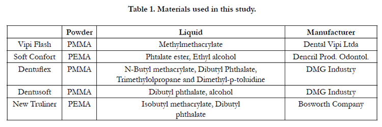

Table 1. Materials used in this study.
Twelve samples of each material were prepared and divided into four groups (n = 3) prior to performing the cytotoxicity test: Group 1: assessment of cytotoxicity immediately after the preparation of the samples; Group 2: assessment of cytotoxicity performed after storage of the samples in distilled water at 37°C for 24 hours; Group 3: assessment of cytotoxicity performed after storage of the samples in distilled water at 37°C for 48 hours; Group 4: assessment of cytotoxicity performed after soaking the samples in water at 55°C for 10 minutes.
Specimens were made in aseptic conditions to avoid contamination of the culture medium. Thus, a single operator, acting on a sterile paper surface, fashioned the specimens using sterilized instruments, protective clothing, gloves, goggles and disposable masks. After being stored and heat-treated prior to its placement in the culture medium to obtain the extracts, the specimens were placed in sterile plastic bags sealed and received ultrasonic bath (Ultrasonic Cleaner 1440D, Odontobrás, Ribeirão Preto - SP - Brazil) for 20 minutes. Then, the samples were exposed to UV light in laminar flow (Veco Brazil, Industry and Trade Equipment Ltda, Campinas - SP - Brazil) for 20 minutes for each side of the samples, aiming to eliminate the possible remaining microorganisms [9].
To analyze the cytotoxic effect of substances released by the samples, were obtained water-soluble extracts of these samples. For this, three specimens of each experimental group, after receiving the heat treatment, were placed in test tubes (Costar, Corning Incorporated, Corning - NY - USA) with 3ml of Eagle's medium, supplemented with 7.5% fetal bovine serum and 80μg/ml gentamicin, and incubated at 37°C for 24 hours. During this incubation period, the toxic substances probably have been diffused into the culture medium, thus forming the extracts which were used in the cytotoxicity test. A test tube containing 9ml of only the culture medium was stored under the same conditions, there by serving as negative control group.
The possible cytotoxic effect of the substances released by the reline was evaluated by cell culture. Thus, hamster fibroblasts (L929-Lutz the Adolfo, São Paulo - SP - Brazil) were propagated in Eagle's culture medium supplemented with 7.5% fetal bovine serum and 80μg/ml gentamicin. The cultivation of cells was done in flasks (Costar, Corning Incorporated, Corning - CA - USA) for cell cultures with a lid containing a filter that allows the passage of CO2. These flasks were incubated in an incubator for cell culture (Forma Scientific, Marietta - OH - USA) with 5% CO2 at a temperature of 37°C and controlled humidity environment. To maintain the culture, the cells were passaged into new flasks after a period of 3 days of incubation. It is noteworthy that all procedures were performed in aseptic area within the laminar flow previously disinfected with 70% alcohol. Furthermore, the materials used, except cells, were previously sterilized by UV light for 20 minutes inside the laminar flow.
For cytotoxicity analysis, the MTT test was used. By this technique, the metiltetrazolium salt is incorporated into cell culture. The succinic dehydrogenase enzyme from viable cells breaks the structure of the tetrazolium salt to produce crystals of formazan blue color, determining thereby the relative values of the intensity of blue color in specific spectrophotometer with a determinate wavelength. The greater is the mitochondrial activity, greater is the intensity of blue light and therefore the greater the number of viable cells. To perform the test, 1.0 x 10⁴ cells/ml were placed in each compartment of a plate with 96 wells, incubated in incubator with 5% CO2 at 37°C for 24 hours. After this incubation period, the culture medium was discarded, the remained cells attached to the bottom plate and 50μl of fresh culture medium were placed in each well of the plate with 50μl of the extract containing the substances released by the specimens. The plate was incubated for 24 hours in an incubator with 5% CO2 at 37°C. For each experimental group were four wells of the plate (analysis in quadruplicate). Four wells of the plate did not receive the extract of released substances and received only 100 μl of fresh culture medium supplemented with 7.5% fetal bovine serum and 80 μg/ml gentamycin (negative control group). After an incubation period of 24 hours, 10μl metiltetrazolium salt (MTT) was added to each well of the plate, which remained incubated for 3 hours at 37°C for the formation of formazan crystals, resulting from mitochondrial activity. Then, 100μl of MTT solubilizing solution were added to each well of the plate which was gently stirred until occurred dissolution of the formazan crystals. Subsequently, the analysis of mitochondrial activity was performed in Multiscan spectrophotometer at a wavelength of 570nm. All procedures were performed three times on different days.
The results were subjected to two-factor factorial analysis of variance (ANOVA) (material and water storage), also including a control group, the level of 5% significance. Moreover, after statistical evaluation, comparing the results with the control of the MTT assay, the materials were classified according to the cytotoxic effect: non-cytotoxic (cell viability above 75% in relation to control group), slightly cytotoxic (cell viability between 50 and 75% in relation to control group), moderately cytotoxic (cell viability between 25 and 50% in relation to control group) and strongly cytotoxic (cell viability below 25% in relation to control group) [10, 11].
Results
Table 2 show the means and standard deviations of absorbance values according to the reline material and treatment adopted: Group 1 to Group 4. The analysis of variance (Table 3) showed significant effect of the main factors, resin (p <0.001) and treatment (p = 0.001), but not the interaction between them. The nonsignificant interaction effect indicates independence between the factors, so that the Tukey test was used for multiple comparisons of average global resin and global averages of treatments separately. The result of these comparisons, the level of significance of 5%, is summarized in Table 2, Table 3 shows the rating of the reline according to the cytotoxic effect, considering the control group with 100% of cell viability.
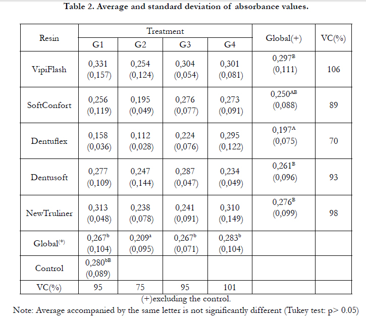
Table 2. Average and standard deviation of absorbance values.
(+)excluding the control.
Note: Average accompanied by the same letter is not significantly different (Tukey test: p> 0.05)
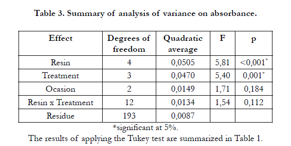
Table 3. Summary of analysis of variance on absorbance.
*significant at 5%.
The results of applying the Tukey test are summarized in Table 1.
It was observed that, regardless of treatment, resins Soft Confort and Dentuflex had the lowest averages, but only the Dentuflex was significantly lower than the others, which were all equivalent. The average number of Dentuflex was only significantly lower than the control, suggesting slightly cytotoxic effect of resin. The other resins, compared to the control group, were classified as non-cytotoxic (Table 4).
As regards treatments, the overall average of absorbance values was lower in the treatment group and the other averages were higher and equivalent , even when compared to control group. This indicates that heat treatment decreased the number of viable cells, and Soft Comfort and Dentuflex resins were classified as slightly and moderately cytotoxic, respectively (Table 4). The water storage of the samples for 24 or 48 hours had no effect on cell viability.
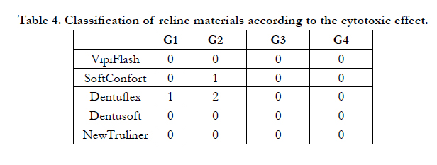
Table 4. Classification of reline materials according to the cytotoxic effect.
Discussion
Biocompatibility studies are needed to evaluate the biological behavior of different materials. Many studies have been conducted to test the cytotoxicity of acrylic resins for denture base and reline [12-14]. However, the current literature reveals limited information about the cytotoxic effect of soft relines materials. Furthermore, studies have found based on imported resins scarcely used by dentists in Brazil. Thus, this study aimed evaluating the cytotoxicity of different types of resilient lining materials, used in Brazil, according to the time of water storage and heat treatment. The hypothesis tested was that the cytotoxicity of these reline could decrease according to the time and treatment. Such factors may reduce the amount of residual monomers by the combined mechanisms of water diffusion and continuously polymerization reaction.
Based on a literature review, Jorge et al. [7] concluded that the cytotoxic effect of the acrylic resins may be influenced by the composition of each material, the proportion powder/liquid or by the methods of polymerization of each resin. The variations in chemical composition and purity of the commercial resin systems, the degree of monomers conversion and their manipulation variables may influence its biological and physical properties [3]. Thus, some technical aspects are relevant and may help reduce the residual monomer, as the conditions and storage time. Usually, the procedures for handling these materials are made empirically by laboratory technicians, and the process of incomplete polymerization or performed with technical failures can provide a high content of residual monomer.
By coming into direct contact with the mucosa of the patients, monomers less toxic are part of the composition of the relining materials [15]. With regard to the relining materials, their formulation is commercially available in powder form, based on poly ethyl methacrylate and liquid, which may contain of monomers such as butyl methacrylate, isobutyl, 2-hydroxyethyl methacrylate, methacryloyloxy ethyl propionate and 1,6 hexanediol dimethacrylate. Furthermore, these materials have in their compositions initiators and plasticizers which are phthalates and benzoyl peroxide added to the polymer. In addition, an activator, which is usually a tertiary amine, is added also, since they are not heat-activated.
After statistical analysis, it was observed that, regardless of treatment, cells exposed to the Soft Confort and Dentuflex showed lower absorbance values and, therefore, were considered more toxic. Only the absorbance values of the Dentuflex material were considered significantly lower than the control group values, suggesting slightly cytotoxic effect of this resin. The other resins, compared to the control group, were classified as non-cytotoxic. These results are in agreement with those found by [8], who observed that the majority of the studied relines had high cell viability and good biocompatibility.
The Dentuflex reline presents in its liquid various components, including n-butyl methacrylate, dibutyl phthalato, trimethylolpropane and dimethyl-p-toluidine. While other resins present in their composition a minor quantity of chemical agents, which could explain the cytotoxicity of Dentuflex resin. The Dentusoft resin liquid which is a tissue conditioner, is composed only of dibutyl phthalate and alcohol. According to its manufacturer, Vipi Flash, consists of methylmethacrylate monomer and polymethylmethacrylate and Soft Confort of ethyl alcohol and plasticizer. The imported reliner New Truliner, selected for this study for comparison with national resins, presents in its composition isobutyl methacrylate monomer [16]. However, studies to evaluate the biocompatibility of the monomers mentioned are necessary. One of the procedures used to reduce the amount of residual monomer and to improve the properties of the acrylic resins is the storage of dentures in water. The storage of samples could reduce the cytotoxicity of acrylic resins because some part of residual monomer found among the polymer chains can diffuse into water. However, the results of this study showed that storage of the samples in water for 24 or 48 hours had no effect on cell viability. The results of this study are consistent with those found by Tay et al. [8], who concluded that the water storage did not reduce the cytotoxicity of some resilient reline. Munksgaard EC [17] concluded that the solubility of phthalates found in reline is 20 times higher in saliva than in water. Thus, studies to evaluate the cytotoxicity of lining materials after storage in saliva are suggested.
Studies show that the hot water immersion, the molecules of residual monomer can be diffused quickly, reaching the remaining free radicals, providing an additional polymerization reaction. Furthermore, during hot water immersion, part of the monomer found between polymer chains diffuses into the water. Urban et al. [18] evaluated the effect of the water bath at 55°C for 10 minutes on the residual monomer into acrylic resins. The authors suggested that, clinically, this treatment could reduce amounts of these components released. On the other hand, in the present study, the heat treatment decreased the number of viable cells. Soft Comfort and Dentuflex resins were classified as slightly and moderately cytotoxic, respectively. One possible explanation for the increased cytotoxicity after heat treatment is that when the resins are immersed in warm water, the content of residual monomer present in the polymerized material can be released and, simultaneously, the water molecules are absorbed by the resin [19]. These phenomena are dependent on the time and thus, the content of residual monomer molecules and water in the polymeric structure was changed during storage until reached the equilibrium. Therefore, the stability between absorption and diffusion of water of the potentially cytotoxic compounds may have occurred more slowly into Soft Comfort and Dentuflex resins with treatment, and toxic substances can have being released from samples to culture medium during of extracts manufacture.
One limitation of this study is the use of only one test to assesscell viability. When a material is assessed, the absence of cytotoxicity does not confer full knowledge of their biocompatibility. Even with standardization of cytotoxicity tests, several aspects should be evaluated, especially in relation to cellular metabolism. The MTT assay was used based on different studies in the literature [12, 13]. However, it is important the application of various tests for the analysis of the biomaterials cytotoxic effect. According to Miret et al. [20], the best way to assess the cytotoxicity of a compound is employing a series of tests that focus on different aspects of cell death. [12], for instance, concluded that the MTT assay was less sensitive than the incorporation of 3H-thymidine, which detected statistically significant difference between experimental groups and control test. Thus, it is considered important the achievement of various analyzes that could detect effects of cellular components in different materials, since the tested substances for example, can not change the mitochondrial activity of the cells but cause damage to its membrane. Moreover, as the results of the initial cytotoxicity tests have limitations as to its direct correlation with clinical situations, clinical researches in vivo are also suggested.
Conclusion
Within the limitations of this in vitro study, it was concluded that: the reliner Dentuflex was slightly cytotoxic; the other resins, compared to the control group, were classified as non-cytotoxic. The storage in water for 24 or 48 hours did not affect the cytotoxicity of reline materials tested and heat treatment reduced the number of viable cells, and Soft Comfort and Dentuflex resins were classified as slightly and moderately cytotoxic, respectively.
Further studies are needed to verify the cytotoxicity of these materials under the same conditions, using other parameters for analysis of cell viability.
Acknowledgment
This work was supported by grants from Sao Paulo State Research Foundation (FAPESP - Grant 2011/18548-7).
References
- Braden M, Wright PS, Parker S (1995) Soft Lining Materials – A Review. Eur. J. Prosthodont Rest Dent 3(4): 163-174.
- Anusavice KJ (1996) Phillip’s science of dental materials. (10th edn), Philadelphia, Saunders. 709p.
- Lefebvre CA, Knoernschild KL, Schuster GS (1994) Cytotoxicity of eluates from light-polymerized denture base resins. J Prosthet Dent 72(6): 644-650.
- Costa CAS (2001) Cytotoxicity testing in cell culture. In: ESTRELA, C. Scientific methodology: teachig and research in dentistry. (1st edn) São Paulo, Artes Médicas LTDA,cap. 9: 145-160.
- Arima T, Murata H, Hamada T (1996) Analysis of composition and structure of hard autopolymerizing reline resins. J Oral Rehabil 23(5): 346-352.
- Tang AT, Li J, Ekstrand J, Liu Y (1999) Cytotoxicity tests of in situ polymerized resins: methodological comparisons and introduction of a tissue culture insert as a testing device. J Biomed Mater Res 45(3): 214-222.
- Jorge JH, Giampaolo ET, Machado AL, Vergani CE (2003) Cytotoxicity of denture base acrylic resins: a literature review. J Prosthet Dent 90: 190-193.
- Tay LY, Herrera DR, Quishida CC, Carlos IZ, Jorge JH (2012) Effect of water storage and heat treatment on the cytotoxicity of soft liners. Gerodontology 29: 275-280.
- Pelka M, Danzl C, Distler W, Petschelt A (2000) A new screening test for toxicity testing of dental materials. J Dent. 28(5): 341-345.
- Ciapetti G, Cenni E, Pratelli L, Pizzoferrato A (1993) In vitro evaluation of cell/biomaterial interaction by MTT assay. Biomaterials 14(5): 359-364.
- Harrison A, Huggett R (1992) Effect of the curing cycle on residual monomer levels of acrylic resin denture base polymers. J Dent 20(6): 370-374.
- Jorge JH, Giampaolo ET, Vergani CE, Machado AL, Pavarina AC, et al. (2004) Cytotoxicity of denture base resins: effect of water bath and microwave postpolymerization heat treatments. Int J Prosthodnt 17(3): 340-344.
- Jorge JH, Giampaolo ET, Vergani CE, Pavarina AC, Machado AL, et al. (2009) Effect of microwave postpolymerization treatment and of storage time in water on the cytotoxicity of denture base and reline acrylic resins. Quintessence Int 40: 93-100.
- Campanha NH. Pavarina AC, Giampaolo ET, Machado AL, Carlos IZ, et al. (2006) Cytotoxicity of hard chair-side reline resins: effect of microwave irradiation and water-bath post-polymerization treatments. Int J Prosthodont 19(2): 195-201.
- Ozdemir KG, Yilmaz H, Yilmaz S (2009) In vitro evaluation of cytotoxicity of soft lining materials on L929 cells by MTT assay. J Biomed Mater Res B Appl Biomater 90: 82–86.
- Upadhyay P, Bhaskar S (2000) Real time monitoring of lymphocyte proliferation by an impedance method. J Immunol Methods 244(1-2): 133-137.
- Munksgaard EC (2004) Leaching of plasticizers from temporary denture soft lining materials. Eur J Oral Sci 112: 101–104.
- Urban VM, Machado AL, Vergani CE, Machado AL (2009) Effect of waterbath post-polymerization on the mechanical properties, degree of conversion, and leaching of residual compounds of hard chairside reline resins. Dent Mater. 25: 662–671.
- Tsuchiya H, Hoshino Y, Tajima K, Takagi N (1994) Leaching and cytotoxicity of formaldehyde and methyl methacrylate from acrylic resin denture base materials. J Prosthet Dent 71(6): 618-624.
- Miret S, De Groene EM, Klaffke W (2006) Comparison of in vitro assays of cellular toxicity in the human hepatic cell line HepG2. J Biomol Screen 11: 184-93.


