Microleakage of Different Pit and Fissure Sealants
Alqarni M1*, Elkwatehy W2
1 Faculty of Dentistry, Umm Al-Qura University, Makkah, Saudi Arabia.
2 Faculty of Dentistry, Department of Public Health and Preventive Dentistry, Mansoura University (Egypt) and Umm Al-Qura University (KSA), Saudi Arabia.
*Corresponding Author
Mansour Alqarni,
Department of Public Health and Preventive Dentistry,
Faculty of Dentistry, Umm Al-Qura University, Makkah, 21514, Saudi Arabia.
Tel: 00966544557554
E-mail: mansour.qarni@yahoo.com
Received: September 15, 2017; Accepted: October 26, 2017; Published: October 28, 2017
Citation:Alqarni M, Elkwatehy W. Microleakage of Different Pit and Fissure Sealants. Int J Dentistry Oral Sci. 2017;4(10):532-536.doi: dx.doi.org/10.19070/2377-8075-17000105
Copyright: Alqarni M©2017. This is an open-access article distributed under the terms of the Creative Commons Attribution License, which permits unrestricted use, distribution and reproduction in any medium, provided the original author and source are credited.
Abstract
Background/Aim: Sealing pits and fissures is considered to be an effective way of preventing caries initiation. The present in vitro study was carried out to evaluate the microleakage and sealing ability of Helioseal F, Seal-it and GCP Glass seal and Compared sealing ability of GCP Glass seal with resin fissure sealants.
Materials and Method: 80 extracted premolars free of caries, cracks or morphogenic diversity were collected and distributed equally in four groups (20 in each). Group-I (Helioseal F), Group-II (Seal-it), Group-III (GCP Glass seal) and Group-IV (GCP Glass seal without etching). Samples were cleaned with pumice slurry and etched with phosphoric acid etchant except Group-IV without etching. Air and water sprays were used for washing and drying, the teeth were treated with sealants followed by thermocycling and immersion in methylene blue dye for 48 hours. The samples were sectioned and examined under a stereomicroscope at x15 magnification then the score was given for microleakage. The sections were photographed to show a score of “0”,“1”,“2” or “3” microleakage and the data was statistically analyzed with Chi-Square test.
Results: Seal-it material was the best sealant material as it was showing significantly least microleakage as compare to Helioseal F and GCP Glass seal and the microleakage was found to be the highest for the GCP Glass seal with etching.
Conclusion: According to the results, the Seal-it material was found to be an excellent dental material for pits and fissures and GCP glass seal should be used without etchant.
2.Introduction
3.Aim of The Work
4.Materials and Methods
4.1 Fissure Sealant Application
4.2 Microleakage Assessment
4.3 Statistical Analysis
5.Results
6.Discussion
7.Conclusions
8.Recommendationsn
9.Acknowledgments
10.References
Keywords
Pit and Fissure Sealants; Stereomicroscope; Microleakage.
Introduction
In spite of numerous prevention methods, dental caries is still a highly prevalent disease in the world [1]. Effects of caries preventive measures are greater on smooth surfaces, while occlusal caries remains a problem. It has been shown that a carious lesion most frequently occurs in pits and fissures of occlusal surfaces, primarily due to their specific anatomy, which is considered to be an ideal site for bacteria and food remnants retention [2, 3].
Sealing pits and fissures is considered to be an effective way of preventing caries initiation. A fissure sealant is defined as a material that is placed in pits and fissures of teeth in order to prevent or arrest the development of dental caries [4].
Microleakage or marginal leakage may be defined as the ingress of oral fluid into the space between the tooth and restorative material. So, the ability of a sealant to prevent microleakage is important [5].
Microleakage is considered as the main problem with direct restorative procedures and one of the main reasons for restoration failure. A dental sealant is successful only if it firmly adheres to the enamel surface, and protects pits and fissures from the oral environment [6].
Many studies have examined microleakage of sealants placed by the conventional method of acid etching of the enamel. All show some microleakage but results vary [7-9].
The present study represents an attempt to detect and compare the microleakage of three dental fissure sealants.
Aim of The Work
The present study was undertaken to evaluate the microleakage and sealing ability of Helioseal F, Seal-it and GCP Glass seal and Compare the sealing ability of GCP Glass seal with resin fissure sealants.
Materials and Methods
The present study was carried out in the Department of Preventive Dentistry, Umm Alqura University, Makkah, Saudi Arabia. This was an in-vitro experimental study.
Eighty extracted maxillary and mandibular Premolars with intact occlusal surfaces were used for the study. The teeth were free of cracks, caries and restorations. Prior to the study, the occlusal surfaces of teeth were cleaned with pumice slurry using brushes at low-speed handpiece.
The Eighty premolars were randomly assigned into four groups of twenty teeth each, according to the material used as pit and fissure sealant. Group-I: Helioseal-F (IvoclarVivadent AG, Schaan / Liechtenstein), Group-II: Seal-it (Spident, Incheon, South Korea), Group-III: GCP Glass seal and Group-IV: GCP Glass seal without etching(GCP, Ridderkerk, Netherlands).
The teeth were etched with 37% phosphoric acid etchant gel for sixty seconds, except group IV without etching, to provide more surface area with micro porosities which allow making materials flow in those areas which will enhance the bonding between material and tooth interface. Oil free air and water sprays were used for twenty seconds to completely rinse the acid and dry the teeth. The sealant was placed over the pit and fissure area respectively. The sealants were cured under the light cure for thirty seconds.
To simulate oral conditions, the sealed teeth were immersed in ready-made artificial saliva in plastic containers for one week in an incubator at 37°C. Prior to testing, the teeth were thermocycled 500 times at 5 ± 2°C to 55 ± 2°C, with a dwell time for thirty seconds.
The surfaces of specimens were coated with two layers of nail polish except for one millimeter around the sealant. The apices of the teeth were sealed with sticky wax. The specimens were stored in 5% methylene blue for 48 hours, then were rinsed with tap water and sectioned bucco-lingually in the mesial and distal surfaces of each tooth with an Isomet low-speed saw (TECHCUT 4TM\Rancho Dominguez, California). Three sections made of each specimen to assess dye penetration under x15 magnification of a stereomicroscope (Meiji Techno\ San Jose, California).The sections were photographed to show scores of microleakage.
The degrees of microleakage were scored by a single observer depending upon leakage level using criteria by Colley et al., [8] as the following:
0 = No dye penetration.
1 = Dye penetration restricted to the outer half of the sealant.
2 = Dye penetration to the inner half of the sealant.
3 = Dye penetration into the underlying fissure.
Data were collected, tabulated and subjected to analysis using SPSS program version 22.0. The comparison between groups carried out using chi-square test. The level of significance was considered at P value ≤ 0.05.
Results
The present study was conducted to evaluate microleakage of different sealant materials. A total number of 240 sectioned surfaced of teeth (60 sections in each group) were examined under stereomicroscope, to evaluate sealant microleakage (Figure 1).
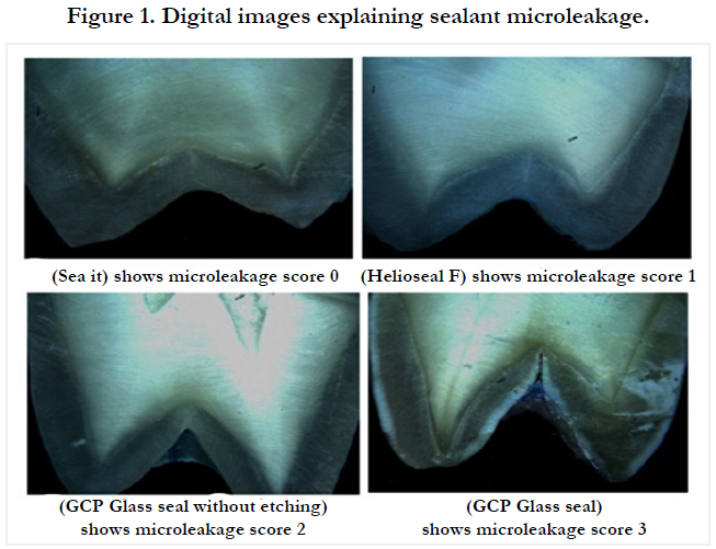
Figure 1. Digital images explaining sealant microleakage.
(Table 1) and (Graph 1) shows the distribution of microleakage scores among different sealant materials. There was statistically significant difference among the different sealant materials (chisquare test) and P-value ≤ 0.05.
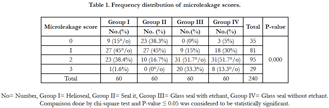

Table 1. Frequency distribution of microleakage scores.
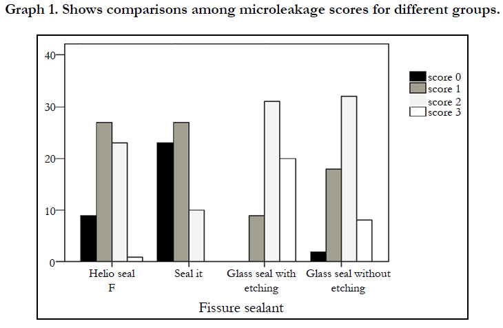
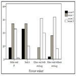
Graph 1. Shows comparisons among microleakage scores for different groups.
(Table 2) shows the comparison among different sealant materials. Sealant materials of all groups were found to be comparable as the degree of microleakage between them were highly significant (p=0.000). On comparison between group I and group-II it showed statistically significant difference (p=0.007).
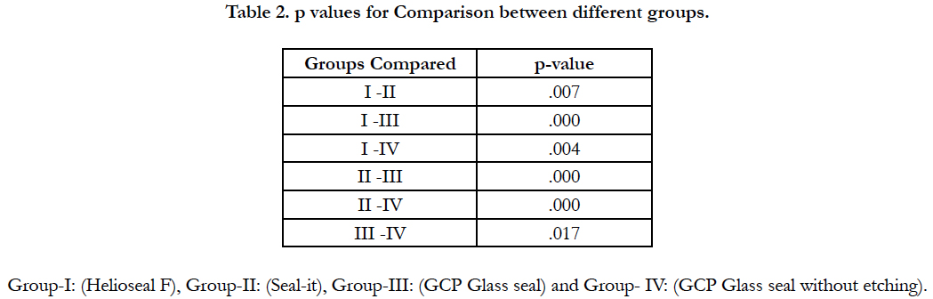
Table 2. p values for Comparison between different groups.
Comparison between Group-I and group-III showed highly significant difference (p=0.000). Also, when group- I was compared with group-IV it showed statistically significant difference (p=.004). Comparison between Group-II vs group-III and group- IV showed the same statistically significant differences (p=.000). Comparison between group-III vs group-IV showed the least statistically significant difference (p=.017). All comparisons were done by chi-square test.
Discussion
Pit-and-fissure sealants have been considered an outstanding adjunct to oral health care preventive strategies in the decrease of occlusal caries onset and/or progression [10]. The properties of an ideal sealing material include biocompatibility, retention, resistance to abrasion and wear [11]. Sealant bonding to enamel is also important because microleakage at the tooth-material interface can lead to treatment failure. The relative effectiveness of different types of sealants has yet to be established. There is rapid raise in the development of restorative materials and techniques in determining the capability of each new material to adapt to the tooth without microleakage.
In the present study, the in vitro microleakage of three materials used as pit-and-fissure sealants were evaluated. All materials were applied without enameloplasty in order to observe the behavior of these materials without removal of tooth substance. The specimens were thermocycled to reproduce the different temperatures to which the teeth are subjected during eating and drinking under clinical conditions.
in vitro microleakage tests carried out with dye penetration technique are stricter than those performed in the oral cavity [12]. This is most likely due to several factors such as the dye being more easily diffused than bacteria and their by-products and the fact that buildup of proteins in the marginal opening/gap may improve the seal. On this basis, they are likely to respond even better on a clinical level [13].
The use of pumice prophylaxis followed by acid etching in all group except group IV was chosen in the present study because it is adopted by most dentists for application of sealants and is also recommended by the manufacturer [14].
Because scarcity of studies has been done to evaluate microleakage of glass carbomer sealant, the present study was performed to evaluate the microleakage of this material in comparison with Helioseal F and Seal-it in vitro. The study results showed significant differences in the sealant penetration among the groups of sealant materials. Seal-it material was found better for sealing ability as it was showing significantly least microleakage as compare to Helioseal F, and GCP Glass seal. On other hands, the microleakage was found to be highest for the GCP Glass seal with etching.
Within the recognized limitation of an in vitro study, the above results are viewed as the theoretical level of leakage which may or may not occur in vivo but may be accepted as an aid for selection of a good sealant material before placement of a pit & fissure sealant.
The sealing ability of resin fissure sealant is depending on the penetration of resin into a morphologically porous enamel layer created by phosphoric acid etching. The high sealing ability of Seal-it may be due to its high flowability which allows with better penetration of etched enamel and formation of resin tags better than Helioseal F. Although phosphoric acid etching effectively increases the bond between the enamel and sealant, it presents problems because of its bad taste and time- consuming procedure. To overcome these problems glass ionomers based compounds were developed to use as fissure sealant [15]. Recently nano-particles developed glass ionomers sealant (GCP Glass seal) used in the present study to improve sealing ability and avoid using of etching. It gives reasonable results compared with Helioseal F and Seal-it but its sealing ability less than them.
The difference was statistically significant. The decreased sealing ability of GCP Glass seal in the present study may be due to complete dryness of the teeth during application of sealant as glass ionomers hydrophilic and wet surface help its retention. Another cause for the decreased sealing ability may be due to thermocycling as glass ionomer more affected by heat than resin [16].
According to manufacturer instructions glass seal should be used without acid etching but in the present study it was used with etching in a separate group to evaluate the effectiveness of etching on sealing ability. The results show statically significant highest leakage in Glass seal with etchant group compared with other groups. The explanation of that may be the flow of glass seal on a smooth surface and chemical bonding with enamel is better than on the rough surface, also, micro-tags and low flowability of glass seal may lead to entrapment of air bubbles which lead to increased microleakage. Another factor may be a formation of minerals or salts due to the reaction between acid and enamel hydroxyapatite which prevent intimate contact between sealant and enamel.
Increased marginal microleakage was observed for group III (GCP Glass seal with etching) compared with group IV (GCP Glass seal without etching), these findings disagreed with that reported by Birkenfeld LH and Schulman A [17]. They reported that etching were significantly reduced microleakage in teeth sealed with a GI bond fissure sealant.
The direction of research will have to be involved in simulating in vitro studies on microleakage with in vivo studies. There is a poor correlation between the extent of microleakage found in vitro & in vivo studies. in vitro studies definitely give a path to compare the sealing abilities of the materials. This elusive ability to prevent microleakage demands controlled clinical studies, which will draw the conclusion about the micromechanical bond and its strength between the fissure sealant and tooth structure [18].
Conclusions
The results observed in the present study suggested that Seal-it was the best material amongst three different materials in terms of least microleakage. Helioseal F gave the promising results where as GCP Glass Seal was the least successful pit and fissure sealant material.
Recommendations
1. The use of glass seal should be according to manufacturer instructions and no need for acid etching before its application.
2. Further studies are needed to evaluate the sealing ability of different sealing materials in vivo.
Acknowledgments
I would like to extend special thanks to Advance Technology Dental Research Center staff at King Abdulaziz University.
References
- Bahrololoomi Z, Soleymani A, Heydari Z. in vitro comparison of microleakage of two materials used as pit and fissure sealant. J Dent Res Dent Clin Dent Prospect. 2011;5(3):83–86. PubMed Central PMCID: PMC3442452.
- Hopcraft MS, Morgan MV. Pattern of dental caries experience on tooth surfaces in an adult population. Community Dent Oral Epidemiol. 2006 Jun;34(3):174-183. PubMed PMID: 16674749.
- Rohr M, Makinson OF, Burrow MF. (1991): Pits and fissures morphology. ASDC J Dent Child. 1991 Apr;58(2):97-103. PubMed PMID: 2050885.
- Feigal RJ, Donly KJ. The use of pit and fissure sealants. Pediatr Dent. 2006 Apr;28(2):143 -150. PubMed PMID: 16708789.
- Trowbridge HO. Model systems for determining biologic effects of microleakage. Oper Dent. 12:164-172. PubMed PMID: 3507000.
- Marković D. Microleakage, adaptation ability and clinical efficacy of two fluoride releasing fissure sealants. Vojnosanit Pregl. 2012 Apr;69(4):320- 325. PubMed PMID: 22624423.
- Ovrebo RC, Raadal M. Microleakage in fissures sealed with resin or glass ionomer cement. Scand J Dent Res. 1990 Feb;98(1):66- 69. PubMed PMID: 2183346.
- Cooley RL, McCourt JW, Huddleston AM, Casmedes HP. Evaluation of a fluoride-containing sealant by SEM, microleakage, and fluoride release. Pediatr Dent. 1990 Feb;12(1):38- 42. PubMed PMID: 2399181.
- Park K, Georgescu M, Scherer W, Schulman A. Comparison of shear strength, fracture patterns, and microleakage among unfilled, filled, and fluoride-releasing sealants. Pediatr Dent. 1993 Dec;15(6):418-421.PubMed PMID: 8153005.
- Simonsen RJ. Pit and fissure sealant: review of the literature. Pediatr Dent. 2002 Oct;24(5):393-414. PubMed PMID: 12412954.
- Pérez-Lajarin L, Cortés-Lillo O, García-Ballesta C, Cózar Hidalgo A. Marginal microleakage of teo fissure sealants: a comparative study. J Dent Child. 2003 Apr;70(1):24-28. PubMed PMID: 12762604.
- Jacobs MS, Windeler AS. An investigation of dental luting cement solubility as a function of the marginal gap. J Prosthet Dent. 1991 Mar;65(3):436- 442. PubMed PMID: 2056466.
- Baldissara P, Comin G, Martone F, Scotti R. Comparative study of the marginal microleakage of six cements in fixed provisional crowns. J Prosthet Dent. 1998 Oct;80(4):417-422. PubMed PMID: 9791787.
- Qadri GW, Noor SN, Mohamad D. Microleakage assessment of a repaired, nano-filled, resin-based fissure sealant. Pediatr Dent. 2009 Oct;31(5):389- 394. PubMed PMID: 19947133.
- Nahid, A, Zahra, B, Yasaman, R. Evaluation of the effect of enamel preparation on retention rate of fissure sealant. Contemp Clin Dent. 2012 Dec;3(4):380–382. PubMed Central PMCID: PMC3636824.
- Subramaniam P, Jayasurya S, Girish Babu KL. Evaluation of glass carbomer sealant and a moisture tolerant resin sealant - A comparative study. Intl J Dental Sci Res. 2015;39(5):429-434.
- Birkenfeld LH, Schulman A. Enhanced retention of glass ionomer- sealant by enamel etching: a microleakage and scanning electron microscopy study. Quintessence Int. 1999 Oct;30(10):712-718. PubMed PMID: 10765856.
- Joshi K, Dave B, Joshi N, Rajashekhara B, Jobanputra L, Yagnik K, et al. Comparative Evaluation of Two Different Pit & Fissure Sealants and a Restorative Material to check their Microleakage – An in vitro Study. Journal of International Oral Health. 2013 Aug;5(4):35–39.


