Towards Bioactive Dental Restorative Materials with Chitosan and Nanodiamonds: Evaluation and Application
S R Grobler1*, V T Perchyonok2, R Mulder1, D Moodley1
1 Faculty of Dentistry, Oral and Dental Research Institute, University of the Western Cape, Private Bag X1, Tygerberg, Cape Town, South Africa.
2 Research and Innovations, VTPCHEM PTY LTD, Glenhuntlyrd, Glenhuntly, Melbourne, Australia.
*Corresponding Author
S R Grobler,
Oral and Dental Research Institute,
Faculty of Dentistry, University of the Western Cape, Private Bag X1,
Tygerberg 7505, Cape Town, South Africa.
Tel: +2721 9373023/4
Fax: +27219373025
E-mail: srgrobler@uwc.ac.za
Received: August 10, 2015; Accepted: September 04, 2015; Published: September 09, 2015
Citation: S R Grobler, V T Perchyonok, R Mulder, D Moodley (2015) Towards Bioactive Dental Restorative Materials with Chitosan and Nanodiamonds: Evaluation and Application. Int J Dentistry Oral Sci. 2(9), 147-154. doi: dx.doi.org/10.19070/2377-8075-1500031
Copyright: S R Grobler© 2015. This is an open-access article distributed under the terms of the Creative Commons Attribution License, which permits unrestricted use, distribution and reproduction in any medium, provided the original author and source are credited.
Abstract
Background/purposes: Recently various articles showed beneficial effects of the addition of different bioactive compounds towards dental materials. Therefore, the aim of this work was to evaluate the effect of the addition of bioactive materials and combination thereof (chitosan/nanodiamond or cyclodextrin/nanodiamond) to a dental composite.
Materials and Methods: The flowable composite Premise by Kerr was used as the standard control dental material. Premise was also modified to contain: 10% nanodiamonds/Premise, 10%chitosan/nanodiamonds/Premise, 10% cyclodextrin/ nanodiamonds/Premise and 10% cyclodextrin/Premise and tested for their dentin bond strength, volumetric shrinkage, Vickers hardness and cytotoxicity.
Results and Conclusion: Beneficial effects of the addition of different bioactive compounds towards dental materials were proved. A higher shear bond strength (p < 5%) was found after 3 months of Premise treated with nanodiamonds, chitosan, cyclodextrin (CD) and combinations thereof than the control Premise. The sequence for the Vickers hardness was: CD (32.5) < nano (34.8) < CD Nano (38.8) < Premise (39) < Chitosan Nano (42.2). Chitosan was found to increase the mouse 3T3 fibroblast cell survival rate (113%), while nanodiamonds (92%) and the combination of chitosan + nanodiamonds (93%) showed little cytotoxicity. The shrinkage was lower for all the additions than for Premise alone. Nanodiamonds and the combination chitosan + nanodiamonds showed little cytotoxicity towards mouse 3T3 fibroblast cells.
2.Introduction
3.Materials and Method
3.1.Shear bond strength tests for dentin bonding
3.2.Volumetric Shrinkage
3.3 Vickers Hardness
3.5 Cytotoxicity
4.Results
4.1.Bond strength testing
4.2.Volumetric Changes and Vickers Hardness of the modified composites
4.3 Cytotoxicity
5.Discussion
5.1 Dentin shear bond strength
5.2 Volumetric Shrinkage and Vickers Hardness
5.3 Cytotoxicity
6.Conclusion
7.Acknowledgement
8.References
Keywords
Nanodiamonds; Chitosan; Cytotoxicity; Dental Material.
Introduction
Traditional dentistry has now evolved to a stage where, under optimal conditions, success rates of up to over 90% have been reported for some procedures (for example, for dental implants) after a period of 10 years [1]. The development of new materials with enhanced properties, careful attention to factors that influence the technique sensitivity of some procedures, and a generally greater awareness of oral health issues among the population have all helped to contribute to these improvements in treatment outcomes [2]. Bioactive materials have evolved over the past three decades from relatively specialized, highly biocompatible, but low-strength dental materials into product compositions for expanded clinical use in restorative dentistry [3].
Chitosan [4] which is produced commercially by de-acetylation of chitin is a natural polysaccharide composed of randomly distributed β- 1, 4-linked D-glucosamine and N-acetyl α-glucosamine. Chitosan is non-toxic, biocompatible, bio-degradable and has muco-adhesive properties and as a result became widely used in the pharmaceutical field as a carrier system for drugs, hormones, proteins, enzymes and genes [5-8]. Chitosan can be successfully used as a drug carrier because it will solubilize and degrade in an acidic environment with the resultant release of the drug [9]. Chitosan is hypoallergenic and has natural antibacterial properties, which further support its use in the army as field bandages [10]. Furthermore, very important findings were that antioxidantchitosan hydrogels (that of resveratrol, propolis and β-carotene) were found to significantly improve the bond strength to dentine with or without phosphoric acid pre-treatment [11] as many other hydrogels do [12].
Nanodiamonds were first synthesized by Soviet scientists [13] in 1962 through the detonation of trinitrotoluene (TNT) with hexogen (RDX) in a closed chamber. Today it can be prepared at room temperature at low cost. There are different types of nanodiamonds [13] namely: single-walled carbon nanotubes, multi-walled carbon nanotubes, carbon black and those with a single-particle size of 2-10 nm. Nanodiamonds (NDs) are carbon nanoparticles with a diamond like octahedral structure of about 2 to 8 nm in diameter [13, 14]. Like diamonds it is chemically stable, stiff, strong and extremely hard. Like nanomaterials, it has a small size, large surface area, and high adsorption capacity.
Thus NDs have superior physical and chemical properties compared to conventional materials and therefore render the ND particles ideal additives to formulation and improvement of conventional dental composites.
Considering that the application of nanoparticles as fillers in polymeric matrices has shown encouraging results in the strengthening of the materials [15, 16] it could be expected that the incorporation of ND nanoparticles into dental polymeric materials could have an enhancing effect on the mechanical properties of the resulting nano-composites. Although the efficiency of using ND in polymeric materials has been suggested [17, 18] controversial findings have been reported in the literature which were mainly attributed to the interactions between the nanoparticles themselves, which tend to form agglomerates which acted as points of stress concentration.
The aim of this study was to evaluate the effect of the addition of ND as well as bio-actives such as chitosan or cyclodextrin and the combination thereof (chitosan/nanodiamond or cyclodextrin/ nanodiamond) to the flowable composite Premise. Therefore, the dentin bond strength, volumetric shrinkage, Vickers hardness and the cytotoxicity were investigated.
Materials and Method
Chitosan (Aldrich, Australia), β-cyclodextrin (Aldrich, Australia) glycerol (Sigma, USA), glacial acetic acid (E. Merck, Germany) were used as received. The degree of de-acetylation of typical commercial chitosan used in this study was 87%. Chitosan with molecular weight 2.5x 10³ KD was used in the study. The isoelectric point was 4.0–5.0. Nanodiamonds were purchased from Ebersoles, (25 carats, 5 grams, size (2-8 microns), Grit 14,000) and used as received.
The flowable composite Premise by Kerr (California, USA) Lot: 4485575 exp 2014-02 shade A3) was used as the standard control material. Furthermore, the mentioned Premise was also modified to 10% nanodiamonds: Premise, 10% Chitosan/nanodiamonds: Premise, 10%CD/nanodiamonds: Premise and 10% CD: Premiseand tested.
Extracted non-carious, intact, human molars stored in water containing a few crystals of thymolat 4°C,were used within two months. Samples were checked before use for any damage caused by their removal. The roots of the teeth were removed with a separating disc and the occlusal enamel removed by grounding wet on 60-grit silicon carbide (SiC) paper. The teeth were embedded in PVC (Consjit Tubing, SA PVC, JHB, RSA) pipe containers with cold cure acrylic resin so that the grounded occlusal surfaces projected well above the resin. The 10mm length pipes were put on a glass surface with one end blocked by the glass and the embedding done through the open end. Immediately after embedding the occlusal surfaces were ground wet with 180-grit followed by 600-grit SiC on a polishing machine to expose the superficial dentin. The samples were washed under a stream of tap water. A standardized zig (Ultradent ISO A2-70) with an internal diameter of 2.5 mm and height of 3 mm was used to shape the modified composite resin studs. Two of these studs were then bonded to the polished dentin surface of each tooth via the bonding agent Premise.
In this way 48 teeth samples (each containing 2 studs) were prepared and divided into 6 groups of 8 each and stored in a solution of artificial saliva. These groups were then treated as outlined in Table 1. After 24 hours one stud of each tooth was tested for shear bond strength and the other one after 3 months. An Instron Universal Testing Machine at a crosshead speed of 0.5mm/minute was used to test the de-bonding strength.
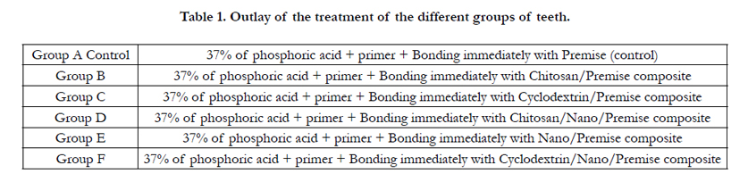

Table 1. Outlay of the treatment of the different groups of teeth.
Volumetric change was measured with an electronic mercury dilatometer [19-21]. Five samples from each material group were light cured for 35.0 seconds at 800mW/cm2 (Dentsply/Caulk Spectrum 800 halogen). The curing output was monitored with a Caulk (Milford, Germany) radiometer to ensure an output of 800mW/cm2 ± 50mW/cm2. Calibration of the electronic mercury dilatometer was done as described previously prior to every specimen test [19-21]. The Teflon specimen holder has a hole with a diameter of 5.0mm and a height of 2.5mm resulting in the specimen volume of 49.087mm3. The dilatometer was kept in a temperature controlled incubator at 25°C ± 1°C. The room temperature was kept constant at 25°C ± 1 during dilatometry testing. Therefore only the effect of polymerization shrinkage from a monomer to a polymer remained.
The surface microhardness was determined with a Vickers Hardness tester (Zwick-Roelldurometer, ZHV1/2 Micro-Vickers, Italy) using a Vickers diamond indenter with a load of HV0.5 (500gf) and a dwell time of 15 seconds [22]. Ten samples were prepared for each material combination under laboratory conditions for the Vickers Hardness. A standard Teflon mould was used with a diameter of 5.0mm and a height of 2.5mm resulting in a specimen volume of 49.087mm3. The light cured samples were placed in the specimen holders on a moist paper towel and kept at 34°C ± 1 for 48 hours in a temperature controlled incubator. The surface of the sample was prepared with 1000 grit silicon carbide paper and then 2000 grit (3M, Massachusets, USA) until about 100μm had been removed from the surface layer. The five indentations of the five samples were taken and an average calculated for each material. Five indentations were made on each of the ten samples in accordance with ASTM E384: Standard Test Method for Knoop and Vickers Hardness of Materials. The distance between indentations was approximately 10μm.
The cytotoxicity was examined as previously reported [23].Briefly, the cells were first grown to near confluency, diluted to a final cell suspension containing approximately 3 ×10⁵ cells/ml and plated out insets of 96 well plates. Chitosan or nanodiamands or a combination thereof (chitosan/nanodiamond) were then added to the growth medium at a concentration of 1 mg/ml. Two hundred μl of each group was added to 20 wells in the 96 well plates. Medium without any gels was used as controls. After 24 hours the well-known MTT colorimetric assay was used to evaluate the cell survival rate. Absorbance was measured at wavelength 540nm on a spectrophotometer to determine the number of viable cells. Three replicates were done in each group.
Results
The shear bond strength values (MPa) of the composite restorative materials were given after 24 hours (Figure 1) as well as after 3 months (Figure 2).
Mean shear bond strength values and differences between the groups were summarized in Figure 1 for bonding to dentin after 24 hours and Figure 2 after 3 month. After 3 months there was a significant (p < 5%) increase in bond strength throughout for Premise treated with nanodiamonds, chitosan, cyclodextrin and combinations thereof relative to the control Premise composite. The highest increase was found with cyclodextrin/nanodiamonds Premise.
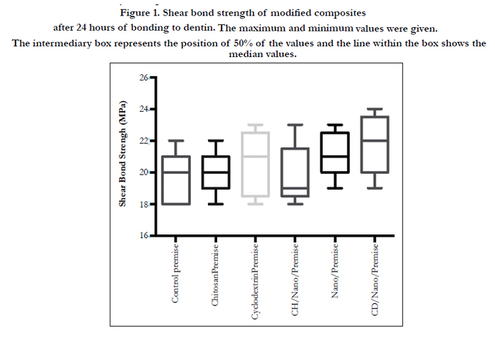
Figure 1. Shear bond strength of modified composites after 24 hours of bonding to dentin. The maximum and minimum values were given. The intermediary box represents the position of 50% of the values and the line within the box shows the median values.

Figure 2. Shear bond strength of modified composites after 3 months of bonding to dentin. The maximum and minimum values were given. The intermediary box represents the position of 50% of the values and the line within the box shows the median values.
Volumetric changes over time (seconds) due to polymerization in modified composites have been explored using an electronic mercury dilatometer and results are summarized in Figure 3.
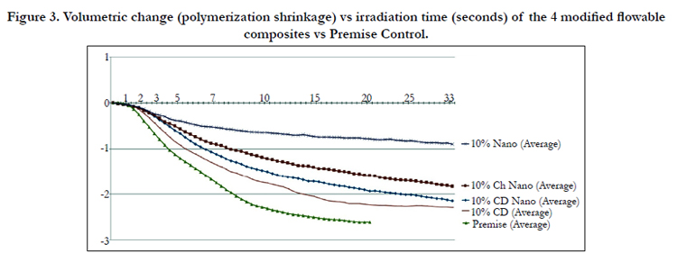
Figure 3. Volumetric change (polymerization shrinkage) vs irradiation time (seconds) of the 4 modified flowable composites vs Premise Control.
The ANOVA test established that there were statistical differences between the mean values (p < 0.05).
Furthermore, the mean Vickers Hardness of the 10%CH nano (VH42.2) was statistically higher than Premise (VH37.7) (p < 0.05). The mean VH of the 10%CD (VH32.5) and 10% nano (VH34.8) was statistically significantly lower (p < 0.05) than the Premise control (VH37.7).
The Friedman ANOVA test by ranks showed significant differences (p < 5%) between the controls and their 3 different gels. The median cell survival rates were found to be: chitosan (113%), nanodiamond (92%), chitosan/nanodiamond (93%). The ANOVA test showed significant differences (<5%) amongst the 3 different gels. Chitosan alone was found to have a significantly (Bonferroni test) higher (p < 0.05) cell survival rate than nanodiamonds or the chitosan + nanodiamond combination. No significant difference (p > 5%) was found between nanodiamonds and the chitosan + nanodiamond combination, although the chitosan + nanodiamond combination was slightly higher, which demonstrates the positive effect of chitosan. The maximum and minimum values were given. The intermediary box represents the position of 50% of the values and the line within the box shows the median values.
The undamaged cells (a) have a spindle shape while the damaged cells (b) show the disappearance of the spindle shape with the formation of globular shaped cells.
Discussion
This study showed that the shear bond strength after 24 hours, amongst Premise and the Premise combinations, were not significantly different (Figure 1). However, significant differences (p < 5%) after a 3 month period between the control Premise and the premise combinations were found (Figure 2). These higher shear bond strength values clearly showed the positive effect of the bio-actives chitosan, nanodiamonds and cyclodextrin over a longer period.
The results of this study (Figure 2) suggest that higher dentine bond strength can be achieved via the immediate interaction with collagen fibers of dentin: chitosan as well as interaction of chitosan: organic core of the flowable material complex with the resulting increase of dentin bond strength. Therefore the newly developed systems support our formerly reported results in addressing at least some of the shortfalls affecting the long-term bonding performance of modern adhesives and the current perspectives for improving bond durability of conventional adhesive systems as demonstrated in our “in vitro” model system. Initial results have proven that this significant increase in bond strength and the durability of resin-dentin bond lasts for a prolonged period (up to 3 months, Figure 2). It is well documented that the hydrostatic pulpal pressure, the dentinal fluid flow, and the increased dentinal wetness in vital dentin can affect the intimate interaction of certain enamel and dentin adhesives with dentinal tissue. However, the newly developed nanodiamond reinforced bio-active composites showed promising results to be able to address at least some shortfalls in the current perspectives in improving bond durability.
Also ionic vs covalent bonding of the chitosan: nanodiamond: composite agent complex may depend on the pH of the environment as the -COOH groups in, for example, naproxen, ibuprofen and/or aspirin ionize at alkaline pH and form covalent "amide" linkage at low pH. The adequate water absorption capacity, together with its cationic nature, which promotes binding to the negative surface of dentin (or skin) can also explain these results. Hydration of the polymer causes mobilization of the polymer chains and hence influences polymeric adhesion [24]. Appropriate swelling is also important to guarantee bio-adhesion. However, over-hydration can form slippery non-adhesive hydrogels [24].
Chitosan and cyclodextrin are potent antioxidants with multiple free hydroxyl groups [25]. These hydroxyl groups can form bridge-type hydrogen bonds within the side chains of hydroxyl, carboxyl, amino or amide groups of the collagen molecules [25]. The formation of these hydrogen bonds most probably ensures the stability of chitosan-collagen or chitosan:collagen:cyclodextrin interaction [25]. By positioning itself between collagen molecules, the host:guest complex formed by chitosan:nanodiamond can potentially also form ionic bonds, as well as covalent bonds with collagen fibrils [26]. Furthermore, in the process of the formation of hydrogen bonds, host:guest complex molecules can replace the water molecules bound to collagen in the extra-complex compartments.
The potential applications of chitosan and cyclodextrin in dentistry include strengthening of the collagen matrix, increasing resin– dentine bond strength, inactivation of collagen-bound proteases and remineralisation of root caries [26]. A detailed investigation of the potential mechanism is currently being conducted in our laboratory.
Research on the various attempts to reduce volumetric change of dental resins will continue until volumetric change due to polymerization is eliminated. Until volumetric change is eliminated, 2mm incremental layers during restoration placement are still advised. Oberholzer et al [20] described and designed an electronically controlled mercury dilatometer for the determination of volumetric change without external influences. We have used this electronic dilatometer extensively for volumetric change studies with excellent accuracy [19-21].
Several attempts have been made to decrease volumetric change during polymerization by changing the chemical constituents of flowable composites. The increase of the molecular weight of the organic component of a dental resin is one way which has been shown as a method of decreasing the volumetric change and improving some physical properties [15, 16]. While it is believed that a smaller filler size should decrease the volumetric change, it was also reported [19] that no clear influence of filler size could be seen for 4 different flowables, although Z250 with a smaller filler size (0.6μm) showed a lower shrinkage relative to SDR with a higher filler size (4.2μm). It was also reported that the rate of polymerization shrinkage of the 4 flowables was higher than that of the composite Z 250, but 3 of the flowables ended with a lower total % shrinkage than Z 250. On the other hand, the addition of nanoparticles as fillers in polymeric matrices have shown [27] encouraging results in the strengthening of the materials. Therefore, it could be expected that the incorporation of ND nanoparticles into dental polymeric materials could have a positive effect on the mechanical properties of the resulting nano-composites. In this study (Figures 3 and 4) we modified the commercially available Premise (Kerr) composite with the addition of nanodiamond powder (size of 2-8 microns, grit of 8000), chitosan or cyclodextrin in order to investigate the effect on the materials. Chitosan and cyclodextrin are both known for their activity as drug carriers and anti-oxidant reaction ability.
Volumetric change without the determination of the degree of conversion, does not provide sufficient insight into the effect on the Premise composite, like the addition of: nanodiamond/ chitosan and nanodiamond/cyclodextrin. From Figure 3 and 4 it is clear that 10% (5% of each) nanodiamonds:Premise have the least volumetric change (0.90%). Unfortunately, it was found that this extremely low volumetric change was mainly due to the insufficient polymer conversion (Figure 5). The surface microhardness of the top surface (34.8VH) was more than the value obtained from the 10% CD:Premise (32.5VH), but the bottom was not polymerised. The microhardness of all the other combination materials indicated adequate conversion.
The control Premise samples had a total volumetric change of 2.62%. However, 10% CD:Premise (2.29%) had the largest volumetric change of all the modified Premise samples. The 10% chitosan/nanodiamonds:Premise had the least volumetric change (1.83%), followed by 10%CD/nanodiamonds:Premise (2.15%). This clearly shows the positive effect on the composite as a result of the various additions.
The volumetric change (Figure 3) for all the materials, including Premise, starts at about 1 to 1.5 seconds after the beginning of irradiation (when the curing unit is switched on). The four modified materials showed more or less a similar rate of polymerization for the first 2.5 seconds (Figure 3). The fastest shrinkage (highest slope) was found for Premise alone which further resulted in also the highest total shrinkage. All three other combinations containing nanodiamonds gave lower total shrinkages (Figure 4) underlying the positive effect of the presence of the small particle size of nanodiamonds on the composite Premise.
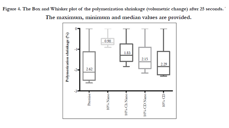
Figure 4. The Box and Whisker plot of the polymerization shrinkage (volumetric change) after 25 seconds. The maximum, minimum and median values are provided.
The low conversion with the addition of only nanodiamonds to Premise could be due to the fact that the light irradiaton particles were blocked and scattered by the darker shade of the very small dark nanodiamond particles. The Vickers hardness of the 10% chitosan/nanodiamonds:Premise (42.2VH) and 10% cyclodextrin/ nanodiamonds:Premise (38.8VH) (Figure 5) would suggest that the polymerization chain formation might be more controlled in a “wave” fashion from the surface to the base of the material, resulting with less side chains and an increased Vickers hardness.
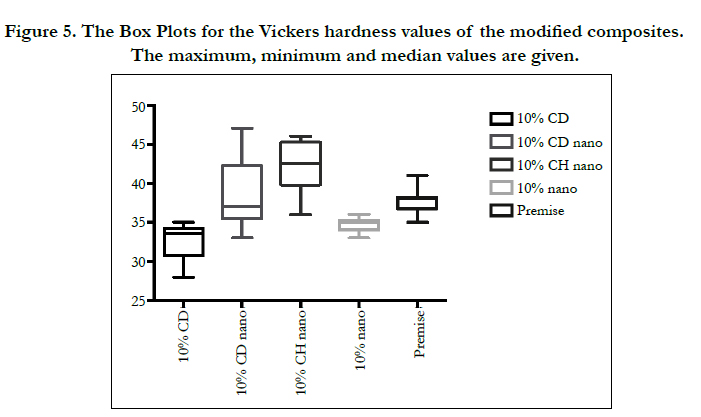
Figure 5. The Box Plots for the Vickers hardness values of the modified composites. The maximum, minimum and median values are given.
The newly formed larger methacrylate molecule in the 10% CH/ nanodiamond:Premise combination has OH and NH2 groups that probably give rise to hydrogen bonding, resulting in attachment between chitosan and the methyl methacrylate molecule through different multipoint linkages [18]. The carbon bond from the nanodiamonds and the darker shade provides a longer linear polymer chain that has been shown to have less polymerization shrinkage [28].
The Kruskal-Wallis multiple-comparison test was used to show statistical significant difference in the total volumetric change among materials.
The degree of conversion should have an important role on the volumetric change data analysis, since conversion has been shown to influence the physical properties of a material [29].
Since the light continuation through the material, intensity of the curing unit as well as other factors will influence the degree of conversion, further investigations towards understanding the detailed mechanism of the action of the nanodiamond as the additive in the bio-active composite should be done.
Due to the overall unique properties (like: hypoallergenic, antibacterial, antioxidant, non-cytotoxic, increased dentine bond strength, etc.) of chitosan and as newly reported research results appear in support of chitosan, it is increasingly accepted in the pharmaceutical field as an excellent carrier system for drugs, hormones, proteins, enzymes and genes.
Nanodiamond powder is one of the promising nanomaterials. It is reported that the nanodiamond (ND) surface becomes modified by biological molecules through the adsorption, non-covalent, and covalent chemical immobilization. Ho [30] demonstrated that nanomaterials can shuttle chemotherapy drugs to cells without producing the negative effects of today's delivery agents. It was also reported that nanodiamonds have multifaceted benefits in transporting drugs [31]. Clusters of the nanodiamonds surround the drug which is released in the cancer cells, while not affecting healthy cells. The diamonds left behind were found not to induce inflammation in cells.
Similar to our findings (Figure 6), nanodiamonds were generally found to be minimally toxic, if at all, to various cell-lines [32]. It was also reported that both 5-nm and 100-nm NDs did not induce cytotoxicity in the A549 cells [33]. However, it was also reported that some nanodiamond particles can be capable of inducing cytotoxic effects on human epidermoid carcinoma cells A431 but without a significant cytotoxic effect on the HaCaT cell line [34]. Nanodiamond toxicity has been studied in vitro and in vivo using various cell cultures and animal models and showed little toxicity looking at cell viability, gene program activity, and in vivo mechanistics [27]. In this study we found chitosan not to be cytotoxic at all and significantly less cytotoxic than nanodiamonds, which are only slightly cytotoxic.
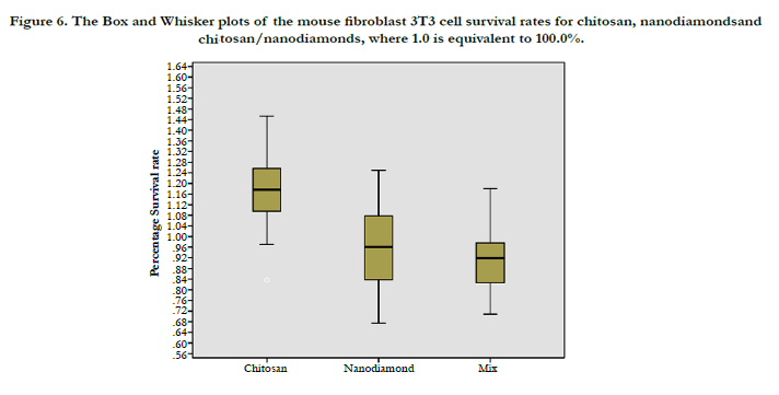
Figure 6. The Box and Whisker plots of the mouse fibroblast 3T3 cell survival rates for chitosan, nanodiamondsand chitosan/ nanodiamonds, where 1.0 is equivalent to 100.0%.
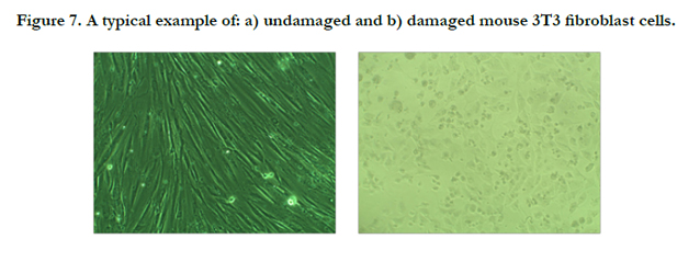
Figure 7. A typical example of: a) undamaged and b) damaged mouse 3T3 fibroblast cells.
In our previous studies [23] on cytotoxicity we found that the presence of chitosan with various substances, like anti-oxidants (resveratrol, propolis and β-carotene) and others (naproxen, ibuprofen) increased the cell survival rate of mouse 3T3 fibroblast cells significantly in comparison to the mentioned substances alone.
The cell survival rate depends on the type of chemical exposed to, its concentration, the type of cell-line used and the exposure period. The fibroblast cells used in this study were exposed to 1mg of chitosan, nanodiamonds or chitosan:nanodiamonds in the growth medium over a 24 hour period. This can be considered a high concentration and well in the top range of concentrations normally tested for other products. Similar concentrations were used (1000μg/ml) to test the cytotoxicity of many medicinal plants [35, 36]. An exposure period of 24 hours used in this study can also be considered a long exposure period (in the top range) [37-39].
Conclusion
A higher shear bond strength (p < 5%) was found after 3 months of Premise treated with nanodiamonds, chitosan, cyclodextrin (CD) and combinations thereof than the control Premise. Furthermore, the shrinkage was lower for all the bio-active additions than for Premise alone. Furthermore, chitosan was found to increase the cell survival rate, while nanodiamonds and the combination chitosan + nanodiamonds showed minimal cytotoxicity towards mouse 3T3 fibroblast cells.
Acknowledgement
We acknowledge financial support from the SADA and UWC. This study forms part of a PhD project for R Mulder.
References
- Schwartz-Arad D, Herzberg R, Levin L (2005) Evaluation of long-term implant success. J Periodontol 76(10): 1623-1628.
- Schrand AM, Hens SAC, Shenderova OA (2009) Nanodiamond particles: properties and perspectives for bioapplications. Critical Reviews in Solid State and Material Sciences 34(1-2): 18-74.
- Danilenko VV (2004) On the history of the discovery of nanodiamond synthesis. Physics of the Solid State 46(4): 595–599.
- Ravi Kumar MNV (2000) A review of chitin and chitosan applications. Reactive and Functional Polymers 46(1): 1-27.
- Palmberger TF, Hombach J, Bernkop-Schnurch A (2008) Thiolated chitosan: Development and in vitro evaluation of an oral delivery system for acyclovir. Int J Pharm 348(1-2): 54-60.
- Macleod GS, Fell JT, Colett JH, Sharma HL, Smith AM (1999)Selective drug delivery to the colon using pectin: chitosan: hydroxypropyl methylcellulose film coated tablets. Int J Pharmacol 187(2): 251-257.
- Bernkop-Schnurch A, Reich-Rohrwig E, Marschutz M, Schuhbauer H, Kratzel M (2004) Development of a sustained release dosage form for alphalipoic acid. II. Evaluation in human volunteers. Drug Dev Ind Pharm 30(1): 35-42.
- Kurosaki Y, Kimura T (2000) Regional variation in oral mucosal drug permeability. Crit Rev Ther Drug Carrier Syst 17(5): 467-508.
- Sadigh-Eteghad S, Talebi M, Farhoudi M, Mahmoudi J, Reyhani B (2013) Effects of Levodopa loaded chitosan nanoparticles on cell viability and caspase- 3 expression in PC12 neural like cells. Neurosciences (Riyadh) 18(3): 281-283.
- Kevin McCue (2003). "New Bandage Uses Biopolymer" Chemistry.org (American Chemical Society). Archived from the original on November 28, 2005. web.archive.org/web/20051128100345/http://www.chemistry.org/portal/a/c/s/1/feature_ent.html?id=401c0f5c4d8511d7f6e36ed9fe800100.
- Perchyonok VT, Grobler S, Olivier A, Zhang S, Oberholzer T (2013) Insights into chitosan hydrogels on dentine bond strength and cytotoxicity.Open Journal of Stomatology 3(1): 75-82.
- Perchyonok VT, Zhang S, Grobler SR, Oberholzer T (2013) Insights into and relative effect of chitosan-H, chitosan-H-propolis, chitosan-H-propolisnystatin and chitosan-H-nystatin on dentine bond strength. Eur J Dent 7(4): 412-418.
- Yevgeny Zababakhin. Detonation nanodiamond. http://en.wikipedia.org/ wiki/Detonation_nanodiamond.
- Shenderova OA, Hens SAC (2010) Detonation nanodiamond particles processing, modification and bioapplications. In Nanodiamonds: Applications in Biology and Nanoscale Medicine. Springer, New York. 79-116.
- Stansbury JW, Dickens SH (2001) Network Formation and Compositional Drift during Photo- Initiated Copolymerization of Dimethacrylate Monomers.Polymer 42(15): 6363-6369.
- Ferracane JL, Greener EH (1986) The effect of resin formulation on the degree of conversion and mechanical properties of dental restorative resins. J Biomed Mater Res 20(1): 121-31.
- Rueggeberg FA (1994) Determination of resin cure using infrared analysis without an internal standard. Dent Mater 10(4): 282-286.
- Halvorson RH, Erickson RL, Davidson CL (2002) Energy dependent polymerization of resin-based composite. Dent Mater 18(6): 463-469.
- Mulder R, Grobler SR, Osman YI (2013) Volumetric change of flowable composite resins due to polymerization as measured with an electronic mercury dilatometer. Oral biology and Dentistry www.hoajonline.com/oralbioldent/2053-5775/1/1.
- Oberholzer TG, Grobler SR, Pameijer CH, Rossouw RJ (2002) A modified dilatometer for determining volumetric polymerization shrinkage of dental materials. Measurement Science and Technology 13(1): 78-83.
- Oberholzer TG, Grobler SR, Rossouw RJ (2001) Polymerization shrink age by 4 different types of dental materials. South African Dental Journal 56(11): 513-516.
- Ramp LC, Broome JC, Ramp MH (2006) Hardness and wear resistance of two resin composites cured with equivalent radiant exposure from a low irradiance LED and QTH light-curing units. Am J Dent 19(1): 31-36.
- Grobler SR, Olivier A, Perchyonok TV, Moodley D, Osman Y (2015) Cytotoxic effect of chitosan-H, resveratrol, β-carotene and propolis and their chitosan hydrogels on Bal b/c mouse 3T3 fibroblast cells. Int J Dent Oral Sci 1(2): 10-14.
- Perchyonok VT, Reher V, Zhang S, Basson NJ, Grobler SR (2015) Bioinspired- Interpenetrating Network (IPNs) Hydrogel (BIOF-INPs) and TMD in Vitro: Bioadhesion, Drug Release and Build in Free Radical Detection and Defense. Open Journal of Stomatology 5(3): 53-61.
- Grobler SR, Perchyonok VT, Zhang S, Reher V, Oliver A (2015) Bioactive- Functionalized Interpenetrating Network Hydrogel (BIOF-IPN): A Novel Biomaterial Transforming the Mechanism of Bio-Repair, Bio-Adhesion and Therapeutic Capability – An In Vitro Study. Journal of Interdisciplinary Medical Dental Science 3:1-9.
- Perchyonok VT, ZhangS, Basson NJ, Grobler SR (2014) Evaluation of tetracycline containing chitosan hydrogels as potential dual action bio-active restorative materials capable of wound healing: in vitro approach. Biointerface Research in Applied Chemistry 4(5): 843-849.
- Mochalin VN, Shenderova O, Gogotsi Y (2012) The Properties and Applications of Nanodiamonds. Nature Nanotechnology 7(1): 11-23.
- Craig RG, Powers JM (2002) Restorative Dental Materials. (11th edtn), Mosby Inc, London. 186.
- Bouschlicher MR, Rueggeberg FA, Wilson BM (2004) Correlation of bottom- to-top surface microhardness and conversion ratios for a variety of resin composite compositions. Oper Dent 29(6): 698-704.
- D Ho. Nanodiamond-Based Therapeutic Delivery Films for the Treatment of Cancer and Inflammation. https://www.google.co.za/webhp?sourceid=chrome-instant&ion=1&espv=2&ie=UTF-8#q=Nanodiamond-Based+Therapeutic+Delivery+Films+For+the+Treatment+of+Cancer+and+Inflammation. Accessed:2015-06-25.
- Turner M (2012) Diamonds deliver on cancer treatment. Cancer Research. J Am Assoc Cancer Res 72(24): 1S-608S.
- Schrand AM, Huang H, Carlson C, Schlager JJ, Omacr Sawa E, et al. (2007) Are Diamond Nanoparticles Cytotoxic? J Phys Chem B 111(1): 2-7.
- Chao JI, Chang CC, Cheng CL (2006) Interaction and toxicity of nanodiamonds in human cells. The FASEB Journal 20(4): A75.
- Burleson T, Yusuf N, Stanishevsky A (2009) Surface modification of nanodiamonds for biomedical application and analysis by infrared spectroscopy. J Ach Mat Manufac Eng 37(2): 258-263.
- Coe FG, Parikh DM, Johnson CA, Anderson GJ (2012) The good and bad: Alkaloid screening and brine shrimp bioassays of aqueous extracts of 31 medicinal plants of eastern Nicaragua. Pharm Biol 50(3): 384-392.
- Ruffa MJ, Ferraro G, Wagner ML, Calcagno ML, Camposa RH, et al. (2002) Cytotoxic effect of Argentine medicinal plant extracts on human hepatocellular carcinoma cell line. J Ethnopharmacol 79(3): 335-339.
- Chen C, Weng M, Wu C, Lin J (2004) Comparison of radical scavenging activity, cytotoxic effects and apoptosis induction in human melanoma cells by Taiwanese propolis from different sources. Evid Based Complement AlternatMed 1(2): 175-185.
- Alija AJ, Bresgen N, Sommerburg O, Siems W, Eckl PM (2004) Cytotoxic and genotoxic effects of ß-carotene breakdown products on primary rat hepatocytes. Carcinogenesis 25(5): 827-831.
- Schwartz J, Shklar G (1992) The selective cytoxic effect of carotenoids and alpha-tocophenol on human cancer cell lines in vitro. J Oral Maxillofac Surg 50(4): 367-373.


