Impact of Er: YAG Laser Irradiation on the Proliferation of Human Periodontal Ligament Fibroblasts
Zhou ZX, Zhang S*, Ge LH
Department of Pediatric Dentistry, School of Stomatology, Peking University, Beijing, China.
*Corresponding Author
Sun Zhang,
Department of Pediatric Dentistry,
School of Stomatology, Peking University, Beijing 100081,
China.
Tel: 0086-10-8219-5361
Fax: 0086-10-62173402
E-mail: zhangsun_1@sina.com
Article Type : Research Article
Received: August 17, 2015; Accepted: September 06, 2015; Published: September 09, 2015
Citation: Zhou ZX, Zhang S, Ge LH (2015) Impact of Er: YAG Laser Irradiation on the Proliferation of Human Periodontal Ligament Fibroblasts. Int J Dentistry Oral Sci. 2(9), 142-146. doi: dx.doi.org/10.19070/2377-8075-1500030
Copyright: Zhang S© 2015. This is an open-access article distributed under the terms of the Creative Commons Attribution License, which permits unrestricted use, distribution and reproduction in any medium, provided the original author and source are credited.
Abstract
Abstract : The key to success of replantation of totally avulsed teeth is periodontal ligament activity. Improving the activity of periodontal ligament cell is beneficial to the success rate of tooth replantation.
Objectives: To observe the changes in proliferation of human periodontal ligament fibroblasts cultured in vitro after Er:YAG laser irradiation.
Methods: Conventional immumohistochemical staining was performed on human periodontal ligament fibroblasts cultured in vitro which was obtained from teeth root surface to identify the cell source. The fifth generation of human periodontal ligament fibroblasts collected were randomly divided into 5 groups, with group A as the control group receiving no laser irradiation, and experimental groups receiving different energies of Er:YAG laser radiation, respectively. The parameters were 50mJ, 10Hz and 1s radiation in B group; 100mJ, 10Hz and 1s radiation in C group; 150mJ, 10Hz and 1s radiation in D group; 200mJ, 10Hz and 1s radiation in E group. Cell growth was detected by CCK-8 method on Day 1, 3, 5, 7 and 9, respectively, and the changes in cell proliferation were analyzed.
Result: The cell proliferation rates in experimental groups were higher than that in the control group, and there were statistical differences.
Conclusion: Er:YAG laser irradiation with low energy and short time can promote the proliferation of periodontal ligament fibroblasts. Further studies are needed to determine whether the success rate of tooth replantation can be improved.
2.Introduction
3.Materials and Method
3.1.Main reagents and apparatus
3.2.hPDLF primary culture
3.4 Cell source identification on hPDLF
3.5 Laser irradiation on hPDLF
3.6 Impact on hPDLF proliferation
3.7 Statistical analysis
4.Results
4.1.Identification of human periodontal ligament fibroblasts
4.2.Impacts of Er:YAG laser irradiation on the proliferation of human periodontal ligament fibroblasts
5.Discussion
6.Conclusion
7.References
Keywords
Er: YAG laser; Human Periodontal Ligament Fibroblast; Cell Proliferation.
Introduction
Total avulsion of teeth, teeth getting completely out of alveolar bone, is a severe teeth trauma in children. As an efficient therapy, replantation of totally avulsed teeth could retain the affected teeth transiently, maintain their functions and beauty and reach periodontal ligament healing through replantation in 15-30 minutes [1]. However, replantation is often conducted after a long time from total avulsion or receiving no proper maintenance clinically. After operation, pulpitis as well as degeneration and necrosis of parodontium and cementoblast will lead to inflammatory or alternative dental root resorptions, causing replantation failure or even early loss of teeth. The key to replantation success is periodontal ligament activity. In order to improve the replantation success rate, related studies have been carried out by many scholars. If avulsed teeth are stored in moist environment especially in biocompatible solution, the survival of periodontal ligament cells can be prolonged. At present, the better preservation solutions are Hank’s balanced salt solution and ViaSpan solution (Du- Pont Pharmaceuticals Co., Wilmington, Del.) [1-4]. A in-vitro trial showed that the prognosis for replantation on dog teeth stored in Ero-Collins solution for 8 hours was the same as that for instant replantation [5]. However, such solutions are difficult be obtained, so the AAE, RCSE and IADT guidelines recommend cool milk as stock solution or that the avulsed teeth be placed in patients’ saliva for at most 30 minutes [6]. Except for the study of preserving medium, preventing dental root resorption and obtaining reattachment through the treatment of root surface is the desire of many scholars. AAE suggests that utilizing sodium fluoride solution, hydroxyapatite is able to be transported into fluorapatite to prevent the dental root resorption [6]. Study of Beagle teeth replantation by Ipbal et al. had verified the positive effect of Emdogin® in promoting periodontal tissue regeneration, which can stimulate the proliferation of activated periodontal ligament cells in damaged root surface and teeth socket, so as to cover root surface and further form acellular cementum, periodontal ligament and alveolar bone, thus inducing tissue regeneration [7].
With the development of study on human body tissue engineering, direct application of personal or external periodontal ligament cells to improve the success rate of tooth replantation is also a hot [8]. Studies have shown implanting of myelomonocyte containing periodontal ligament cells into periodontal clearance is able to improve the periodontal ligament healing of replanted teeth in dog [9]. There are plenty of evidences indicating that PDL-derived stem/progenitor cells are the best choice for periodontal ligament tissue regeneration. Together with the totally avulsed dog teeth, the platelet-rich fibrin (PRF) as scaffold and cell sheet fragments of periodontal ligament stem cells (PDLSCs) were given with replantation by Yin-Hua Zhao et al. In consequence, the periodontal ligament healing was achieved after 8 weeks; meanwhile, the periodontal ligament regeneration tissues were discovered, and teeth ankylosis and inflammation was reduced. The author considered that PDLSCs/PRF can improve the success rate of the periodontal ligament of replanted teeth [10]. Although studies in vitro have obtained many results, there also exist problems such as ethical issues and uneasy acquisitions for replantation using periodontal ligament cells. Since tooth replantation success is relevant to the activity and proliferation of periodontal ligament, we suspect that the amount and the proliferation ability of periodontal ligament cells can be increased directly. It has been widely known that low-level laser treatment (LLLT) is conducive to the proliferation of mesenchymal cells and periodontal ligament fibroblasts [11, 12]. Li Mo and Li Mei found that the proliferation of periodontal ligament fibroblasts could be promoted through Nd: YAG laser irradiating at proper energy parameters [13, 14]. Impact of Er:YAG laser irradiation on human periodontal ligament fibroblasts were ever studied by Pourzarandian et al. The energy density used in the study was 1.68 - 5.0 J/cm2 and the results indicated that compared with the control group without laser irradiation, the cell proliferation rates in laser irradiation group were significantly faster, and a statistical difference was found [15]. On teeth root and in periodontal clearance of totally avulsed teeth, there are residual periodontal ligament cells which played a very important role in the replanted teeth healing process and are easily acquired. The hypothesis in this study is that Er:YAG laser irradiates directly on the periodontal ligament cells in teeth root or periodontal clearance, and the proliferation of periodontal ligament cells could be improved, further improving the success rate of replantation of avulsed teeth. The objective of the present study is to obtain human periodontal ligament fibroblasts cultured in vitro and observe their proliferation changes under the Er:YAG laser irradiations of different energy densities. Thus, the mechanism of Er:YAG laser in impacting periodontal ligament cells was preliminarily discussed, and application of laser in the periodontal ligament tissues reconstruction was prospected. This study has been checked and approved by Biomedical ethics committee in Peking University School of Stomatology, with the Register Number PKUSSIRB -201417105.
Materials and Method
αMEM culture medium (GIBOCO, America), 15% Fetal calt serum (Hyclone, America), Penicillin and Streptomycin (GIBOCO, America), Dispase enzyme (Sigma, America), Type I collagenase (Sigma, America) and CCK-8 (DOJINDO, Japan).
Anti-human vimentin antibody (Boster, China), Anti-human keratin antibody (Boster, China), DAB developing reagent kit (Boster, China) and ready-to-use SABC-POD reagent kit (Boster, China).
Super clean bench (Thermo Formal, America), CO2 incubator (Thermo Formal, America) and Inverted phase contrast microscope (Nikon, America).
Er:YAG laser (DEKA Smart 2940D) ( Electronic Engineering, Italy) providing pulse Er:YAG laser at 2940 nm, 5-20 Hz frequency rage, with the lowest energy of 50 mJ/pulse, the largest energy of 700 mJ/pulse and the largest pulse width of 450 μs. Phone light mouth was sapphire glass window, with 1 mm phone output spot, light guide articulated arm as optical transmission system and the phone equipped with compressed air and sprinkler system. Diode laser was the guide laser, with 680 nm wave length.
Two first premolars removed due to orthodontic treatment in maxillofacial surgery clinics in Peking University School of Stomatology were obtained. The removed teeth were put immediately into sterile α MEM medium with penicillin and streptomycin, stored at 4°C and used on the same day. 1/3 of periodontium tissues of in-vitro teeth root were scraped in the Super Clean Bench, avoiding scraping root tip or cement-enamel junction tissues. Primary culture was performed with the combination of enzyme digestion and tissue block method. When cells grew until they occupied 80% of bottom wall area of culture dish, passage was conducted by 1:3, while culture solution was changed every 2-3 days. The cells of the fifth passage were taken for the following experiments.
Aseptic round cover glass was placed into 24-well plate, and 3×10^3 cells were inoculated into each well. 95% ethyl alcohol was used for fixation. According to conventional ABC method, staining for vimentin and keratin antibody was conducted. Then, cytoplasm staining was observed with the help of an inverted microscope after hematoxylin redyeing to ascertain cell source.
The fifth generation of hPDLF cells was taken and placed into a centrifuge tube. Laser irradiations were performed after centrifuging. More specific, the control group did not receive laser irradiation and was set as group A. According to output energy 50 mJ, 100 mJ, 150 mJ and 200 mJ, experimental groups were took as group B, C, D and E respectively, with 1s radiation time, 10 Hz frequency and corresponding energy power of 0.5 W, 1W, 1.5 W and 2 W, respectively. After laser source and the laser handle were placed on the right above centrifuge tube for fixation in the center, irradiation was performed in Super Clean Bench, with 11.5 cm irradiation distance. hPDLF was inoculated into a 96-well plate (6×10^4 per/ml, 100μl/well).
24 h after inoculating, CCK-8 experiments were conducted on Day 1, 3, 5, 7 and 9, respectively. hPDLF was detected and the data were recorded.
SPSS 21.0 software was adopted. One-way ANOVA analysis was used for the obtained experiment data. P < 0.05 was considered as statistically significant.
Results
After immunohistochemical staining, the tissues were observed under a microscope. The results showed positive staining for vimentin (blue karyon and dark cytoplasm) (Figure. 1), negative staining for keratin (blue karyon, no positive staining in cytoplasm (Figure. 2). Mixed epithelial cells were excluded and no positive staining results were seen in the control group. This suggested that the cultured cells were mesoblast-derived fibroblasts.
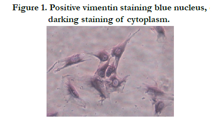
Figure 1. Positive vimentin staining blue nucleus, darking staining of cytoplasm.
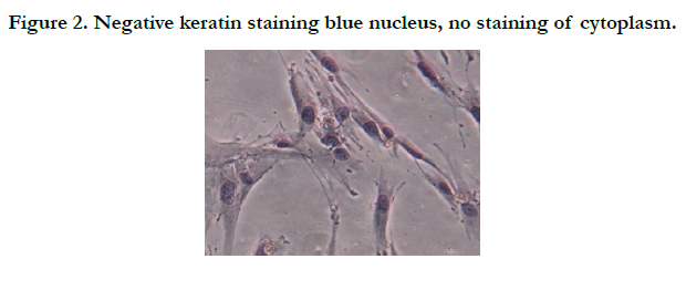
Figure 2. Negative keratin staining blue nucleus, no staining of cytoplasm.
Growth curve obtained after cell counting (Figure.3) was shaped as “S”. Cell count peaked on day 7 and then decreased, which conformed to cell growth rhythm. As shown in Table 1, after Er:YAG laser irradiation, the proliferation of periodontal ligament fibroblasts was quickened, and the values in experimental groups were higher than those in the control group. In cellular proliferative stage, group C at 1w, group D at 1.5 w and group E at 2 w, there were statistical differences between each experimental group and the control group (P < 0.05).
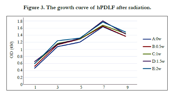
Figure 3. The growth curve of hPDLF after radiation.
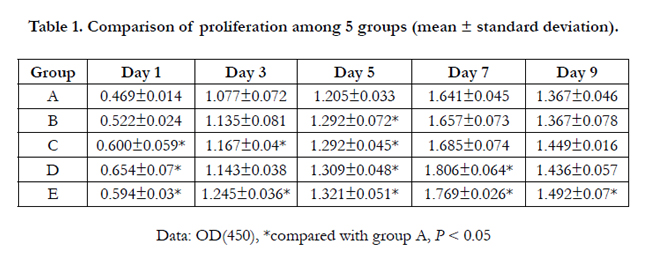

Table 1. Comparison of proliferation among 5 groups (mean ± standard deviation).
Discussion
Effects of laser on tissues include photothermal effect, photochemical effect, electromagnetic effect, pressure effect, biostimulation effect of weak laser. For those characteristics, the effects of laser on tissues are different from those of traditional mobile phones or scalpel on tissues. Therefore, laser has a wide prospect in clinical practice.
Er:YAG laser, with a wave length of 2940 nm, is able to be absorbed by water and hydroxyapatite. The moisture in teeth tissues and soft tissues could absorb Er:YAG laser to generate micro explosion for tissue ablation. Meanwhile, the energy is transformed into kinetic energy, which cause very little thermal damage, and Er:YAG laser penetrates slightly into the tissues. Therefore, it has a little effect on the surrounding tissues. Besides, Er:YAG laser can play a sterilization and disinfection role, comfort the patients and cause slight postoperative reactions. Currently, Er:YAG laser has a wide application in removing necrotic tissues, cavity preparation, pit and fissure sealing, endodontic treatment, periodontal treatment, dental trauma, soft tissue surgery, etc [16-23].
In periodontal ligament, hPDLF is the largest in the number and the most important in the function. hPDLF belongs to mesenchymal cell, whose principal function is to synthetize new collagen and degrade old collagen fibers. This is closely related to the periodontal ligament reconstruction and remodeling.
Attention is paid to the impact of laser on cell activity and proliferation. Low-power laser is able to promote cell metabolism, improve gene expression and strengthen the enzyme activity. Among them, one function manifests as promoting cell proliferation. Low-intensity lasers have the ability to promote fibroblasts proliferation, mainly reflecting in acceleration of wound healing and regeneration of transplanted tissues [24].
In this study, the cultured human periodontal ligament fibroblasts were irradiated by Er:YAG laser in groups, and the cell growth of experimental groups and the control groups were detected by CCK-8 method on Day 1, 3, 5, 7 and 9, respectively. The results showed that growth curves in experimental groups were consistent with that in the control group, indicating that the growth cycle of periodontal ligament cells was not changed after Er:YAG laser irradiation. As shown in Table 1, compared with the control group, the cell proliferations in experimental groups showed different degrees of increase. On Day 1, 3, 5 and 7, the higher the energy was, the more obvious the differences were, and statistical differences existed between the control group and group C, D, E (P < 0.05). However, there was no obvious change in cell form after irradiation. These suggested that within the setting range of the parameter, Er:YAG laser could promote the periodontal ligament fibroblasts proliferation and improve cell activity. The intergroup comparison showed statistical differences between group E and group B with group C on Day 3, 5, 7 and 9, but no statistical difference between group E and group D. In addition, by comparing experimental groups and the control group, we found that proliferation effect in group E had the longest during. Therefore, we speculated that, within a certain energy range, the higher the energy was, the longer the laser effect lasted.
Er:YAG laser has 15000 times the absorption rate of Nd:YAG laser in water [25], while the depth of tissue penetration is also less than that by Nd: YAG laser. With very little energy, the tissues containing much water can be removed, and the thermal side effects can be minimized. So, the laser at appropriate parameters and lower energy and power would lead to small thermal damage to cells and can improve cell activity. It was reported that when Nd:YAG laser irradiated onto human periodontal ligament cells, even at less than 0.5w, the cell activity was decreased, which was associated with thermal damage and deeper penetration of Nd: YAG laser [26]. The photothermal effect of Er:YAG laser may enhance the catalytic activity of intracellular enzyme, improve cell functions and result in lower thermal effects and temperature rise without damage to cells.
The molecular mechanism of laser in promoting cell proliferation effects has been controversial. One of the classical theories is that the laser energy is absorbed by intracellular chromophore and converted into metabolic energy, which is consistent with the improvement of the intracellular ATP levels after laser irradiation [27]. Experiments in vitro confirmed that laser irradiation was able to increase the proton electrochemical potential and ATP synthesis [28, 29]. APT can not only regulate the DNA, RNA and protein synthesis, but also regulate cell oxygen consumption and mitochondrial membrane potential [30]. Under the transmission electron microscopy, the same phenomenon has been also observed by Pourzarandian [15]. After laser irradiation, rough endoplasmic reticulum, golgi apparatus and mitochondria in cells become more prominent. The possible mechanism is that laser has the ability to increase the production of reactive oxygen, alter the redox potential of cells and induce the generation of redox state-regulated transcription factors [31].
In this study, laser irradiation adopted one-time shock dose which was consistent with that clinically. Moreover, cells were placed in the centrifuge tube and then inoculated into 96-well plate after irradiation, which was distinct from the majority of experiments using direct irradiation after placed into 96-well plate or culture dishes. This avoided damage to the surface structure of the 96- well plate or culture dishes, thus affecting the attachment and proliferation of periodontal ligament fibroblasts. As it was difficult to perform the laser irradiating in super clean bench due to smaller well in the 96-well plate, the operation difficulty and error were reduced to a certain extent by using this experimental method.
In this experiment, the previous hypothesis that Er:YAG laser was available for promoting the proliferation of human periodontal ligament fibroblasts was confirmed and a certain improvement was made in experimental methods. However, there still are some limitations. The oral environment for survival of human periodontal ligament fibroblasts is more complex compared with the environment of culture medium in-vitro in this experiment. Therefore, animal experiments are still needed to explore the optimal laser parameters before clinical use and to investigate the impact of laser irradiation of the totally luxated teeth root surface or the periodontal ligament cells in teeth socket on success rate of replantation of totally luxated teeth.
Conclusion
The results of this study have confirmed that Er:YAG laser irradiation at appropriate parameters are able to improve the activity and accelerate the growth rate of human periodontal membrane fibroblasts. Thus, we speculate that Er:YAG laser irradiation can accelerate the proliferation of the periodontal ligament fibroblasts and promote new attachment formation of periodontal tissues. In order to determine whether the success rate of tooth replantation can be improved by Er:YAG laser irradiation, however, animal experiments and further researches are still needed.
References
- Andreasen JO, Andreasen FM, Andersson L (2007) Traumatic injuries to the teeth. (4th edtn), Blackwell Munksgaard, England.
- Mcdonald RE (2004) Dentistry for the child and adolescent. (8th edtn), Mosby Inc., America.
- Hiltz J, Trope M (1991) Vitality of human lip fibroblasts in milk, Hanks balanced salt solution and Viaspan storage media. Endod Dent Traumatol 7(2): 69-72.
- Trope M, Friedman S (1992) Periodontal healing of replanted dog teeth stored in Viaspan, milk and Hank’s balanced salt solution. Endod Dent Traumatol 8(5): 183-188.
- Sottovia AD, Filho DS, Poi WR, Panzarini SR, Luize DS, et al. (2010) Tooth Replantation after use of Euro-Collins Solution or Bovine Milk as Storage Medium: A Histomorphometric Analysis in Dogs. J Oral Maxillofac Surg 68(1): 111-119.
- McIntyre JD, Lee JY, Trope M, Vann WF Jr (2009) Permanent tooth replantation following avulsion: Using a decision tree to achieve the best outcome. Pediatr Dent 31(2): 137-144.
- Iqbal MK, Bamaas N (2001) Effect of enamel matrix derivative (EMDOGAIN) upon periodontal healing after replantation of permanent incisors in beagle dogs. Dent Traumatol 17(1): 36-45.
- Zhou YF, Li YS, Mao L, Peng H (2012) Periodontal healing by periodontal ligament cell sheets in a teeth replantation model. Arch Oral Biol 57(2): 167-176.
- Reichert da Silva Assuncao L, Colenci R, Ferreira do-Amaral CC, Sonoda CK, Mogami Bomfim SR, et al. (2011) Periodontal tissue engineering after tooth replantation. J Periodontol 82(5): 758-766.
- Zhao YH, Zhang M, Liu NX, Lv X, Zhang J, et al. (2013) The combined use of cell sheet fragments of periodontal ligament stem cells and plateletrich fibrin granules for avulsed tooth reimplantation. Biomaterials 34(22): 5506-5520.
- Alghamdi KM, Kumar A, Moussa NA (2012) Low-level laser therapy: a useful technique for enhancing the proliferation of various cultured cells. Lasers Med Sci 27(1): 237-249.
- Kreisler M, Christoffers AB, Willershausen B, d’Hoedt B (2003) Effect of low-level GaAlAs laser irradiation on the proliferation rate of human periodontal ligament fibroblasts: an in vitro study. J Clin Periodontol 30(4): 353-358.
- Liu M, Ruan Y, Pan CHB (2008) Effects of the Nd: YAG laser radiation on the proliferation and alkaline phosphatase activity of human periodontal ligament fibroblasts. Chin J Stomatol Res (Electronic Version) 2(3): 234-239.
- Li M, Zeng XQ, Zhang JC (2004) Effect of Pulsed Nd: YAG Laser on cell proliferation of Human Periodontal Fibroblasts in vitro. Chin J Laser Med Surg 13(2): 98-100.
- Karu T, Pyatibrat L, Kalendo G (1995) Irradiation with He-Ne laser increases ATP level in cells cultivated in vitro. J Photochem Photobiol B 27(3): 219-223.
- Cozean C, Arcoria CJ, Pelagalli J, Powell GL (1997) Dentistry for the 21st century? Erbium: YAG laser for teeth. J Am Dent Assoc 128(8): 1080-1087.
- Zhang S, Chen T, Ge LH (2012) Scanning electron microscopy study of cavity preparation in deciduous teeth using the Er: YAG laser with different powers. Lasers Med Sci 27(1): 141-144.
- Zhang S, Chen T, Ge LH (2013) Evaluation of clinical outcomes for Er: YAG laser application in caries therapy of children. Journal of Peking University(Health Science) 45(1): 87-91.
- Krmek SJ, Miletic I, Simeon P, Mehicić GP, Anić I, et al. (2009) The temperature changes in the pulp chamber during cavity preparation with the Er: YAG laser using a very short pulse. Photomed Laser Surg 27(2): 351-355.
- Dewsnup N, Pileggi R, Haddix J, Nair U, Walker C, et al. (2010) Comparison of bacterial reduction in straight and curved canals using erbium,chromium:yttrium-scandium-gallium-garnet laser treatment versus a traditionalirrigation technique with sodium hypochlorite. J Endod 36(4): 725-728.
- de Moura AA, Moura-Netto C, Barletta FB, Vieira-Júnior ND, Eduardo Cde P (2010) Morphological assessment of dentine and cementum following apicectomy with Zekrya burs and Er: YAG laser associated with direct and indirect Nd: YAG laser irradiation. Oral Surg Oral Med Oral Pathol Oral Radiol Endod 109(4): 77-82.
- Inamoto K, Horiba N, Senda S, Naitoh M, Ariji E et al. (2009) Possibility of root canal preparation by Er: YAG laser. Oral Surg Oral Med Oral Pathol Oral Radiol Endod 107(1): e47-e55.
- Zhan Z, Wu W, Zhao H, Zhang X, Xie S (2012) Shear bond strength of a self-etch adhesive to caries-affected dentin after caries removal by Er: YAG laser. Lasers in Dentistry 8208(8): 10-12.
- Chen HX, Gao GH, Qian HW (2001) Research of low power laser on cell proliferation. Chin J Phys Med Rehabil 23(4): 246-247.
- Ishikawa I, Aoki A, Takasaki AA, Mizutani K, Sasaki KM, et al. (2009) Application of lasers in periodontics: true innovation or myth? Periodontology 2000 50: 90-126.
- Chen YJ, Jeng JH, Jane Yao CC, Chen MH, Hou LT, et al. (2005) Long- Term Effect of Pulsed Nd:YAG Laser Irradiation on Cultured Human Periodontal Fibroblasts. Lasers Surg Med 36(3): 225-233.
- Karu T, Pyatibrat L, Kalendo G (1995) Irradiation with He-Ne laser increases ATP level in cells cultivated in vitro. J Photochem Photobiol B 27(3): 219-223.
- Silveira PC, Silva LA, Fraga DB, Freitas TP, Streck EL, et al. (2009) Evaluation of mitochondrial respiratory chain activity in muscle healing by lowlevel laser therapy. J Photochem Photobiol B 95(2): 89-92.
- Lane N (2006) Cell biology: power games. Nature 443(7114): 901-903.
- Greco M, Guida G, Perlino E, Marra E, Quagliariello E (1989) Increase in RNA and protein synthesis by mitochondria irradiated with helium-neon laser. Biochem Biophys Res Commun 163(3): 1428-1434.
- Chen AC, Arany PR, Huang YY, Tomkinson EM, Sharma SK, et al. (2011) Low-level laser therapy activates NF-kB via generation of reactive oxygen species in mouse embryonic fibroblasts. PLoS One 6(7): e22453.


