Cytotoxic Effect of Chitosan-H, Resveratrol, β-Carotene and Propolis and their Chitosan Hydrogels on Balb/C Mouse 3T3 Fibroblast Cells
S R Grobler1*, A Olivier1, T V Perchyonok2, D Moodley1, Y Osman1.
1 Faculty of Dentistry, Oral and Dental Research Institute, University of the Western Cape, Private Bag X1, Tygerberg 7505, Cape Town, South Africa.
2 VTPCHEM PTY LTD, Glenhuntly, Melbourne, 3163, Australia and School of Dentistry and Oral Health, Griffith University, Southport, 4215, QLD, Australia.
*Corresponding Author
Sias R. Grobler,
Faculty of Dentistry, Oral and Dental Research Institute,
University of the Western Cape, Private Bag X1,
Tygerberg 7505, Cape Town,
South Africa.
E-mail: srgrobler@uwc.ac.za
Article Type: Case Report
Received: November 07, 2014; Accepted: December 02, 2014; Published: December 11, 2014
Citation:Citation: S R Grobler et al., (2014) Cytotoxic Effect of Chitosan-H, Resveratrol, β-Carotene and Propolisand their Chitosan Hydrogels On Balb/C Mouse 3T3 Fibroblast Cells. Int J Dentistry Oral Sci. 1(2), 10-14. doi: dx.doi.org/10.19070/2377-8075-140003
Copyright: S R Grobler © 2014. This is an open-access article distributed under the terms of the Creative Commons Attribution License, which permits unrestricted use, distribution and reproduction in any medium, provided the original author and source are credited.
Abstract
Background/purposes: The beneficial effect (bond strength and longevity) of the addition of different chitosan/antioxidant hydrogels to dental restoratives was reported. However, it still needs to be verified whether their presence would not damage the pulp cells. Therefore, the purpose of this study was to determine the relative cytotoxic effect of resveratrol, propolis and β-carotene in relation to their respective chitosan/antioxidant hydrogels on mouse Balb/c mouse 3T3 fibroblast cells.
Materials and Methods: The mentioned hydrogels were prepared by the dispersion of the corresponding components in glycerol and acetic acid with the addition of the chitosan polymer. The cell survival rate was determined over a 24 hour period according to the standard MTT assay.
Results and Conclusion: The Kruskal-Wallis Multiple-Comparison Test and Bonferroni test showed significant differences in the cell survival rates (p<0.05) amongst resveratrol (31%), propolis(64%) and β-carotene (95%): Also amongst chitosan(114%), chitosan/resveratrol(87%) and resveratrol(31%); chitosan(114%), chitosan/β-carotene(100%) and β-carotene(95%); chitosan(114%), chitosan/propolis(95%) and propolis(64%). To conclude, this study showed: 1) that chitosan-H has a positive effect on the cell survival rate of Balb/c mouse 3T3 fibroblast cells and therefore most probably also on human pulp fibroblast cells, 2) these chitosan hydrogels are safe to be used to improve the bond strength and longevity of a tooth dental material, and 3) it further proved that chitosan in itself improved the cell survival rate of various antioxidants. The mean cell survival rate was found to be: resveratrol (31%); propolis (64%); β-carotene (95%), chitosan+resveratrol (87%), chitosan+propolis (95%), chitosan+ β-carotene (100%) and chitosan (114%).
2.Introduction
3.Materials and Methods
3.1.Cell maintenance and culture
4.Results
5.Discussion
6.Conclusion
7.Acknowledgement
8.References
Keywords
Chitosan Hydrogels; Cytotoxicity; Fibroblast Cells; Bond Strength.
Introduction
In the last decade there has been increased interest in the role which reactive oxygen species and antioxidants may play in the aging process and in the development of diseases. This lead to various studies [1-4] where the possible beneficial effects of antioxidant supplementation were investigated. In this context are the compounds resveratrol, propolis and β-carotene and many others known for their antioxidant content [5].
Over the years the 3T3 mouse fibroblast cell-line has extensively been used as a standard cell line to test the cytotoxic effect of various dental materials and constituents thereof [6-9]. Cytotoxiceffects on this cell line give one a good indication of what would happen towards human pulp fibroblast cells. In one study, for example, it was reported that the cytotoxicity of the standard 3T3 fibroblast cell-line was in between that of 4 different human pulp fibroblast cell-lines [8]. Therefore, we used the Balb/c mouse 3T3 fibroblast cell-line to provide a good indication of the cytotoxicity of the mentioned anti-oxidants and chitosan gels thereof.
Chitosan [10] which is produced commercially by de-acetylation of chitin is a natural polysaccharide composed of randomly distributed β- 1, 4-linked D-glucosamine and N-acetyl α -glucosamine.
Chitosan is non-toxic, biocompatible, bio-degradable and has muco-adhesive properties and as a result became widely used in the pharmaceutical field as a carrier system for drugs, hormones, proteins, enzymes and genes [11-14]. Chitosan is positively charged in acid medium because the amino groups become protonated which leads to an increase in its solubility. Chitosan can be successfully used as a drug carrier because it will solubilize and degrade in an acidic environment with the resultant release of the drug [15]. Chitosan is hypoallergenic and has natural antibacterial properties, which further support its use in the army as field bandages [16]. However, chitosan has not been approved by the FDA as drug carrier but it has been approved in some combinations for wound healing, although not in every form [17]. Furthermore, very important findings were that antioxidant-chitosan hydrogels (that of resveratrol, propolis and β-carotene) were found to significantly improve the bond strength to dentine with or without phosphoric acid pre-treatment [18] as many other hydrogels do [19].
Resveratrol (3,5,4'-trihydroxy-trans-stilbene) [20] is produced by several plants when damaged by bacteria or fungi. There is limited evidence of its health effects in humans and the long-term effects of resveratrol on humans are currently unknown [21]. However, the presence of resveratrol mainly in red wine was suggested as the explanation for the cardio-protective effects of wine [22].
β-Carotene (beta-carotene) is a red-orange pigmented hydrocarbon and a carotene with beta-rings. Among the general class of carotenes, β-carotene is distinguished by having rings at both ends of the molecule, is fat soluble and therefore better absorbed in the body when consumed with fat. There is no scientific evidence that β-carotene can be used for any medical treatment, but high levels of β-carotene have also been associated with lung cancer in smokers [23].
On the other hand propolis has no definite composition like resveratrol or β-carotene and its composition varies even within a hive [24]. This is because it is a sort of resin which is used to seal the hive and is collected from any plant near the hive wherever it is located. Therefore, it makes sense that different medical claims can be made from one propolis which will not necessarily be true for another and therefore the composition also varies from region to region. Propolis which is a product of bees has been used as an ointment since 350 BC, the time of Aristotle [25]. It has been used for abscesses; for healing wounds and tumors; and Egyptians have used it for mummification [25]. Furthermore, a positive effect of propolis on enamel micro-hardness was also reported due to particular components of mineralization activity in propolis [26].
The beneficial effect of various chitosan hydrogels was reported as antioxidant and drug carrier also in tooth bonding agents [27-30]. However, their cytotoxic effects towards fibroblast cells were not investigated and if found to be cytotoxic their positive gain became less important in bonding agents.
Therefore, the purpose of this study was to determine the relative cytotoxic effect of resveratrol, propolis and β-carotene in relation to their respective chitosan hydrogels. Thus, the parameters studied were: the cytotoxicity of propolis, resveratrol, β-carotene and chitosan. Relative to this, the cytotoxicity of the chitosan-propolis hydrogel, the chitosan-resveratrol hydrogel and the chitosan-β-carotene hydrogel were determined.
Materials and Methods
Chitosan (Aldrich, Australia), glycerol (Sigma, USA) and glacial acetic acid (E. Merck, Germany) were used as received from the manufacturers. The degree of de-acetylation of typical commercial chitosan used in this study is 87%. Chitosan with molecular weight 2.5x 10³ KD was used in the study. The iso-electric point was 4.0–5.0.
Chitosan hydrogels have been prepared using a methodology previously described [31]. The corresponding antioxidant mixture was incorporated by dispersion of 0.2 grams of the antioxidant powder (propolis, resveratrol, or β-carotene) in 200µL glycerol (5% w/w) using a mortar and pestle and 10 milliliters of glacial acetic acid (3% w/w) added with continuous mixing and finally the chitosan polymer (0.2grams) was spread on the surface of the dispersion and mixed well for 12 hours to form the required gel. Gels were poured into petri-dishes and were dried at room temperature.
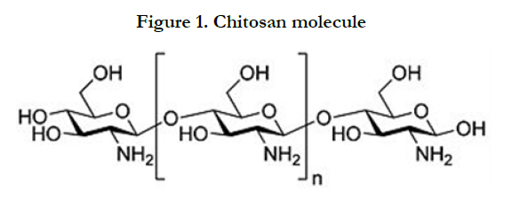
Figure 1. Chitosan molecule
A Balb/c 3T3 mouse fibroblast cell line (The National Repository for Biological Materials, Sandringam) was maintained and cultured in standard conditions (37°C under 5% carbon dioxide and 95% humidity) in Dulbecco’s Modified Eagles Medium (DMEM). The medium was supplemented with 10% fetal bovine serum, penicillin (10.000U/ml) and streptomycin (10.000 µg/ml) mix (Biochrom Ltd), changed every second day and cells sub-cultured using routine trypsin/EDTA procedures. The pH of this growth medium was adjusted when necessary to 8.0 [18].
To test the cytotoxicity towards 3T3 cells, the cells were first grown to near confluency. Then the 3T3 cells were diluted to a final cell suspension containing approximately 3 ×10⁵ cells/ml and plated out in seven 96 well plates, a plate for each compound. Resveratrol, propolis, β-carotene,chitosan-H, chitosan+resveratrol, chitosan+propolis and chitosan+β-carotene were added to the growth medium at a concentration of 1 mg/ml. Two hundred µl of each group (7 groups) was added to 20 wells in the 96 well plates. Medium without any chitosan gels was used as controls. After 24 hours the widely used MTT colorimetric assay was used to evaluate cell growth. This assay involves the ability of viable cells to use mitochondrial dehydrogenase enzymes to convert MTT (a soluble tetrazolium salt) to a blue/violet formazan end-product [32]. Twenty µl MTT (5mg/ml in phosphate- buffered solution) was added to each well and left for a further 3 hours to incubate at 37°C. The medium was discarded to eliminate the MTT and the precipitated formazan crystals were solubilized with 100 µl/ well of dimethylsulfoxide (DMSO). Absorbance was measured at wavelength 540 nm on a spectrophotometer to determine the number of viable cells. Three replicates were done in each group.
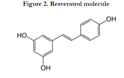
Figure 2. Resveratrol molecule
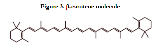
Figure 3. β-carotene molecule
Results
Figure 4 shows the Box & Whisker plot of the % survival rate of mouse Balb/c 3T3 fibroblast cells when exposed to chitosan, resveratrol, and chitosan+resveratrol. The maximum and minimum values were given. The intermediary box represents the position of 50% of the values and the line within the box shows the median values.
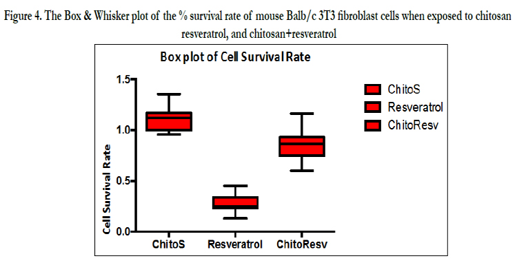
Figure 4. The Box & Whisker plot of the % survival rate of mouse Balb/c 3T3 fibroblast cells when exposed to chitosan, resveratrol, and chitosan+resveratrol
The Kruskal-Wallis Multiple-Comparison Test (Dunn's Test) and Bonferroni test showed significant differences amongst the 3 groups (p<0.05).
Figure 5 shows the % survival rate of mouse Balb/c 3T3 fibroblast cells when exposed to chitosan, propolis and chitosan+propolis.
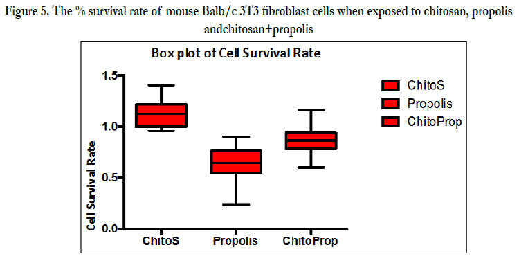
Figure 5. The % survival rate of mouse Balb/c 3T3 fibroblast cells when exposed to chitosan, propolis and chitosan+propolis
The Kruskal-Wallis Multiple-Comparison Test (Dunn's Test) and Bonferroni test showed significant differences amongst the 3 groups (p<0.05).
Figure 6. Percentage survival rate of the 3T3 cells when exposed to chitosan, β-carotene and chitosan+β-carotene.
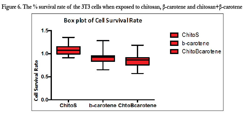
Figure 6. The % survival rate of the 3T3 cells when exposed to chitosan, β-carotene and chitosan+β-carotene
The Kruskal-Wallis Multiple-Comparison Test (Dunn's Test) and Bonferroni test showed significant differences amongst chitosan and β-carotene and chitosan and chitosan+β-carotene (p<0.05). It was found that chitosan-H significantly improved the cell survival rate to 114% in comparison to the control (100%) without chitosan.
The mean cell survival rate was found to be: resveratrol (31%); propolis (64%); β-carotene (95%), chitosan+resveratrol (87%), chitosan+propolis (95%), chitosan+ β-carotene (100%) and chitosan (114%).
Discussion
The results clearly showed (Fig. 4, 5 and 6) that chitosan in its own capacity stimulated the cell survival rate to a value of 114% which is 14% higher than that of the control value (only growth medium). From this it might also be deduced that chitosan actually stimulates cell growth to a significantly higher value. It was reported [33] that a higher degree of chitosan de-acetylation gave rise to lower uptake and cytotoxicity of chitosan nanoparticles towards A549 cells. The degree of de-acetylation of chitosan used in our study was obtained as 87%.However it should be remembered that the cytotoxicity towards different cell lines could differ between different chemicals and derivatives thereof [6,7,9,34].
The overall summary of the cell survival rates showed that when chitosan was present (chitosan/resveratrol; chitosan/propolis; chitosan/β-carotene)(Figure. 4-6) better survival rates (improvements) were found than when the antioxidants were used on their own. In this context it was found that the cell survival rate for chitosan/resveratrol was 87% while that of resveratrol was only 31%. For chitosan/propolis it was 95% in comparison to 64% for propolis and for chitosan/β-carotene it was 100% in comparison to 95% for β-carotene.
Our low survival rate with resveratrol (31% survival rate, Figure 4) indirectly supported the findings that resveratrol could suppress lymphocyte proliferation, development of cell-mediated cytotoxicity [35] and that it triggers CD95 signaling-dependent apoptosis in human tumor cells [36]. Furthermore, it caused a 70% growth inhibition onCACo-2 cells [37]. It was also reported [38] that resveratrol could be an anti-cancer agent for breast cancer and furthermore, that resveratrol suppressed cancer metabolism [39].
Propolis(Fig. 5) was reported to be cytotoxic when exposed to 3T3 cells with a mean cell survival rate of only 64% [18]. This is in agreement with various reports which showed the cytotoxic activities of propolis [40] on various cell types. However, as stated before this could only be true for the 2 kinds of propolisas they all differ from area to area.
Relative to our high concentration (1 mg/ml) a low β-carotene concentration (~40 mg/l) was reported to substantiate a selective cytotoxic effect on human tumor cells [41]. However, we could not see an influence of β-carotene (93%) on 3T3 cells at a much higher concentration (1 mg/ml). In another study, Alija [42] could also not find any cytotoxic effect on hepatocytes at a relatively low concentration of ~5 mg/l. This might differ between different cell lines. Normally, the cell survival rate will depend on a number of factors: type of substance to be tested, the concentration of the substance, the type of cell-line used and the time of cell exposure to the substance to be tested. All gel systems utilized in this study (1mg of chitosan:antioxidant/ml growth medium over a 24 hour period) were tested for their survival rates. This can be considered a high concentration and well in the top range of concentrations normally tested for other products. This is similar to the highest concentration used (1000µg/ml) to test the cytotoxic effect of many medicinal plants [43,44]. An exposure period of 24 hours used in this study can also be accepted as a long exposure period (in the top range) [43-47].
Conclusion
To conclude, this study clearly showed: 1) that chitosan-H (87% de-acetylated) has a positive effect on the cell survival rate of Balb/c mouse 3T3 fibroblast cells, and therefore most probably also on human pulp fibroblast cells, 2) these chitosan hydrogels are safe for use to improve the strength and longevity of a dental material, and 3) it further proved that chitosan in itself improved the cell survival rate of various antioxidants.
Acknowledgement
This study was financially supported by the SA Dental Association and The University of the Western Cape.
References
- Subash V, Saritha G, FareedullahM (2010) Role of antioxidants and oxidative stress in cardiovascular diseases. Ann Biomed Res 3: 158-73.
- Guo RF, Ward PA (2007) Role of oxidants in lung injury during sepsis. Antioxid Redox Signal 9(11): 1991-2002.
- Johansen JS, Harris AK, Rychly DJ, Ergul A (2005) Oxidative stress and the use of antioxidants in diabetes: Linking basic science to clinical practice. Cardiovasc Diabetol 4(1): 5.
- Yadav SB, Suryakar AN, DurgawalePP (2006) Antioxidant treatment, a new therapeutic approach to reversible male infertility. Biomed Res 7: 75-178.
- Carlesen MH, Halvorsen BL, Holte K, Bohn SK, Dragland S (2010) The total antioxidant content of more than 3100 foods, beverages, spices, herbs and supplements used worldwide. NutrJ 9: 3.
- Geurtsen WF, Lehmann F, Spahl W, Leyhausen G (1998) Cytotoxicity of 35 dental resin composite monomers/additives in permanent 3T3 and three human primary fibroblast cultures.J of Biomed Mat Res 41(3): 474-80.
- Grobler SR, Olivier A, Moodley DS, VW Kotze TJ (2008) Cytotoxicity of recent dentin bonding agents on mouse fibroblast cells. Quintessence Int 39: 511-16.
- Moodley D, Grobler SR, Olivier A (2004) Cytotoxicity of a dentine bonding agent on four different cell-lines. SA Dent J 60: 101-5.
- Olivier A, Grobler SR, Osman Y (2012) Cytotoxicity of seven recent dentine bonding agents on mouse 3T3 fibroblast cells. Open Journal of Stomatology 2: 244-250.
- Ravi Kumar MNV (2001) A review of chitin and chitosan applications. Re-active and Functional Polymers 46: 1-27.
- Palmberger TF, Hombach J, Bernkop-Schnurch A (2008) Thiolated chitosan: Development and in vitro evaluation of an oral delivery system for acyclovir. Int J Pharm 348(1-2): 54-60.
- Macleod GS, Fell JT, Colett JH, Sharma HL, Smith AM (1999) Selective drug delivery to the colon using pectin:chitosan:hydroxypropyl methylcellulose film coated tablets. Int J Pharm 187(2): 251-7.
- Bernkop-Schnurch A, Reich-Rohrwig E, Marschutz M, Schuhbauer H, Kratzel M (2000) Development of a sustained release dosage form for alphalipoic acid. II. Evaluation in human volunteers. Drug DevInd Pharm 30(1): 35-42.
- Kurosaki Y, Kimura T (2000) Regional variation in oral mucosal drug permeability. Crit Rev Ther Drug Carrier Syst 17: 467-508.
- Sadigh-Eteghad S, Talebi M, Farhoudi M, Mahmoudi J. Reyhani B (2013) Effects of Levodopa loaded chitosan nanoparticles on cell viability and caspase-3 expression in PC12 neural like cells. Neurosciences (Riyadh) 18(3): 281–3.
- Kevin McCue (March 3 2003). "New Bandage Uses Biopolymer" Chemistry. org (American Chemical Society). Archived from the original on November 28, 2005. http://web.archive.org/web/20051128100345/http://www. chemistry.org/portal/a/c/s/1/feature_ent.html?id=401c0f5c4d8511d7f6e36ed9fe800100. Retrieved 2006-07-10.
- Rodriques S, Dionisio M, Lopez CR, Grenha A (2012) Biocompatibility of Chitosan Carriers with Application in drug delivery. J FunctBiomater 3:615-41;
- Perchyonok VT, Grobler SR, Zhang S, Olivier A, Oberholzer T (2013) Insights into chitosan hydrogels on dentine bond strength and cytotoxicity. Open J Stomatol 3: 75-82.
- Perchyonok VT, Zhang S, Grobler SR, Olivier A, Oberholzer T (2013) Insights into and relative effect of chitosan-H, chitosan-H-propolis, chitosan-H-propolis-nystatin, chitosan-H-nystatin on dentine bond strength. Eur J of Dent 7(4): 412-418.
- Karre P, Helfenstein A,Wehrli H, Wettstein A (2004) "Pflanzenfarbstoffe XXV. Über die Konstitution des Lycopins und Carotins". Helvetica ChimicaActa 13(5): 1084–99.
- Athar M, Back JH, Tang X, Kim KH, KopelovichL, et al. (2007) "Resveratrol: a review of preclinical studies for human cancer prevention". Toxicol. Appl. Pharmacol 224: 274–83.
- Baur JA, Sinclair DA (2006) "Therapeutic potential of resveratrol: the in vivo evidence". Nature Reviews Drug Discovery 5: 493–506.
- American Cancer Society. Vitamin A, Retinoids, and Provitamin A Carotenoids. http://www.cancer.org/treatment/treatmentsandsideeffects/complementaryandalternativemedicine/ herbsvitaminsandminerals/vitamin-a-andbeta-carotene.
- Toreti VC, Sato HH, Pastore GM, Park YK (2013) "Recent progress of propolis for its biological and chemical compositions and its botanical origin". Evid Based Complement Alternat Med; Article ID 697390, 13
- Medline Plus, “Propolis,” 2012. http//www.nlm.nih.gov/medlineplus/druginfo/natural/390.html
- Giamalia I, Steinberg D, Grobler S, Gedalia I (1999) The effect of propolis exposure on microhardness of human enamel in vitro. J Oral Rehabil 26(12):4725.
- Perchyonok VT, Zhang S, Grobler SR, Oberholzer T, Massey W (2014). Insights into functional tea infused-chitosan hydrogels as potential bio-active restorative materials. Eur J Gen Dent 3: 22-8.
- Perchyonok VT, Zhang S,Basson NJ, Grobler SR, Oberholzer T, et al. (2013).Insights into chitosan based gels as functional restorative biomaterials prototypes: in vitro approach. Open J of Stomatology 3: 22-30 .
- Perchyonok VT, Zhang S, Grobler SR, Oberholzer TG, Massey W (2014) Insight and relative effect of aspirin, naproxen and ibuprofen containing hydrogels: from design to performance as a functional dual capacity restorative material and build in free radical defence: in-vitro studies. Open J of Stomatology 4(2): 73-83
- Perchyonok VT, Zhang S,Basson NJ, Grobler SR, Oberholzer T, et al. (2014).Insights into functional tetracycline/antioxidant containing chitosan hydrogels as potential bio-active restorative materials: structure, function and antimicrobial activity. Open J of Stomatology 4: 99-108.
- Perchyonok VT, Zhang S, Oberholzer TG (2012) Alternative chitosan based drug delivery system to fight oral mucositis: synergy of conventional and bioactives towards the optimal solution. Current Nanoscience 8: 541-7.
- Mosmann T. (1983) Rapid colorimetric assay for cellular growth and survival: Application to proliferation and cytotoxicity assays. J Immunol Methods 65(1-2): 55-63.
- Huang M, Khor E, Lim L-Y (2004) Uptake and Cytotoxicity of Chitosan Molecules and Nanoparticles: Effects of Molecular Weight and Degree of Deacetylation. Pharm Res 21(2): 344-53.
- Grobler SR, Olivier, A, Moodley A, Kotze TJ Van W (2004) Cytotoxicity of two concentrations of a dentine bonding agent on mouse 3T3 and human pulp fibroblast cell-lines. SA Dent J 59(9): 368-72.
- Gao X, Xu YX, Janakiraman N, Chapman RA, Gautam SC (2001) Immunomodulatory activity of resveratrol: Suppression of lymphocyte proliferation, development of cell-mediated cytotoxicity, and cytokine pro-duction. BiochemPharmacol 62: 1299-1308.
- Clement M, Hirpara JL, Chawdhury S, Pervaiz S (1998) Chemopreventive agent resveratrol, a natural product derived from grapes, triggers CD95 signalling-de-pendent apoptosis in human tumor cells. Blood 92(3): 996-1002.
- Loha JW, Saunders M, Lima L-Y (2012) Cytotoxicity of mono-dispersed chitosan nanoparticles against the Caco-2 cells. Toxicol Appl Pharmacol 262(3): 273–82.
- Nakagawa H, Uemura Y, Shikata H, Hioki K, Tsubura A (2001) Resveratrol inhibits human breast cancer cell growth and may mitigate the effect of linoleic acid, a potent breast cancer cell stimulator. J Cancer Res ClinOncol 126(4): 258-64.
- Iqbal MA, Bamezai RNK (2012) Resveratrol inhibits cancer cell metabolism by down regulating pyru-vate kinase M2 via inhibition of mammalian target of rapamycin.7(5): e36764.
- Banskota AH, Tezuka Y, Prasain JK, Matsushige K, Saiki I (1998) Chemical constituents of Brazilian propolis and their cytotoxic activities. J Nat Prod 61(7): 896-900.
- Schwartz J, Shklar G (1992) The selective cytotoxic effect of carotenoids and α-tocopherol on human cancer cell lines in vitro. J. Oral MaxilofacSurg 50(4): 367-73.
- AlijaAJ, Bresgen N, Sommerburg O, Siems W, Eckl PM (2003) Cytotoxic and genotoxiceffects of β-carotene breakdown products on primary rat hepatocytes. Oxford Journals 25: 827-31.
- Coe FG, Parikh DM, Johnson CA, Anderson GJ (2012) The good and bad. Alkaloid screening and brine shrimp bioassays of aqueous extracts of 31 medicinal plants of eastern Nicaraqua. Pharmaceutical Biology 50(3): 384-92.
- Ruffia MJ, Ferraro G, Wagner ML, Calcagno RH, Campo L (2002) Cavallaros cytotoxic of Argentine medicinal plant extracts on human hepatocellular carcinoma cell line. Journal of Ethnopharmacology 79: 335-330.
- Chen C, Weng M, Wu C, Lin J (2004) Comparison of radical scavenging activity, cytotoxicity effects and apoptosis induction in human melanoma cells by Taiwanese propolis from different sources. Evidence-Based Complementary and Alternative Medicine 1: 175-85.
- Alija AJ, Bresgen N, Sommerburg O, Siems W, Eckl PM (2004) Cytotoxic and genotoxiceffects of β-carotene breakdown products on primary hepatocytes. Oxford Journals 25(5): 827-31.
- Schwartz J, Shklar G (1992) The selective cytoxic effect of carotenoids and alpha-tocophenol on human cancer cell lines in vitro. J Oral Maxillofac Surg.50(4): 367-73


