Inhibitory Effect of Vernonia amygdalina on Dimethylnitrosamine (DMN)-induced Liver Fibrosis in Rats
Usunomena U1*, Okolie N. Paulinus2, Eze I. Gerald3
1 Department of Basic sciences (Biochemistry unit), Faculty of Basic and Applied sciences, Benson Idahosa University, Benin City, Nigeria.
2 Department of Biochemistry, Faculty of Life sciences, University of Benin, Benin City, Edo State, Nigeria.
3 Department of Anatomy, College of Medicine, University of Benin, Benin City, Edo State, Nigeria.
*Corresponding Author
Usunobun Usunomena,
Department of Basic sciences (Biochemistry unit),
Faculty of Basic and Applied sciences,
Benson Idahosa University, P.M.B. 1100, Benin City,
300001, Edo state, Nigeria.
Tel: +2348034174871
E-mail: uusunobun@biu.edu.ng
Article Type: Research Article
Received: June 26, 2015; Accepted: August 19, 2015; Published: August 21, 2015
Citation: Usunomena U, Okolie N. Paulinus, Eze I. Gerald (2015) Inhibitory Effect of Vernonia amygdalina on Dimethylnitrosamine (DMN)- induced Liver Fibrosis in Rats. Int J Clin Pharmacol Toxicol, 4(4) 179-184. doi: dx.doi.org/10.19070/2167-910X-1500030
Copyright: Usunomena U© 2015. This is an open-access article distributed under the terms of the Creative Commons Attribution License, which permits unrestricted use, distribution and reproduction in any medium, provided the original author and source are credited.
Abstract
In this study, the protective effect of Vernonia amygdalina ethanolic leaf extract against hepatic fibrosis induced by repeated intermittent administration of Dimethylnitrosamine (DMN) was investigated in rats. A total of 48 rats divided into 4 groups were used. Group 1 served as control, Group 2 received 200mg/kg Vernonia amygdalina only for 14days, Group 3 received 200mg/kg Vernonia amygdalina for 14 days followed by intraperitoneal administration of 10mg/kg DMN on first three days of each week for 2 weeks, while Group 4 received intraperitoneal administration of 10mg/kg DMN on the first three days of each week for 2 weeks. Oral administration of Vernonia amygdalina led to recovery of body/liver weight losses and significantly reversed elevation of total collagen, AST, ALT and ALP in DMN-exposed rats. Severe oxidative stress induced in fibrotic rats was evidenced by elevation in MDA levels associated with a fall in the activities of GSH, SOD and CAT in the DMN-treated group. These were significantly reversed by simultaneous treatment with Vernonia amygdalina. Recovery of rat liver tissue from DMN-induced hepatocellular necrosis and hepatic fibrosis by Vernonia amygdalina treatment was confirmedby histopathological evaluation of liver tissue. In conclusion, Vernonia amygdalina exhibits hepatoprotective, antioxidant, free radical-scavenging, and anti-fibrotic activity against DMN-induced hepatic fibrosis.
2.Introduction
3.Materials and Methods
3.1.Collection, Identification and Preparation of plant materials
3.2.Extraction of the plant leaves
3.3.Experimental animals, DMN and extract administration
3.4.Collection of tissue samples and preparation of liver homogenates
3.5.Biochemical assays
3.6.Histology
3.7.Statistical analysis
4.Results
5.Discussion
6.Conclusion
7.References
Keywords
Collagen; Dimethylnitrosmine; Fibrosis; Liver; Rat; Vernonia Amygdalina.
Introduction
The liver is the largest visceral organ in the human body and the chief site for metabolism and detoxification. Liver diseases often progress from sub-clinical icteric hepatitis to necro-inflammatory hepatitis, fibrosis, cirrhosis, and hepatocellular carcinoma [1]. Liver cirrhosis often results in high mortality [2] and is also a risk factor in the development of hepatocellular carcinoma (HCC) [3], which ranks the fifth of the cancer incidence worldwide [4].
Several experimental and clinical evidence have shown a common link between chronic liver injury, oxidative stress, activation of hepatic stellate cells (HSCs) and their transformation to myofibroblast- like cells, associated with increased production of extracellular matrix proteins during hepatic fibrosis [5, 6]. Repeated administration of dimethylnitrosamine (DMN), a potent hepatotoxin, carcinogen and mutagen has been demonstrated to induce bridging fibrosis, necrosis and collapse of parenchymal framework of liver [7].
Despite remarkable advances in the field of modern medicine, hepatic diseases remain a major public health problem, thus the search for new effective medicines without side effects is still on-going [8]. Hepatic cells are involved in a variety of metabolic events; therefore the establishment of liver protective/therapeutic agents is of paramount importance in the protection of the liver from damage. Natural remedies from traditional plants are seen as effective and safe alternative treatments for hepatotoxicity. The present investigation was undertaken with the objective of evaluating the anti-fibrotic, antioxidant and protective efficacy of ethanolic leaf extract of Vernonia amygdalina against repeated DMN-induced hepatic fibrosis in rats.
Materials and Methods
Fresh leaves of Vernonia amygdalina were purchased from a local market in Benin City, Edo state, Nigeria. The leaves were identified by Dr. Chris Akoma, a Botanist in the Department of Basic Sciences, Faculty of Basic and Applied Sciences, Benson Idahosa University, Benin city, Edo State. The Vernonia amygdalina leaves were separated from the stalk, washed and air-dried at room temperature (24°C) and then pulverized, crushed into fine powder and weighed.
Ethanolic extracts of the plant leaves was prepared by soaking 200g of the dry powdered plant leaves in one (1) litre of absolute ethanol at room temperature for 48hrs (for thorough extraction). The extract was then filtered first through a Whatmann filter paper No. 42 (125mm) and then through cotton wool. The extract was thereafter concentrated using a rotary evaporator with the water bath set at 40°C to one-tenth its original volume and then finally freeze dried. The dried residue (crude extract) was then stored at 4°C. Aliquot portions of the crude plant extract residue were weighed and dissolved in distilled water for use on each day of our experiments.
Male Albino wistar rats weighing 150-225 g were obtained from the Animal Unit of the University of Ibadan, Ibadan, Oyo state, Nigeria. The animals were housed in controlled environmental conditions (temperature - 24±2°C; relative humidity-50-70%; 12 h light/dark cycle) at the animal house of the Department of Biochemistry, University of Benin, Benin city, Edo state. The animals were provided standard pellet diet and water ad libitum. Institutional Animal Ethical Committee permission was obtained before performing the experiments.
DMN used in this work was synthesized in a fume chamber at the Department of Biochemistry, University of Ibadan, Oyo state, Nigeria, according to the method of Vogel [9].
A total of 48 rats divided into 4 groups were used. Group 1 served as control and was given normal saline, Group 2 received 200mg/kg Vernonia Amygdalina. only for 14 days, Group 3 received 200mg/kg Vernonia Amygdalina. for 14 days followed by intraperitoneal administration of 10mg/kg DMN (dissolved in 0.15M NaCl) on first three days of each week for two weeks, while Group 4 received intraperitoneal administration of 10mg/kg DMN (dissolved in 0.15M NaCl) on the first three days of each week for two weeks. Before use, the Vernonia Amygdalina. leaf extract was reconstituted in distilled water and administered orally via gastric intubation.
Body weights of animals were taken on first and last day while morphological and behavioural changes were monitored on a daily basis. 24 hours after the end of treatment, rats were sacrificed by cardiac puncture and blood collected via the ocular vein in plain tubes and allowed to stand for 45 min before it was centrifuged at 4,000 rpm for 30 min. Serum was stored at -20°C until analyzed.
Following sacrifice, liver samples were quickly harvested, rinsed with normal saline and weighed to obtain the absolute organ weights.
Relative weights were calculated using the formula:
Relative Organ Weight = Absolute Organ Weight/Body Weight at Sacrifice X 100%
A section of each liver sample was fixed in 10% phosphate-buffered formalin for histological examination while the remaining section were stored at –20°C for biochemical analysis. 10% liver homogenate was prepared in physiological saline. The homogenate was centrifuged at 5000 x g for 15 minutes and the clear supernatant obtained used for biochemical analysis.
Serum AST and ALT activities were estimated colorimetrically according to the method of Reitman and Frankel [10], while ALP assay was done by monitoring change in absorbance at 405nm due to liberation of para-nitrophenol, using Randox kits (UK) according to manufacturer’s instructions. Liver total collagen was assayed as hydroxyproline derivatives following hydrolysis using QuickZymeR kits (Biosciences, Netherlands). Reduced glutathione was estimated colorimetrically by measuring the reduction of Ellman’s reagent (5, 5’di-thio-bis-2-nitrobenzoic acid) at 412nm as described by Ellman [11]. Superoxide dismutase (SOD) was assayed based on the ability of the enzyme to inhibit the autooxidation of epinephrine according to the method of Misra and Fridovich [12]. The assay of catalase was carried out colorimetrically based on the measurement of the rate of decomposition of H2O2 after the addition of the sample containing the enzyme by reacting it with excess KMnO4 and then measuring the residual KMnO4. spectrophotometrically at 480nm [13]. MDA was estimated in a colorimetric reaction with thiobarbituric acid [14].
Liver sections fixed in formol-saline were processed for light microscopy at the Department of Anatomy, School of Basic Medical Sciences, College of Medicine, University of Benin. The resultant slides were read and interpreted by one of us, G. I. Eze., a consultant pathologist.
Numerical data obtained from the study were expressed as mean value ± standard deviation. Differences between means of control and tested groups were determined using Statistical Package for social scientist (SPSS). A probability level of less than 5% (p < 0.05) was considered significant.
Results
Table 1 shows the effect of ethanolic leaf extract of Vernonia Amygdalina. on body and relative liver weight in the DMN-treated rats and controls. DMN alone-treated rats were docile, weak and showed significant fall in body weight (p < 0.05), liver weight (p < 0.05) and their ratio (p < 0.05), when compared to saline-treated control. This adversity was significantly protected and reversed back toward normalcy in rats simultaneously treated with Vernonia amygdalina after DMN treatments. The viscera showed various degrees of ascites in rats receiving DMN alone. However, Vernonia Amygdalina. alone treated rats did not show any change in body weight and liver weight when compared to control.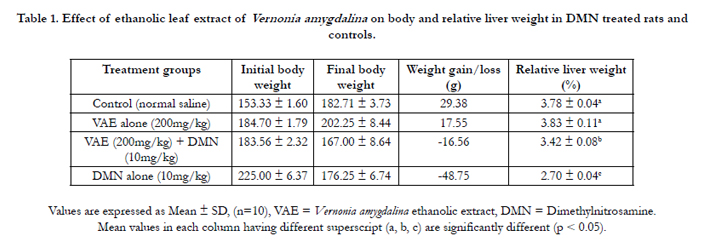

Table 1. Effect of ethanolic leaf extract of Vernonia amygdalina on body and relative liver weight in DMN treated rats and controls.
Table 2 shows the effect of Vernonia Amygdalina. on liver total collagen and serum AST, ALT, and ALP in the DMN-treated rats and controls. Total liver collagen of the DMN group was significantly higher than in each of the other groups (p < 0.05). However, the extract administration led to significant decrease in liver total collagen (p < 0.05). The DMN group also had significantly higher serum levels of ALT, AST and ALP when compared with corresponding values for each of the other two treatment groups and controls (p < 0.05).
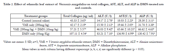
Table 2. Effect of ethanolic leaf extract of Vernonia amygdalina on total collagen, AST, ALT, and ALP in DMN-treated rats and controls.
Table 3 shows the effect of ethanolic leaf extract of Vernonia Amygdalina. on liver MDA, GSH, SOD and CAT in DMN treated rats and controls. The mean liver MDA levels of the DMN treated rats were significantly higher than those in the rats that received VAE and intraperitoneal DMN injections as well as in the control and the group that received the extract alone (p < 0.05). The mean hepatic SOD and CAT levels of rats in the DMN group were significantly lower than corresponding values for DMN + extract group, extract-only group and control group (p < 0.05). The DMN group also had significantly lower liver GSH concentration than any of the other groups (p < 0.05). Histological examination of the liver sections revealed that while the control and Vernonia Amygdalina.-alone groups had normal features (Plate A and B), DMN caused marked pathologic changes such as hemorrhagic necrosis, bridging fibrosis, extensive collagen deposition and early cirrhosis (Plate D). These changes were significantly mitigated in the rats given Vernonia Amygdalina. and DMN (Plate C).
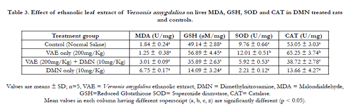
Table 3. Effect of ethanolic leaf extract of Vernonia amygdalina on liver MDA, GSH, SOD and CAT in DMN treated rats and controls.
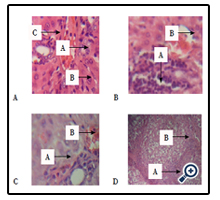
Figure
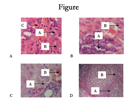
Plate A: Photomicrograph of liver section from control rats showing liver parenchyma with central vein and radiating column of hepatocytes, with portal triad (A), hepatocytes (B) and sinusoid (C), portal tracts appear normal (H&E staining; x 40). Plate B: Liver section from rats treated with Vernonia amydalina alone (200mg/kg) and sacrificed after day 14 showing mild vascular congestion A and mild periportal infiltrates of lymphocytes B (H&E x 40). Plate C: Liver section from rats treated with Vernonia amydalina (200mg/kg) and DMN (10mg/kg) and sacrificed after 14 days showing extensive areas of normal hepatocytes A and focal hemorrhagic necrosis B (H&E x 40). Plate D: Liver section from rats treated with DMN alone (10mg/kg) and sacrificed after 14 days showing well developed bridging fibrosis, cirrhosis and extensive deposition of collagen (A); and hemorrhagic necrosis (B) (H&E x 40).
Discussion
Fibrosis as a scarring response to liver damage contains the injurious process (Poynard et al., 2003) that can ultimately lead to impairment of liver function, development of hepatocellular carcinoma, and portal hypertension with all its associated complications.
The fall in liver weight, body weight and their ratio observed in DMN treated rats are in acceptance with previous reports [7, 15-18]. The decrease could be attributed to the reduction in protein synthesis, massive cell necrosis and collapse of parenchymal cells [16, 17, 7]. Body weight changes serve as a sensitive indication of the general health status of animals. The organ-to-body weight ratio can be used as an index for assessing the state of an organ, as significant reduction in organ-to-body weight ratio can be traced to organ or tissue necrosis, while a significantly high ratio is a possible indication of tissue inflammation [19]. Thus the observed decrease in the weight of the liver in this study indicates DMN toxic effect on liver organ. The drastic reduction in food and water intake caused by DMN induction may have also contributed to the decrease in liver weight. It is possible that the pain due to liver injury caused by DMN administration affected the appetite and a concomitant decrease in food intake, thus resulted in reduction in the body weight of the rats. However, treatment of rats with 200mg/kg Vernonia Amygdalina. simultaneously with DMN administration significantly restored (p < 0.05) the body weight and organ to weight ratio compared to DMN alone rats. It may therefore be inferred that Vernonia Amygdalina. has the potential to induce the activation of cellular enzymes responsible for increased metabolic activity in liver and the regeneration of hepatic tissues. In agreement with these findings, in a study by Shin and Moon [20], DMN-induced decrease in body and liver weight were significantly increased by grape skin or seeds ingestion. This study also agrees with findings by Ahmad et al [17] that DMN-induced decrease in body and liver weight showed a considerable increase in body weight and liver weight during the course of treatment with Vitamin B12. Our result is also in accordance with Qing-Wei and Geng-Tao, [21] who reported that bicyclol attenuated the decrease of body weight in mice treated with DMN.
It is postulated that DMN treatment will induce inflammation of hepatocytes resulting in necrosis and this is indicated by an increase in marker enzymes of hepatotoxicity (AST, ALT, ALP) in the serum, accompanied by their fall in liver tissue [22-24]. Our results are in agreement with these reports which were corroborated with histopathological observations. However, oral administration of Vernonia Amygdalina. significantly reversed AST, ALT and ALP in the DMN-exposed rats. Thus the extract significantly prevents liver damage by maintaining the integrity of the plasma membrane, thereby suppressing the leakage of enzymes. In a similar study by George et al [25], higher enzyme activity levels of AST and ALT were observed in DMN-induced group while treatment with silymarin and curcumin restored the increased activity levels to near normal. Similarly, Sharma and Singh [26] reported that ethanolic root extract of Operculina turpethum manifested therapeutic effects by significantly restoring enzymatic levels of AST, ALT and ALP, thereby reducing DMN-induced hepatic damage in mice. Also in a study by Shin and Moon [20], DMN-induced increase in serum AST, ALT, and ALP levels were significantly suppressed by grape skin or seed ingestion. Similarly, Jung et al., [27], reported that Serum ALP and AST were significantly raised by DMN treatment (p < 0.05) and the levels significantly decreased by 1-O-Hexyl-2,3,5-Trimethylhydroquinone (HTHQ) treatment.
There exists convincing evidence to support the contention that oxidative stress and liberation of reactive oxygen species (ROS) play vital role in the etiology and progression of liver fibrosis [5,28,6]. In this study, oxidative stress, marked by elevation in the status of malondialdehyde (MDA) was noticed in the liver of DMNtreated rats similar to previous reports [22, 29]. Also, oxidative stress induced by DMN injection is demonstrated by a highly significant fall in the activities of GSH, SOD and CAT which were also in agreement with previous reports [22-24, 30]. The decrease in the activities of these enzymes could possibly be due to their over-utilization toward suppression of ROS that are liberated during metabolism of DMN in the liver. The reduction in liver SOD might be due to a continuous higher production of superoxide radical by the mitochondria of the damaged liver cells. The reduction in the activity of CAT may result in a number of deleterious effects due to accumulation of highly toxic metabolites and hydrogen peroxide, thus the high MDA levels on DMN treated rats. Glucose 6-phosphate dehydrogenase, the rate-limiting enzyme of pentose phosphate pathway is the principal intracellular source of NADPH which in turn is used as reducing equivalent to maintain GSH stores, which are used to scavenge ROS. In this study, the significant decrease in GSH in DMN-treated rats compared to the control and extract-treated rats may be attributable to reduction in glucose 6-phosphate dehydrogenase which in turn might decrease NADPH levels, thereby unable to maintain GSH stores. Iwalokun et al [31] reported that reduced glucose-6-phosphate dehydrogenase activity is associated with increased cellular ROS accumulation resulting in depletion of glutathione stores and enhanced oxidative stress. However supplementation of rats with ethanolic leaf extract of Vernonia Amygdalina. (200mg/kg), decreased liver MDA and increased liver SOD, CAT and GST compared to rats given DMN alone. The effect of Vernonia Amygdalina. possibly involves the inhibition of neutrophil infiltration and lipid peroxidation, thus, their biological significance in eliminating reactive free radicals and significant restoration of antioxidant status in the tissue. Increased levels of SOD, CAT and GSH due to treatment of rats with ethanolic leaf extracts of Vernonia Amygdalina. leaves indicates that the extract contain compounds such as flavonoids, saponins, tannins, Vitamin C and beta-carotenes that converted free radicals to more stable products. This may also be due to direct stimulatory effect of the extract on the antioxidant enzymes studied. In a similar work by Sharma and Singh [26], Operculina turpethum significantly restored SOD, CAT and GSH in the liver as well as decreased lipid peroxidation, exhibiting significant curative effect against DMN-induced toxicity.
Accumulation of connective tissue proteins, especially collagen has been reported in DMN- induced fibrotic rats and measurement of these parameters is suggested as a valuable tool in the quantification of fibrosis as well as assessment of potency of anti- fibrotic drugs during therapeutic trials [32]. In the present study, repeated administration of DMN alone caused a linear increase in collagen, with a near 3-fold increase observed in total collagen content, indicating establishment of hepatic fibrosis which is in agreement with previous reports [7, 33-37]. Ito cells also called hepatic stellate cells (HSCs) are primarily responsible for the increased collagen synthesis in the injured liver [38, 2, 7]. During hepatic necrosis, these cells become activated and behave like myofibroblasts, initiating vigorous collagen synthesis [7]. No specific identification of Ito cells were made in the present study, but the enhanced collagen deposition is presumed to reflect increased Ito cell activity. Also decreased synthesis of collagenolytic enzymes by the impaired hepatocytes may have further contributed to the accumulation of collagen in the liver. The near 3-fold increase in DMN alone rats coincided with H & E stained histopathological studies of DMN treated rats as fibrosis and deposition of fibrous proteins were observed in the perisinusoidal space. However, simultaneous administration of Vernonia Amygdalina. (200mg/kg) along with DMN (10mg/kg each) showed protective anti-fibrogenic effect of Vernonia Amygdalina. expressed by the decrease of total collagen level. The significant restoration of collagen levels may be by way of blocking the pathways to collagenesis or by increasing the levels of collagenolytic enzymes. In agreement with these findings, Shin and Moon [20] reported that DMN-induced increase in liver total collagen was significantly suppressed by ingestion of grape skin or seeds. Similarly, Yan et al [39] reported that Sorafenib significantly decreased collagen deposition by 58.31% as compared to DMN alone treated rats. Earlier on, George et al [25] reported that the significant increase in liver total collagen observed in DMN-treated fibrotic rats was significantly restored by silymarin and curcumin.
Hepatic fibrosis induced by DMN is well demonstrated in the H & E stained histopathological studies of DMN treated rats as fibrosis and deposition of fibrous proteins were observed in the sinusoidal space similar to previous results of Ahmad et al [16], George and Chandrakasan [35]; George et al [7] and George, [25] as staining of liver sections revealed inflammation, disruption of normal liver architecture, hemorrhage and distinct deposition of thick collagen fibers. The liver necrosis found in animals caused collapse of the parenchymal framework of the liver. The high mortality rate, increased portal pressure and decreased liver blood flow as well as ascites observed in DMN-alone rats are consistent with previous reports [40, 41, 7]. However, simultaneous administration of ethanolic leaf extract of 200mg/kg Vernonia Amygdalina. with DMN significantly reduced the inflammation, necrosis and fibrotic area compared to DMN-induced hepatic fibrosis. Thus, in the sections of the rats intoxicated with DMN and thereafter treated with ethanolic leaf extract of Vernonia Amygdalina., the normal cellular architecture appeared to have less damage compared to DMN-induced fibrotic rats, indicating some form of restoration to the DMN-induced liver injury and confirming the protective and inhibitory effect of the extract against liver fibrosis. The hepatoprotective effects observed in histological evidence in the liver tissues of rats simultaneously treated with Vernonia Amygdalina. and DMN is most likely a consequence of extract-induced reduction in lipid peroxidation and elevation/stabilization of tissue antioxidant enzyme activities. This is so because, leaves of Vernonia amygdalina have been reported to contain bioflavonoids such as luteolin, luteolin 7-O-β-glucoside and luteolin 7-O-β-glucuronoside besides several stigmastine type saponins such as vernoniosides A1, A2, B1, B2, D3, A4 and C as well as sesquiterpene lactones such as vernolide and vernodalol [42-44].
Conclusion
Our present results demonstrate that treatment with Vernonia Amygdalina. protects liver against DMN-induced hepatic necrosis, inflammatory changes and hepatic fibrosis by way of its antioxidant, free radical scavenging, membrane stabilizing and suppression of fibrosis.
References
- Lima CF, Fernandes-Ferreira M, Pereira-Wilson C (2007) Drinking of Salvia officinalis tea increases CCl4-induced hepatotoxicity in mice. Food Chem Toxicol 45(3): 456-464.
- Friedman SL (2000) Molecular regulation of hepatic fibrosis, an integrated cellular response to tissue injury. J Biol Chem 275(4): 2247-2250.
- Bataller R, Brenner DA (2005) Liver fibrosis. J Clin Invest 115(2): 209-218.
- Caldwell S, Park SH (2009) The epidemiology of hepatocellular cancer: from the perspectives of public health problem to tumor biology. J Gastroenterol 44(suppl 19): 96-101.
- Poli G (2000) Pathogenesis of liver fibrosis: role of oxidative stress. Mol Aspects Med 21(3): 49-98.
- Vendemiale G, Grattagliano I, Caruso ML, Serviddio G, Valentini AM, et al. (2001) Increased oxidative stress in dimethylnitrosamine-induced liver fibrosis in the rat: effect of N-acetylcysteine and interferon-alpha. Toxicol Appl Pharmacol 175(2): 130-139.
- George J, Rao KR, Stern R, Chandrakasan G (2001) Dimethylnitrosamineinduced liver injury in rats: the early deposition of collagen. Toxicology 156(2-3): 129-138.
- Choi JH, Kim DW, Yun N, Choi JS, Islam MN, et al (2011) Protective effects of hyperoside against carbon tetrachloride-induced liver damage in mice. J Nat Prod 74:1055-1060.
- Vogel AI (1971) A textbook of practical organic Chemistry including qualitative organic analysis. Longman group limited, London. 426.
- Reitmans S, Frankel S (1957) A colorimetric method for the determination of SGOT and SGPT. Am J Clin Path 28: 56-58.
- Ellman GL (1959) Tissue sulfhydryl groups. Arch Biochem Biophys 82(1): 70-77.
- Misra HP, Fridovich I (1972) The role of superoxide anion in the autooxidation of epinephrine and a simple assay of superoxide dismutase. J Biol Chem 247(10): 3170-3175.
- Cohen G, Dembiec D, Marcus J (1970) Measurement of catalase activity in tissue extracts. Anal Biochem 34: 30-38.
- Ohkawa H, Ohishi N, Yogi K (1979) Assay for lipid peroxides in animal tissues by thiobarbituric acid reaction. Anal Biochem 95(2): 351-358.
- Lee HS, Jung KH, Park IS, Kwon SW, Lee DH, et al. (2009) Protective effect of Moringa on dimethylnitrosamine-induced hepatic fibrosis in rats.Dig Dis Sci 54: 782-788.
- Ahmad R, Ahmed S, Khan NU, Hasnain A (2009) Operculina turpethum attenuates N-nitrosodimethylamine induced toxic liver injury and clastogenicity in rats. Chem Biol Interact 181(2): 145-153.
- Ahmad A, Afroz N, Gupta UD, Ahmad R (2014) Vitamin B12 supplement alleviates N-nitrosodimethylamine induced hepatic fibrosis in rats. Pharm Biol 1-8.
- Shimizu I (2000) Sho-saiko-to: Japanese herbal medicine for protection against hepatic fibrosis and carcinoma. J Gastroenterol Hepatol 15: D84-D90.
- El-Hilaly J, Israili ZH, Lyoussi B (2004) Acute and chronic toxicological studies of Ajuga iva in experimental animals. J Ethnopharmacol 91: 43-50.
- Shin M, Moon J (2010) Effect of dietary supplementation of grape skin and seeds on liver fibrosis induced by dimethylnitrosamine in rats. Nutr Res Pract 4(5): 369-374.
- Hu QW, Liu GT (2006) Effects of bicyclol on dimethylnitrosamine-induced liver fibrosis in mice and its mechanism of action. Life Sci 79(6): 606-612.
- Priya S, Vijayalakshmi P, Vivekanandan P, Karthikeyan S (2011a) N-Acetylcysteine attenuates dimethylnitrosoamine induced oxidative stress in rats. Eur J Pharmacol 654(2): 181-186.
- Priya S, Vijayalakshmi P, Vivekanandan P, Karthikeyan S (2011b) Influence of N-acetylcysteine against dimethylnitrosamine induced hepatotoxicity in rats. Toxicol Ind Health 27(10): 914-922.
- Wang JH, Shin JW, Son JY, Cho JH, Son CG (2010) Antifibrotic effects of CGX, a traditional herbal formula, and its mechanisms in rats. J Ethnopharmacol 127(2): 534-542.
- George J, Suguna L, Jayalakshmi R., Chandrakasan G (2006) Efficacy of Silymarin and Curcumin on Diethylnitrosamine induced liver fibrosis in rats. Biomedicine 26(3-4): 18-26.
- Sharma V, Singh M (2014) Attenuation of N-nitrosodimethylamine induced hepatotoxicity by Operculina turpethum in Swiss Albino mice. Iran J Basic Med Sci 17(1): 73-80.
- Jung YR, Lee YJ, Lee NJ, Lin CM, Moon JH, et al. (2010) Inhibitory Effect of 1-O-Hexyl-2,3,5-Trimethylhydroquinone on Dimethylnitrosamineinduced Liver Fibrosis in male SD rats. Toxicol Res 26(3): 193-201.
- Parola M, Robino G (2001) Oxidative stress-related molecules and liver fibrosis.J Hepatol 35(2): 297-306.
- Tahan V, Ozaras R, Canbakan B, Uzun H, Aydin S, et al. (2004) Melatonin reduces dimethylnitrosamine-induced liver fibrosis in rats. J Pineal Res 37(2): 78-84.
- Hong SW, Jung KH, Zheng HM, Lee HS, Suh JK, et al. (2010) The protective effect of resveratrol on dimethylnitrosamine-induced liver fibrosis in rats. Arch Pharm Res 33(4): 601-609.
- Iwalokun BA, Efedede BU, Alabi-Sofunde JA, Oduala T, Magbagbeola OA, et al. (2006) Hepatoprotective and antioxidant activities of Vernonia Amygdalina. on acetaminophen-induced hepatic damage on mice. J Med Food 524-530.
- Kusunose M, Qiu B, Cui T, Hamada A, Yoshika S, et al. (2002) Effect of Sho-saiko-to extract on hepatic inflammation and fibrosis in Dimethylnitrosamine- induced liver injury in rats. Biol Pharm Bull 25(11): 1417-1421.
- George J, Chandrakasan G (1996) Molecular characteristics of dimethylnitrosamine- induced fibrotic liver collagen. Biochim Biophys Acta 1292(2): 215-222.
- George J, Chandrakasan G (1997) Lactate dehydrogenase isoenzymes in dimethylnitrosamine induced hepatic fibrosis in rats. J Clin Biochem Nutr 22(1): 51-62.
- George J, Chandrakasan G (2000) Biochemical abnormalities during the progression of hepatic fibrosis induced by dimethylnitrosamine. Clin Biochem 33(7): 563-570.
- Iimuro Y, Fujimoto J (2003) Strategy of gene therapy for liver cirrhosis and hepatocellular carcinoma. J Hepatobiliary Pancreat Surg 10(1): 45-47.
- Mu YP, Liu P, Liu Y, Li FH., Chen GF, et al. (2006) Inhibiting action of Xiayuxue decoction on hepatic fibrosis at progressive stage in rats and study on the prescription and syndrome. J Tradit Chin Med 47: 215-218.
- Brenner DA, Waterboer T, Choi SK, Lindquist JN, Stefanovic B, et al. (2000) New aspects of hepatic fibrosis. J Hepatol 32(1 suppl): 32-38.
- Wang Y, Gao J, Zhang D, Zhang J, Ma J, et al. (2010) New insights into the antifibrotic effects of sorafenib on hepatic stellate cells and liver fibrosis. J Hepatol 53(1): 132-144.
- Jenkins SA, Grandison A, Baxter JN, Day DW, Taylor I, et al. (1985) A dimethylnitrosamine-induced model of cirrhosis and portal hypertension in the rat. J Hepatol 1(5): 489-499.
- Chowdhury SA, Taylor R (1989) Insulin sensitivity in experimental cirrhosis. Mol Cell Biochem 89(1): 69-72.
- Igile GO, Oleszek W, Jurzysta M, Burda S, Fafunso M, et al. (1994) Flavonoids from Vernonia Amygdalina. and their antioxidant activities. J Agric Food Chem 42: 2445-2448.
- Jisaka M, Ohigashi H, Takagaki T, Nozaki H, Tada T, et al. (1992) Bitter steroid glucosides, Vernoniosides A1, A2 and A3 and related B1 from a possible medicinal plant, Vernonia Amygdalina., used by wild chimpanzees. Tetrahedron 48(4): 625-632.
- Erasto P, Grierson DS, Afolayan AJ (2006) Bioactive sesquiterpene lactones from the leaves of Vernonia Amygdalina.. J Ethnopharmacol 106(1): 117- 120.


