Pigmented Basal Cell Carcinoma
Niema Aqil*, Aicha Nassiri, Fatima Zahra Mernissi
Dermatology, University Hospital Hassan II, Fès, Morocco.
*Corresponding Author
Niema Aqil,
Dermatology, University Hospital Hassan II, Fès, Morocco.
E-mail: niemaaqil90@gmail.com
Received: July 24, 2018; Accepted : September 24, 2018; Published: September 25, 2018
Citation: Niema Aqil, Aicha Nassiri, Fatima Zahra Mernissi. Pigmented Basal Cell Carcinoma. Int J Clin Dermatol Res. 2018;6(6):184. doi: dx.doi.org/10.19070/2332-2977-1800042
Copyright: Aqil N© 2018. This is an open-access article distributed under the terms of the Creative Commons Attribution License, which permits unrestricted use, distribution and reproduction in any medium, provided the original author and source are credited.
2.Through the Dermoscope
3.References
Abbreviations
BCC: Basal Cell Carcinoma.
Through the Dermoscope
A 55-year-old man, phototype IV in Fitzpatrick scale, presented with a 5-year history of a slowly extending papule of the nose. The physical examination revealed a 7-mm, well-defined, black papule with a hyperkeratotic surface was observed, and a differential diagnosis between seborrheic keratosis, angioma, angiokeratoma, basal cell carcinoma (BCC), and nodular melanoma was considered (Figure 1). On dermoscopy, the lesion showed leaflike areas, white shiny lines, rosettes, white veil and scales (Figure 2). Based on the dermoscopic features, the diagnosis of a pigmented nodular BCC was taken into consideration, and the lesion was therefore excised. The histopathological study confirmed the diagnosis of nodular BCC (Figure 3). Dermoscopy is a noninvasive diagnostic tool useful for BCC diagnosis. Dermoscopic features associated with pigmented BCCs have been well characterized [1, 2]. Pigmented BCCs should not have a pigment network or streaks (pseudopods) since the presence of these structures are seen primarily in melanocytic tumors (e.g., melanocytic nevi or melanoma). In addition, the presence of one or more of the following structures is required for the diagnosis: Large bluegray ovoid nests, multiple blue-gray globules, leaf-like structures, spoke-wheel-like structures, arborizing telangiectasia and ulceration [3].
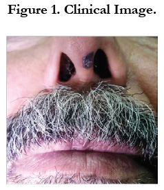
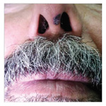
Figure 1. Clinical Image.
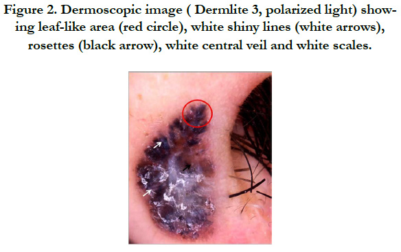
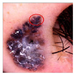
Figure 2. Dermoscopic image (Dermlite 3, polarized light) showing leaf-like area (red circle), white shiny lines (white arrows), rosettes (black arrow), white central veil and white scales.
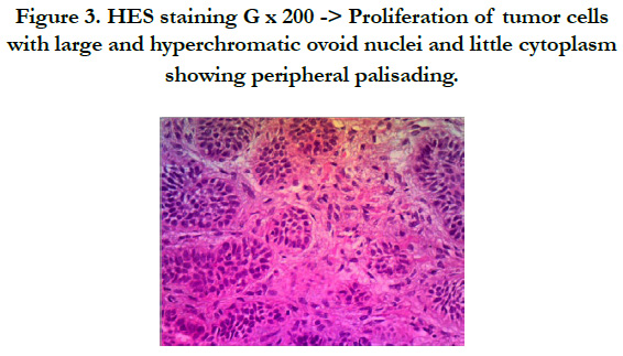
Figure 3. HES staining G x 200 -> Proliferation of tumor cells with large and hyperchromatic ovoid nuclei and little cytoplasm showing peripheral palisading.
References
- Menzies SW, Westerhoff K, Rabinovitz H, Kopf AW, McCarthy WH, Katz B. Surface microscopy of pigmented basal cell carcinoma. Arch Dermatol. 2000 Aug;136(8):1012-6. PubMed PMID: 10926737.
- Menzies SW. Dermoscopy of pigmented basal cell carcinoma. Clin Dermatol. 2002 May-Jun;20(3):268-9. PubMed PMID: 12074864.
- Marghoob AA, Braun R, editors. An atlas of dermoscopy. CRC Press; 2012 Jul 26.


