Pilomatricoma of the Arm in a Child: A Rare Localization
K Moustaide1*, A Mazti2, O Eljouari1, H BayBay2, N Hammas2, S Gallouj1, FZ Mernissi1
1 Dermatology Department, University Hospital Hassan II, Morocco.
2 Anatomopathology Department, University Hospital Hassan II, Morocco.
*Corresponding Author
K. Moustaide,
Dermatology Department University Hospital Hassan II, Morocco.
E-mail: kmoustaide@gmail.com
Received: May 22, 2018; Accepted : June 11, 2018; Published: June 13, 2018
Citation: K Moustaide, A Mazti, O Eljouari, H BayBay, N Hammas, S Gallouj, et al., Pilomatricoma of the Arm in a Child: A Rare Localization. Int J Clin Dermatol Res. 2018;6(3):172-173. doi: dx.doi.org/10.19070/2332-2977-1800039
Copyright: K Moustaide© 2018. This is an open-access article distributed under the terms of the Creative Commons Attribution License, which permits unrestricted use, distribution and reproduction in any medium, provided the original author and source are credited.
Abstract
Pilomatrixoma mummified epithelioma of Malherbe, is a skin tumor, benign and rare. It is most commonly seen in the head and neck areas, during the first two decades of life. We report a 8-year-old child with a rare localisation of pilomatrixoma in the right arm. The pilomatrixoma was excised and at one year follow-up there has been no evidence of recurrence.
2.Case Report
3.Discussion
4.Conclusion
4.References
Keywords
Skin Aging; Anti Aging Cream; Fitzpatrick Skin Types; Anti Aging Attributes; Benchmarking Data.
Introduction
Pilomatricoma or mummified epithelioma of Malherbe is a skin tumor, benign, rare, whose frequency is lessthan 2% of all primary tumors of the skin. It is a tumor of the young subject of less than 20 years old. The most frequent locations are the head and the neck, the localization at the level of the limbs are main exceptional.
Case Report
We report the case of an 8-year-old patient who was seen for a tume faction of the external edge of the right arm, evolving for 6 months. The clinical examination revealed a tumor of three centimeters in diameter, hard, painless, adherent to the skin but mobile from the deep plane. The skin was normal (Figure 1). The radiography of the arm showed a calcification of the soft tissues. The child had undergone total excision of the tumor under local anesthesia. The tumor nodule was encapsulated, indurated, measuring three centimeters of major axis. The anatomopathological study confirmed the diagnosis of pilomatricoma (Figure 2). At one year of decline, no recurrence was noted.
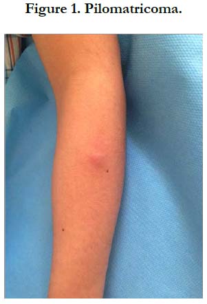
Figure 1. Pilomatricoma.
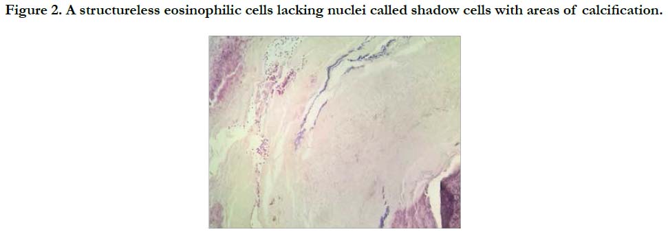
Figure 2. A structureless eosinophilic cells lacking nuclei called shadow cells with areas of calcification.
Discussion
Originally described in 1881 by Malherbe and Chenantais, as a benign, calcified tumor of the sebaceous glands, the mummified epithelioma of Malherbe or pilomatricoma was confirmed later by Forbis and Helwing [1]. In its typical form, the pilomatricoma is clinically translated by a small subcutaneous nodule, solitary, asymptomatic some times painful, and willingly affecting the woman with a sex ratio of 3/2. It occurs mainly during the first two decades of life, rarely beyond [1]. The usual locations are the neck and the head [1, 2] only some exceptional isolated locations in the limbs as the case of our patient have been reported in the literature [3, 4]. Several clinical forms have been described in pilomatricoma, including familial form soften associated with systemic diseases such as myotonic dystrophy [5].
In our patient, the skin was normal. However, there is an an ectodermal form where the skin is flaccid, consequence of a compression of the skin by the tumor resulting in the disappearance of the elastic fibers [6, 7]. The evolution can be towards the ulceration and can pose a problem of differential diagnosis with cutaneous carcinoma.
Standard radiography is useful only when there is suspicion of a pilomatricoma when it is significantly calcified as is the case in our patient [8].
In all cases, the diagnostic confirmation is histological. This will also eliminate other differential diagnoses, mainly squamous and pilar cysts but especially the malignant pilomatricoma or trichomatricial carcinoma whose more aggressive. The immunohistochemical study can facilitate this distinction. The carcinoma to us degeneration of the pilomatricoma remains controversial [2, 9]. The prognosis of the pilomatricoma is generally good. Healing without recurrence is the rule after total surgical excision [6, 10].
Conclusion
Pilomatricoma is a benign tumor, from the hair matrix ,very rare in children. The location in the members remains exceptional. The histological diagnosis will eliminate a malignant pilomatricoma. The treatment is surgical excision to avoid any recurrence.
References
- Forbis R, Helwig EB. Pilomatrixoma (calcifying epithelioma). Arch Dermatol. 1961 Apr;83:606-18. PubMed PMID: 13700704.
- Yoshimura Y, Obara S, Mikami T, Matsuda S. Calcifying epithelioma (pilomatrixoma) of the head and neck: analysis of 37 cases. Br J Oral Maxillofac Surg. 1997 Dec;35(6):429-32. PubMed PMID: 9486450.
- Hughes J, Lam A, Rogers M. Use of ultrasonography in the diagnosis of childhood pilomatrixoma. Pediatr Dermatol. 1999 Sep-Oct;16(5):341-4. PubMed PMID: 10571829.
- Sari A, Yavuzer R, Isik I, Latıfoǧlu O, Ataoǧlu Ö. Atypical presentation of pilomatricoma: a case report. Dermatol Surg. 2002 Jul;28(7):603-5. Pub- Med PMID: 12135516.
- Geh JL, Moss AL. Multiple pilomatrixomata and myotonic dystrophy: a familial association. Br J Plast Surg. 1999 Mar;52(2):143-5. PubMed PMID: 10434894.
- Aissoudi M, Doss N, Bouzaiene A. A case for diagnosis: pilomatrixoma. Ann Dermatol Venereol. 1995;122(11-12):797-8. PubMed PMID: 8729830.
- Karpuzoglu T, Elpek GÖ, Alpsoy E, Gelen T, Aksoy NH, Karpuzoglu G. Multiple familial pilomatrixomas. J Eur Acad Dermatol Venereol. 2003 May;17(3):358-9. PubMed PMID: 12702091.
- Haller JO, Kassner EG, Ostrowitz A, Kottmeier PK, Pertschuk LP. Pilomatrixoma (calcifying epithelioma of Malherbe): radiographic features. Radiology. 1977 Apr;123(1):151-3. PubMed PMID: 847140.
- Lever WF. Histopathology of the skin. Masson et Cie; 1969.
- Khammash MR, Todd DJ, Abalkhail A. Concurrent pilomatrix carcinoma and giant pilomatrixoma. Australas J Dermatol. 2001 May;42(2):120-3. PubMed PMID: 11309036.


