A Morphometric Study of Incisura Fibularis in South Indian Population with its Clinical Implications
Gupta C1*, Nayak N1, Palimar V2
1 Department of Anatomy, Kasturba Medical College, Manipal Academy of Higher Education, Manipal, India.
2 Department of Forensic Medicine, Kasturba Medical College, Manipal Academy of Higher Education, Manipal, India.
*Corresponding Author
Dr. Chandni Gupta,
Associate Professor, Department of Anatomy,
KMC Manipal, 576104, India.
E-mail: chandnipalimar@gmail.com
Received: January 16, 2018; Accepted: January 27, 2018; Published: January 30, 2018
Citation: Gupta C, Nayak N, Palimar V. A Morphometric Study of Incisura Fibularis in South Indian Population with its Clinical Implications. Int J Anat Appl Physiol. 2018;4(1):84-86. doi: dx.doi.org/10.19070/2572-7451-1800014
Copyright:Gupta C© 2018. This is an open-access article distributed under the terms of the Creative Commons Attribution License, which permits unrestricted use, distribution and reproduction in any medium, provided the original author and source are credited.
Abstract
Background: The distal tibiofibular joint is formed by the medial convex surface of the distal end of the fibula and the lateral concave surface of the fibular incisura of the tibia. The bond between the tibia and the fibula at the level of the ankle is of utmost significance for the appropriate functioning of the distal tibiofibular joint. So, this study was done to measure various dimensions of fibular incisura of tibia and width of distal end of fibula.
Materials and Method: 47 adult fully ossified dry tibias and 37 fibulae were taken for the study. Various measurements of fibular incisura of tibiae and width of distal end of fibulae were measured using vernier caliper.
Results: The total (right + left) mean values of the width and depth of the incisura fibularis of tibia was 2.33 and 1.08cm. The total mean values of the length of anterior and posterior facet of incisura fibularis of tibia was 1.34 and 1.28cm. The total mean width of distal end of fibula was 2.44cm.
Conclusion: The results of this study will be useful for biomedical peoples who make implants of lower end of tibia and fibula for orthopaedic surgeons during ankle reconstruction surgeries.
2.Introduction
3.Materials and Method
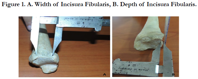
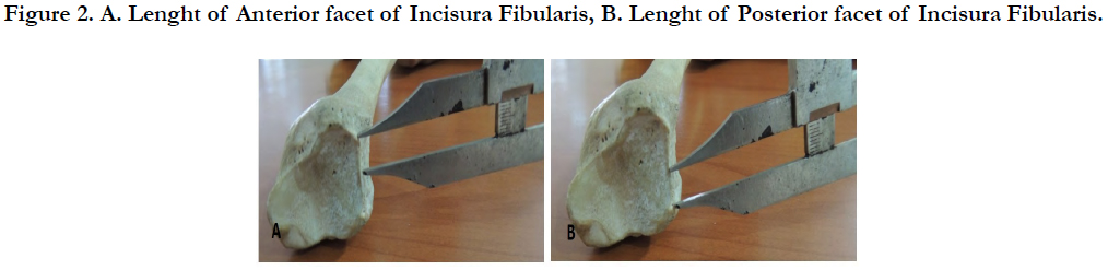
3.1 Incisura Fibularis (Figure 1 and 2)
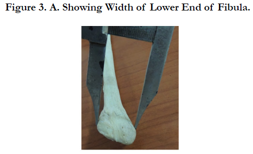
3.2 Fibula (Figure. 3)
4.Results
5.Discussion
6.References
KeyWords
Tibia; Fibula; Incisura Fibularis; Syndesmosis; Implants.
Introduction
The lateral surface of the distal end of tibia has a triangular fibular notch called incisura fibularis. This fibular notch is for articulation with the distal end of fibula to form distal tibiofibular joint. This distal tibiofibular joint is a syndesmosis type of fibrous joint. The association between the tibia and the fibula near the ankle is of principal significance for the appropriate functioning of the distal tibiofibular joint. During fracture or dislocation of the ankle joint it is very important to look for the syndesmosis for disruption and injury because it is important for the treatment and prognosis. It is very difficult to look for syndesmotic injury by radiographic criteria because of discrepancies in the extent of rotation, the wide anatomic unevenness in the depth of the fibular incisura and the shape of the tibial tubercle [1].
The morphometry of the distal tibia is significant when the stability of these articulations is put into account. Ankle is one of the most commonly injured joint and the morphometry of incisura fibularis and the distal articular surfaces of fibula will help in the reconstruction surgeries after dislocation fractures and in the construction of implants in South Indians [1].
So, this study was undertaken to measure various parameters of incisura fibularis and the distal articular surfaces of fibula.
Materials and Method
The study was carried out on 47 dry tibiae (22 right and 25 left) and 37 fibulae (20 right and 17 left). All bones were adult type and without any signs of erosion. Following parameters were studied on tibia and fibula:
a) Width: Anteroposterior length between the anterior and the posterior tubercles.
b) Depth: Height measured from the deepest point of the fibular incisura to a line between the tips of the anterior and posterior tubercles.
c) Length of anterior facet: Anteroposterior length measured between the tip of the anterior tubercle and the deepest point of the fibular incisura.
d) Length of posterior facet: Anteroposterior length measured between the tip of the posterior tubercle and the deepest point of the fibular incisura.
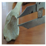
a) Width of the fibula was measured between the medial and lateral borders of the fibula.
All measurements were taken with the help of vernier caliper in centimeters.
Statistical analysis was done using Spss software 16 for all the parameters. Paired sample T test was done to see the statistical significance (P value <0.05) between right and left side.
Results
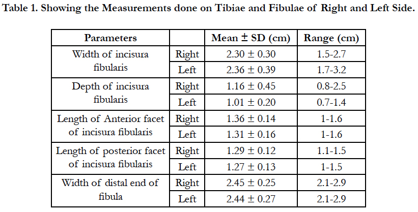
The mean and standard deviation of all the parameters of right, left tibia are shown in Table 1.
The total (right + left) mean values of the width and depth of the incisura fibularis of tibia was 2.33 and 1.08cm. The total mean values of the length of anterior and posterior facet of incisura fibularis of tibia was 1.34 and 1.28cm. The total mean width of distal end of fibula was 2.44cm.
There was no statistical significance in any parameter of right and left side as P value in all was >0.05.

Discussion
An undisrupted tibiofibular syndesmosis joint is significant in the normal functioning of ankle joint. The purpose of the syndesmosis is to retain the normal articulation of the ankle joint by determining the specific relationship between the distal tibia and the fibula. The measurements of the incisura fibularis and the distal end of fibula are vital in considering the stability of ankle joint, in designing of prostheses for use in ankle arthroplasty and in interpretation of diagnostic images of the ankle joint [2].
Taşer F et al., found the total mean values of the width and depth of the incisura fibularis as 2.32 and 0.36cm while in our study we got the values as 2.33 and 1.08cm. Results of width of their study were almost equal to our study [1]. A wider incisura indicates a greater separation of the anterior and posterior incisural tubercles. This leads to a shallower incisura which predisposes an individual to instability of the tibiofibular syndesmosis [3].
Taşer F et al., got the total length of anterior and posterior facet of incisura fibularis of tibia as 1.08 and 1.32cm while in our study the values were 1.34 and 1.28cm. Results of length of posterior facet of their study were almost comparable to our study [1]. Dimensions of these facets can be used to locate the fibula in the fibular incisura. There is an association between the location of the fibula and frequent ankle instability. In syndesmoses with a more posteriorly located fibula, there is less structural stability and so, they are more vulnerable to sprains [3]. The total mean width of distal end of fibula as 1.27cm while in our study the value was 2.44 cm [1].
Yildirim H et al., found that in males and females the length of the anterior and posterior facet was same it was 1.04 and 0.89 cm. The depth of the fibular incisura of the tibia was 0.36cm in males and 0.29cm in females. They found that all parameters of men were significantly higher than the parameters of women [4].
Kulkarni RR et al., found the total mean values of the width, depth of the incisura fibularis on right and left side as 2.35, 0.62cm and 2.31, 0.61cm while in our study we got the values as 2.30, 1.16cm and 2.36, 1.01cm. Results of width of their study were almost equal to our study. They got the total length of anterior, posterior facet of incisura fibularis of tibia on right and left side as 1.15, 1.47cm and 1.10, 1.46cm while in our study the values were 1.36, 1.29cm and 1.31, 1.27cm. The total mean width of distal end of fibula as 1.27cm while in our study the value was 2.44 cm [2].
Musa M et al., total mean values of the width and depth of the incisura fibularis as 2.15 and 0.34cm while in our study we got the values as 2.33 and 1.08cm. Results of width of their study were almost equal to our study. They got the total length of anterior and posterior facet of incisura fibularis of tibia as 1.14 and 1.61cm while in our study the values were 1.34 and 1.28cm [5].
Ebraheim NA et al., found that the length of anterior syndesmotic facet (1.12cm) is shorter than the posterior (1.48cm). In our study we found the length of anterior and posterior facet as 1.34 and 1.28cm. They found the depth of fibular incisura of the tibia as 0.42 while in our study we got the depth as 1.08cm. This may be because of racial differences because we have done the study in South Indian population [6].
These results of the study will help in the reconstruction operations following fracture dislocation of the ankle joint in South Indian population.
References
- Taser F,Toker S, Kilincoglu V. Evaluation of morphometric characteristics of the fibular incisura on dry bones. Joint diseases and related surgery. 2009;20(1):52-58.PMID:19522692.
- Kulkarni RR, Rao CP, Nidhi S. Importance of fibular incisura measurements in ankle reconstructive surgeries. Int J AJ Inst Med Sci. 2012 Nov;1(2):80- 85.
- Hermans JJ, Beumer A, de Jong TA, Kleinrensink GJ. Anatomy of the distal tibiofibular syndesmosis in adults: a pictorial essay with a multimodality approach. J Anat. 201 0 Dec;217(6):633-45. PMID:21108526. PMCID:PMC3039176. doi: 10.1111/j.1469-7580.2010.01302.x.
- Yildirim H, Mavi A, Buyukbebeci O, Gumuflburun E. Evaluation of the fibular incisura of the tibia with magnetic resonance imaging. Foot Ankle Int.2003 May;24(5):387-91. PMID:12801193.DOI:10.1177/1071100703 02400502.
- Musa M, Pamela M, Moses O, Beda O, Gichambira G. Morphometric Characteristics of the Fibular Incisura in Adult Kenyans. Anatomy Journal of Africa. 2014;3(1):243–249.
- Ebraheim NA, Lu J, Yang H, Rollins J. The fibular incisure of the tibia on CT scan: a cadaver study. Foot Ankle Int. 1998 May;19(5):318-21. PMID:9622423.DOI:10.1177/107110079801900509.


