Technological Approaches to Isolation of Biologically Active Substances from the Tissues of Pancreas, Duodenum, and Gastric Mucosa
Kashinova EB, Kotenkova EA, Ertikeeva EA, Akhremko AG, Chernukha IM*
The V.M. Gorbatov All-Russian Meat Research Institute, Moscow, Russia.
*Corresponding Author
Chernukha I.M,
TheV.M. Gorbatov All-Russian Meat Research Institute, Moscow, Russia.
E-mail: imcher@inbox.ru
Received: May 26, 2016; Accepted: June 09, 2016; Published: June 10, 2016
Citation: Kashinova EB, Kotenkova EA, Ertikeeva EA, Akhremko AG, Chernukha IM (2016) Technological Approaches to Isolation of Biologically Active Substances from the Tissues of Pancreas, Duodenum, and Gastric Mucosa. J Translational Diagn Technol. 1(2), 7-10.
Copyright: Chernukha IM© 2016. This is an open-access article distributed under the terms of the Creative Commons Attribution License, which permits unrestricted use, distribution and reproduction in any medium, provided the original author and source are credited.
Abstract
This paper presents the results of technology optimization for isolation of target BAS by extraction. The objects of the study were the tissue of pork pancreas, duodenum, and gastric mucosa. The studies have shown that the optimum stirring speed for maximum protein yield was 600 rpm, whereas 800 rpm caused excessive foaming, which increased costs for defoamers. Extraction at the speed of 400 rpm resulted in decreased protein yield and increased extraction time. Electrophoretic study showed that the rate of extraction did not qualitatively affect the profile of protein and peptide extracts, but quantitatively affected the yield of specific protein fractions. Comparison of electrophoregrams with international protein databases showed high tissue specificity of investigated internal organs, while the intensification of protein bands on the tracks was observed predominantly in the range less than 70 kDa. The obtained results allowed to identify the technological modes of target biologically active substances isolation from the pork tissues that are recommended for use in animal feed.
2.Introduction
3.Materials and Methods
4.Results and Discussion
5.Conclusions
6.Acknowledgement
7.References
Keywords
Optimization; Extraction; Electrophoresis.
Introduction
Recent publications indicate the relevance of slaughter by-products as a source for the development of feed additives aimed at the treatment and prevention of gastrointestinal tract diseases of farm animals, and as optimal substitution for antibiotics. But, unfortunately, almost all newly developed or existing feed additives are produced using traditional technological workflow not including detailed study of protein and peptide profile and isolation of target biologically active substances that may be involved in biological and physiological processes in the body of animals. The aim of the work was to determine the technological approaches to isolation of BAS from the tissues of pork pancreas, duodenum, and gastric mucosa [1], as well as description of their protein and peptide profile.
Materials and Methods
Objects of the study were saline extracts of pork pancreas, duodenum, and gastric mucosa. Extraction of raw material was performed as follows: grinding → extraction → obtaining of supernatant (centrifugation or filtration). Raw material was deep-frozen at minus 40°C, then ground in a meat grinder (KENWOOD, England) with a hole diameter of 3-5 mm. Next, the extraction was performed using laboratory dispersing unit (Laboteks, Russia) at a ratio of tissue and 0.9% sodium chloride volumes as 1:5 and temperature of 4-5°C, while stirring speed was varied (400, 600, and 800 rpm). Selection of extract samples for determination of protein concentration was performed prior to extraction, every 5 minutes during the first 30 minutes and subsequently every hour during 8 hours. The selected samples were centrifuged in CM-6M centrifuge (ELMI, Latvia) at 3500 rpm for 5 minutes, then the supernatant was collected, in which the protein concentration was determined by biuret method using BioChemSA photometer (HTI, USA).
Pancreas and gastric mucosa extracts were separated from the suspension by filtration through a cloth filter, and the duodenum extract was separated by centrifugation at 2000 rpm for 2 minutes.
To perform electrophoretic studies, 2 samples for each extract were selected: 1- point of maximum protein concentration at given rotation, and 2- extract after the extraction.
Analysis of protein composition was performed by denaturing gradient electrophoresis in 7-25% PAGE, using Fermentas marker (Fermentas, Lithuania) as a tracking dye.
Results and Discussion
At the speed of 800 rpm, maximum protein concentration in samples of pancreas (Figure 1, A) was detected for the extraction during 6 hours (32.45 g/L). At 600 rpm, the protein concentration reached 32.15 g/L during the first 1.5 hours and stayed almost unchanged. During the extraction of pancreas at the speed of 400 rpm, the protein concentration within the first 10 minutes dramatically increased up to 25.0 g/L, and then varied sinusoidally for the next 3 hours not exceeding 31.3 g/L. This is supposedly due to the enzymatic hydrolysis typical for low extraction rate.
It is worth noting that before the extraction of pancreas tissues, "zero samples" have already contained protein (5.8 to 7.9 g/L). The protein concentration in "zero sample" from the extract of the gastric mucosa (Figure 1, B) accounted for only 0.7 to 1.02 g/L, and then the protein concentration increased with no major changes. With the stirring speed of 800 rpm for 7 hours, the protein concentration increased up to 11.9 g/L. Maximum protein yield was observed during fourth hour of extraction (12.7 g/L), and with further extraction reduction of protein concentration was observed down to 11.9 g/L. With the stirring speed of 600 rpm, there was more intense protein yield. After three hours of extraction, the protein concentration reached the maximum value of 12.7 g/L (at 800 rpm, such value was observed after 4 hours), then the protein concentration reduced to 12.3 g/L due to autolysis and enzymatic hydrolysis typical for a long time extraction. With the stirring speed of 400 rpm, the protein concentration increased to 11.75 g/L (6 hours), and after 8 hours this value did not reach 12 g/L.
The protein concentration in "zero samples" of duodenum were 0.64 to 0.90 g/L. Extraction at the speed of 800 rpm was characterized by an increase in protein content up to 13.35 g/L (Figure 1, B). The maximum concentration of protein (16.28 g/L) was found during the third hour of extraction, after which a sharp decline was observed. With the extraction rate of 600 rpm for 2 hours, more steady increase in the protein content (up to 16.55 g/L) was shown with further reduction.
Extraction at the speed of 400 rpm for 6 hours was characterized by increase of protein concentration up to the maximum value of 16.13 g/L, followed by reduction.
Usage of high speed stirring (800 rpm) was characterized by rich foaming resulting in increased costs for defoamers. At the stirring speed of 400 rpm, the protein concentration in the extracts reached maximum values at longer extraction time, which involves extra energy costs.
Electrophoregrams analysis showed the presence of large amounts of low molecular weight and high molecular weight proteins in studied extracts. The composition of each tissue extracts were identical for all applied speed rates. The highest intensity of protein bands was observed on the tracks at 600 rpm (Figures 2, 3, and 4). Proteomic profiles of pancreas, gastric mucosa and duodenum extracts obtained at different stirring speeds were analyzed in accordance with the UniProt Protein DataBase. The pancreatic extracts (400 rpm - tracks 1 (3 hours) and 2 (8 hours), 600 rpm - tracks 3 (2 hours) and 4 (8 hours), 800 rpm - tracks 5 (6 hours) and 6 (8 hours)), along with major structural proteins, presumably contain a number of minor proteins that are characterized by the tissue specificity (Figure 2).
Low molecular weight protein fractions, along with major structural proteins, presumably contain (MW 35-10 kDa) gastrin stimulating the secretion of pancreatic enzymes, insulin involved in glucose metabolism [2], pancreatic polypeptide that inhibits pancreatic secretion and stimulates secretion of gastric juice [3], phospholipase A2, AIF-1 involved in humoral immunity; glucagon (21 kDa), which plays a key role in glucose metabolism; glutathione- S-transferase omega 1, and so on. Fractions in a range of 70 to 38 kDa presumably contain the following proteins and enzymes: ISL1 (40 kDa) and MNX1 (40 kDa) involved in the restoration of pancreatic exocrine function and formation of cellular immunity; carboxypeptidase A1 (47 kDa) and B (47 kDa), protein (WLS gene) (52 kDa) involved in regulation of exocrine pancreatic function and intercellular protein transport; ATP-binding cassette transporter of G subfamily (72 kDa) responsible for transmembrane transport [4], and so on. Electrophoretogram analysis of gastric mucosa extracts (Figure 3) at 400 rpm - track 7 (9 hours), 600 rpm - tracks 8 (3 hours) and 9 (8 hours), 800 rpm - tracks 10 (4 hours) and 11 (8 hours) also showed no significant differences between them. All extracts in the range of 6 to 35 kDa presumably contain the following functional peptides and proteins: cardiac phospholamban involved in membrane transport, pepsin B involved in digestion, secretin stimulating the secretion of gastric juice; lysozymes C-1, C-2, C-3, aquaporin-3 (31 kDa) [5], and beta-subunit of H(+)/R(+)-ATPase involved in water, salts, and ions transport [6]. The range of 75 to 35 kDa presumably contain interferon stimulating protein involved in the mechanism of innate immunity and induction of interferon genes expression [7], anion-exchange protein involved in the transport of carbon dioxide and maintaining the acid-base balance [4].
Proteomic analysis of duodenum extracts (Figure 4) at the stirring speed of 400 rpm - tracks 12 (6 hours) and 13 (8 hours), 600 rpm - tracks 14 (2 hours) and 15 (8 hours) and 800 rpm – tracks 16 (3 hours) and 17 (8 hours) revealed the presence of protein fractions with low intensity in the ranges of 40 to 25 kDa and 15 to 7 kDa that might correspond to cytochrome B oxidase and cytochrome C oxidase [8], phospholipase A2, inhibitor of Na+/K+ - ATPase (20 kDa) involved in the activation of peptidase inhibitors, heparin-binding growth factor, protein 88 of myeloid differentiation of primary gene response (MyD, 88.33 kDa) involved in the reactions of the innate immune response, inflammation [9] as well as in the regulation of transcription factor NF-κB, and so on [4].
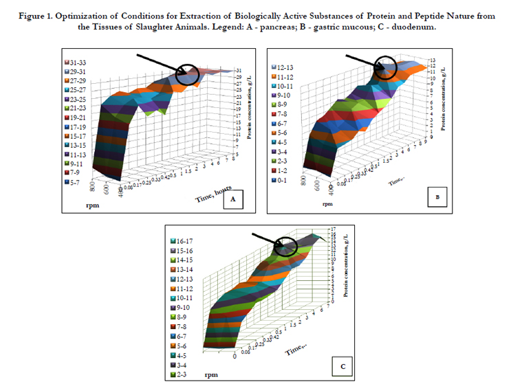
Figure 1. Optimization of Conditions for Extraction of Biologically Active Substances of Protein and Peptide Nature from the Tissues of Slaughter Animals. Legend: A - pancreas; B - gastric mucous; C - duodenum.
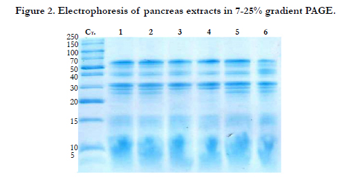
Figure 2. Electrophoresis of pancreas extracts in 7-25% gradient PAGE.
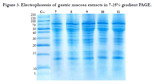
Figure 3. Electrophoresis of gastric mucosa extracts in 7-25% gradient PAGE.
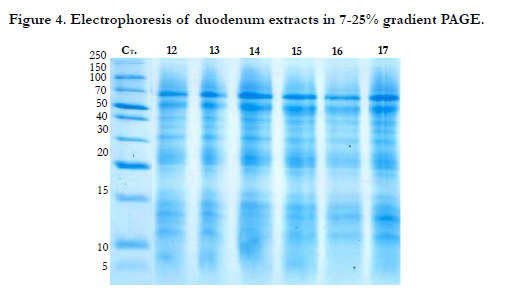
Figure 4. Electrophoresis of duodenum extracts in 7-25% gradient PAGE.
Conclusions
The obtained results allowed to identify the optimal technological modes of pancreas, duodenum, and gastric mucosa extraction. For duodenum and pancreas, the optimal stirring speed is 600 rpm for 2 hours, and for gastric mucosa this value is 600 rpm for 3 hours. Analysis of proteomic profile in accordance with the international protein databases led to the assumption that studied pork tissues contained proteins and peptides involved in the immune response, regulation of cellular and humoral immunity, stimulation of digestion and nutrients digestibility. These results support the usage of duodenal, pancreatic, and gastric mucosa tissues as a promising source of BAS for the development of feed additive technology to stimulate immunity and normalize the functioning of farm animals gastrointestinal tract.
Acknowledgement
This work was supported by the Russian Science Foundation, project № 15-16-00008.
References
- Chernukha IM, Usha BV, Makarenko AN, Fedulova LV, Elizarova TS, et al. (2012) Development of enzyme-tissue preparation for the treatment of gastrointestinal disorders based on the extracts of pork duodenal, pancreatic, and gastric mucosa tissues. Veterinary and feeding 4: 12-14.
- Van Laere AS, et al., (2003) Аregulatorymutation in IGF2 causes a major QTL effect on muscle growth in the pig. Nature 425(6960): 832–836.
- Hamid Said M (2012) Physiology of the Gastrointestinal Tract: Two Volume Set 5th Edition.
- UniProtProteindatabase [electronic resource]. – Access mode: www.uniprot.org/.
- Li X, Lei T, Xia T, Chen X, Feng S,et al. (2008) Molecular characterization, chromosomal and expression patterns of three aquaglyceroporins (AQP3, 7,9) from pig. Comp Biochem Physiol B Biochem Mol Biol. 149(3): 468–476.
- Abe K, et al., (2011) Conformationalrearrangementofgastric H(+),K(+)-ATPaseinducedby an acidsuppressant. Nature Communications 2: 155.
- Xie L, et al.(2010) Molecular cloning and functional characterization of porcine stimulator of interferon genes (STING). Developmental & Comparative Immunology 34(8): 847–854.
- DNA Data bank of Japan [electronic resource]. – Access mode: www.ddbj.nig.ac.jp/intro-e.html.
- Matveeva VG, Golovkin AS, Grigoriev EV, Ponasenko AV (2011) Role of Triggering Receptor Expressed on Myeloid Cells in the Activation of Innate Immunity General Reanimatology.General resuscitation 7(3):1- 70.


