Control of Recurrent Pyoderma with PYOspot® in an Atopic Dog
Lorente-Méndez C*, Ruíz-Tapia P
1 ADERVET Dermatología Veterinaria. Madrid C/ Corazón de Maria, 6428002 Madrid - Spain.
2 Hospital Veterinario Universitario Universidad de Extremadura, Av. de la Universidad s/n. 1300 Cáceres - Spain.
*Corresponding Author
Carmen Lorente-Méndez, DVM, PhD, DipECVD.
ADERVET Dermatología Veterinaria.
Madrid, Spain.
Tel : 00 34 609200018
email: clorente@adervet.com
Received: March, 2016; Accepted: April 13, 2016; Published: April 19, 2016
Citation: Lorente-Méndez C, Ruíz-Tapia P (2016) Control of Recurrent Pyoderma with PYOspot® in an Atopic Dog. Int J Vet Health Sci Res. 4(2), 110-115.DOI : dx.doi.org/10.19070/2332-2748-1600024
Copyright: Lorente-Méndez C© 2016. This is an open-access article distributed under the terms of the Creative Commons Attribution License, which permits unrestricted use, distribution and reproduction in any medium, provided the original author and source are credited.
Abstract
Recurrent pyoderma is not an uncommon complication of canine atopic dermatitis and the use of frequent courses of antibiotic for its control is of concern, related to the possible developing of resistances. This report describes a case where the use of a topical spot-on product PYOspot® avoids the recurrence of pyoderma in a dog with severe canine atopic dermatitis.
A female French bulldog severely affected by canine atopic dermatitis was suffering from recurrent episodes of pyoderma, 5 in the last year. Pyoderma was manifested as bacterial folliculitis with alopecia and papule-crusted lesions and as intertrigo in facial folds and paws. The infection was controlled with courses of antibiotic lasting at least for 1 month, but it recurred once the treatment was stopped. The dog was in treatment with cyclosporine to control clinical signs of atopic dermatitis. The administration of a weekly treatment with a topical spot-on formulation containing a natural complex with antimicrobial properties, PYOspot®, prevents pyoderma recurrence in this dog.
2.Introduction
3.Case Report
3.1.Medical History
3.2.Clinical examination
3.3.Diagnosis
3.4.Treatment
4.Discussion
5.Conclusion
6.References
Keywords
Recurrent Pyoderma; Canine Atopic Dermatitis; PYOspot
Introduction
Bacterial resistance generates great concern in human and veterinary medicine. Canine pyoderma, bacterial skin infection, is a very common process in veterinary medicine and atopic patients are at increased risk for the development of superficial pyoderma. The use of antibiotics to control recurrent pyoderma increases the risk of developing Methicillin resistant S. pseudintermedius (MRSP) or multidrug resistant (MDR) S. pseudintermedius strains. Topical antiseptics are of extreme importance as they rarely stimulate the development of resistance and their use is highly recommended in order to prevent bacterial infections and as complementary treatment for pyoderma. In this case report we realize the efficacy of a new topical not antibiotic product with antimicrobial properties, PYOspot®, in the control of recurrent pyoderma in a severe case of atopic dermatitis. The product was easy to apply and well accepted by the owner and the dog. The use of topical antimicrobial products is highly recommended and this result suggest that PYOspot® is a very good option as topical treatment in cases of atopic dermatitis in order to control the development of superficial pyoderma.
Atopic dermatitis (AD) is considered to be one of the most common chronic skin diseases in dogs [1], affecting approximately 10% of dog population [2]. Dogs with AD often suffer secondary bacterial infection most commonly due to Staphylococcus pseudintermedius [3, 4]. Atopic inflamed skin harbours more bacteria in the cornified layer than the skin of healthy dogs [5, 6], which, joined with the skin impairment and inflammation, favours the adherence and proliferation of bacteria (Staphylococcus pseudintermedius) resulting in bacterial infection (pyoderma) [3, 7]. Moreover, pyoderma contributes to increase pruritus and inflammation, enhancing the poor skin condition. All these factors generate a vicious cycle where the control of pruritus, inflammation and infection is imperative to maintain the animal free of clinical signs.
Resolution of pyoderma requires systemic antibiotic therapy, but the use of frequent courses of antibiotic treatments can induce the development of bacterial resistances. All the efforts must be done to avoid the recurrence of bacterial infections. While the first approach is to control the primary disease, yet there are many cases in which the recurrence of pyoderma is present although the treatment is implemented to control the atopic dermatitis. Antibiotic treatment of recurrent pyoderma in dogs with AD may represent a potential risk for an increase of bacterial resistance in the future. The use of products able to restore the skin barrier and increase the natural defenses against pathogenic organisms
would be a great option to control proliferation of bacteria [8].
This report describes how a topical spot-on formulation that contains an antimicrobial plant extract helps a dog with severe atopic dermatitis, to control her episodes of recurrent pyoderma.
Huma is a female French Bulldog of 2,5 years old, weighing 8kg (Figure 1).
She lives in a flat in Madrid without any other pet and she was fed a high quality prescription diet for atopic dermatitis.
She has been presented to our consultation 1 year before with a severe pruritic dermatitis, lasting for more than 6 months, that had made her life nearly impossible. High level pruritus and pyoderma were the two problems of concern. Systemic antibiotic therapy and a diagnostic protocol for allergic diseases were established. After a negative response to two strict elimination diets, based on protein hydrolysates, and a continuous and strict monthly control of fleas, atopic dermatitis was diagnosed.
At this stage, her pruritus had been more or less controlled with 50mg cyclosporine every other day for the last 3 months (previously on daily basis), but pyoderma had been a concern with 5 recurrent episodes during the last year. The episodes of pyoderma were resolved with courses of cephalosporin treatment for 3-4 weeks, but it recurred in 1-2 months in spite of weekly baths with chlorhexidine shampoo.
During the last year, she had also had two corneal ulcerations secondary to facial pruritus, and she developed calcinosis cutis after switching glucocorticoids to cyclosporine, we have seen this occur in other cases. The calcinosis cutis was resolved in 2 months.
At presentation, the owner reported an increase in pruritus over the last 2 weeks. The pruritus was more intense on legs and face. Huma was losing hair in large amounts and the owner had seen areas of alopecia.
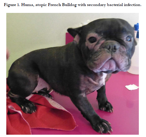
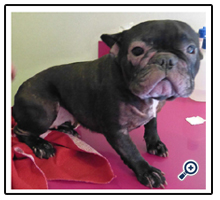
Figure 1. Huma, atopic French Bulldog with secondary bacterial infection
Physical examination revealed multifocal partial alopecia, with crusting-papules on dorsal trunk, ventral neck and inner part of limbs. (Figure 2)
Foci of alopecia with excoriation, erythema and acanthosis were evident on the inner part of rear legs. There were erythema and acanthosis interdigital and between pads. Chin, periocular area and facial folds exhibited marked alopecia, erythema and acanthosis with inflammation, xerosis and desquamation (Figure 3). Moderate to severe erythema with alopecia and lichenification of the area between the metatarsal pad and the tarsus were found. Isolated pustules could be found on the abdomen. Both pinnae presented crusting desquamation, acanthosis and erythema. Otoscopy revealed erythema and hyperplasia but external ear canals were permeable with no important amount of exudate. Trunco-podal pruritic reflex was induced by rubbing the animal trunk. The dog exhibited facial pruritus in the examination room.
Clinical evaluation could be summarized as diffuse partial alopecia with papular-pustular-crust lesions and facial dermatitis in a pruritic atopic dog treated with cyclosporine.
Figure 2. Multifocal alopecia of the trunk with papular-crusts in bacterial folliculitis.
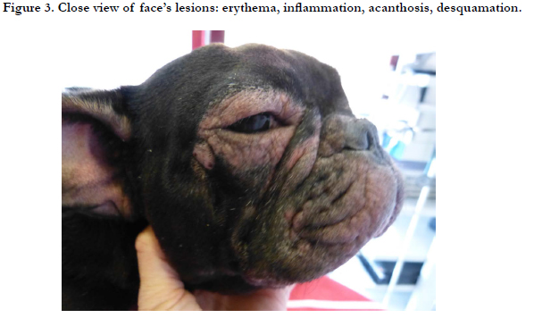
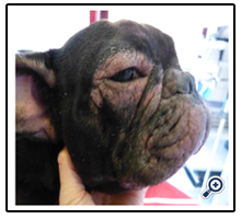
Figure 3. Close view of face’s lesions: erythema, inflammation, acanthosis, desquamation.
The principal differential diagnosis was bacterial folliculitis (trunk) and bacterial overgrowth (facial and podal) secondary to a flare of the atopic disease. Nevertheless, demodicosis and dermatophytosis must be included in the differential diagnosis of the alopecia and Malassezia dermatitis in differential diagnosis of facial and podal dermatitis.
Wood lamp was negative and trichography revealed neither the presence of demodex, nor the presence of spores, nor hair alterations compatible with dermatophytosis.
Superficial cytology of chin, periocular area, facial folds, paws and distal limbs revealed the presence of huge amounts of cocci and scattered groups of neutrophils. (Figur 4). Cytology of pustule content showed the existence of neutrophils and intra- and extracellular cocci (Figure 5).
The final diagnosis was bacterial folliculitis and bacterial overgrowth.
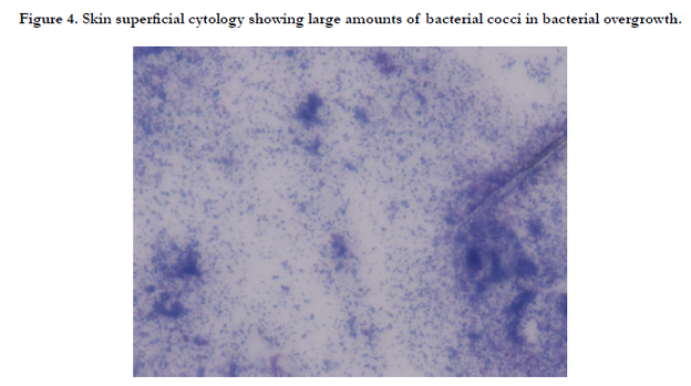
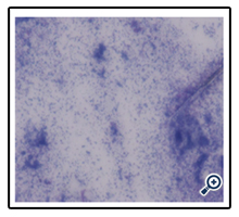
Figure 4. Skin superficial cytology showing large amounts of bacterial cocci in bacterial overgrowth.
Cefovecin 8mg/kg given subcutaneously (SC) was chosen as treatment for pyoderma after bacterial identification and antibiotic sensitivity testing to guarantee the success of treatment and to avoid possible gastrointestinal effects in this dog. Frequency of chlorhexidine shampoo was increased to three weekly applications. To control the outbreak of the atopic dermatitis, cyclosporine was prescribed daily instead of every other day.
Two weeks later the dog´s skin condition had improved markedly: no papules, crusts or pustules over the body and the hair was growing. There was no trunco-podal pruritic reflex and the owner only observed a mild facial pruritus and pruritus in distal limbs. A second subcutaneous Cefovecine shot was administered. At this point, it was decided to add to the treatment a weekly application of PYOspot®, a spot-on product containing different active ingredients with antimicrobial properties as well as essential fatty acids to repair skin barrier. Frequency of shampoos was reduced to once a week and PYOspot® should be put on 2 days after the shampoo.
At the next recheck, 14 days later, hair growth was evident and the pruritus was under control. The treatment was maintained with Cyclosporine 50mg daily and topical PYOspot® application, 2 days after chlorhexidine shampooing, every week.
Two month after beginning PYOspot®, the hair has grown back completely and pruritus was minimal, restricted to distal limbs and face (Figure 6 & 7).
During the next 10 months, treatment with cyclosporine every other day and PYOspot® once a week has been maintained and Huma has not had a new pyoderma episode since.
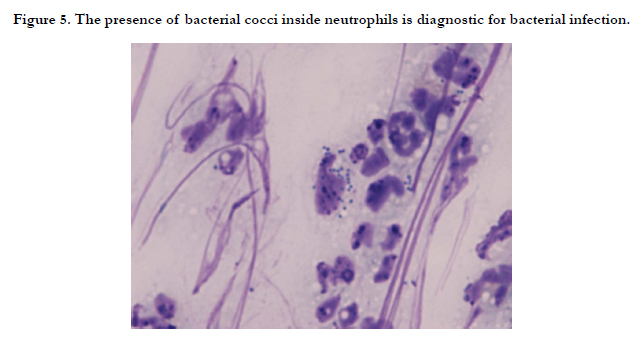
Figure 5. The presence of bacterial cocci inside neutrophils is diagnostic for bacterial infection.
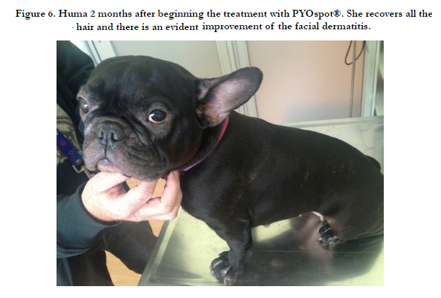
Figure 6. Huma 2 months after beginning the treatment with PYOspot®. She recovers all the hair and there is an evident improvement of the facial dermatitis.
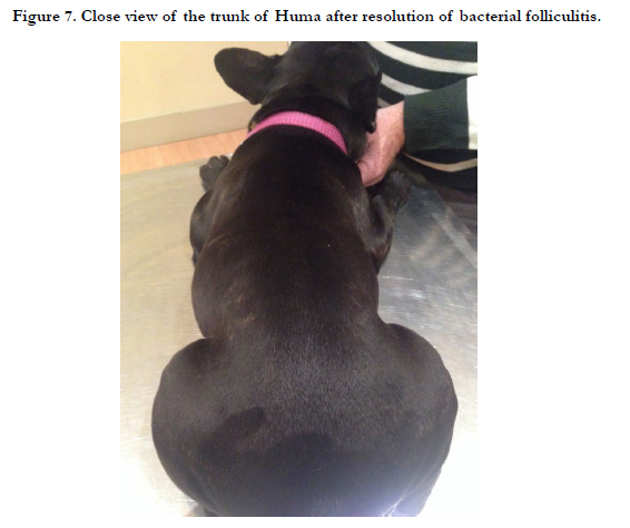
Figure 7. Close view of the trunk of Huma after resolution of bacterial folliculitis.
Discussion
Canine atopic dermatitis is a complex and multifactorial disease in which different aspects play an important role: genetics with a hyper-reactive immunitary status, skin barrier alterations, and environmental factors. Recurrent secondary pyoderma is a frequent complication in affected dogs. The bacterial infection increases the clinical signs by its direct effects over the skin, but also indirectly on the immune-system [9]. It is known that some staphylococci can produce superantigens that can possibly alter the skin barrier, stimulate the immune system and trigger atopic lesions, as it has been demonstrated in humans [3,10,11]. During the recurrent episodes of pyoderma that Huma suffered, the skin condition and the pruritus clearly impair, and once the infection was declared it was needed to initiated an antimicrobial treatment .
Staphylococcus pseudintermedius is the common pathogen implicated in canine pyoderma and its sensitivity has been well described [12, 13]. In this case Huma had previous episodes of gastroenteritis during antibiotherapy, SQ Cefovecine 8mg/kg every 14 days was choosed as treatment for her skin infection.
Treatment of pyoderma use to need is systemic antibiotic treatment for long periods of a minimun 3- 4 weeks. Antimicrobials are among the most important treatment options available in veterinary and human medicine, but antimicrobial resistance has progressively compromised their efficacy.
As the consensus say, “any use of antimicrobials, whether considered therapeutic or not, and prudent or otherwise, exposes bacterial pathogens and the commensal microbiota to varying concentrations of antimicrobial drug for variable times. This creates a selection pressure that can result in emergence of resistance or, if a resistant subpopulation is present, an increase in the abundance of resistant bacteria” [14]. The use of topical antiseptics or antimicrobial non antibiotic treatments for the prevention and treatment of pyoderma to minimize the likelihood of antimicrobial resistance, is highly recommended and thoroughly investigated. In this case, the recurrent pyoderma episodes were impairing the global condition of the atopic dermatitis and the possibility of developing resistance was of great concern. It was decided to introduce a new topical antimicrobial therapy in the hope to control the recurrence of pyoderma. PYOspot®, a spoton formulation, that combines neem and ajowan plant extracts, several essential oils along with an antimicrobial plant extracted complex (PhytoC-2®), was added to Huma treatment, 14 days after initiating the systemic antibiotic, as soon as an improvement of the condition was reached.
In an atopic condition, the control of the inflammation and the pruritus is crucial to avoid the episodes of pyoderma. According to the owner, the pruritic condition of Huma was well controlled with 50mg cyclosporine every other day and the life quality was good, but pyoderma recurred despite it. The development of this new episode of pyoderma could be due to an outbreak of allergy or to an inadequate control of the atopic condition, so it was decided to intensify the administration of cyclosporine daily. Two months later the good condition of Huma allowed the reduction of cyclosporine administration to every other day.
PYOspot® is easy to apply and its introduction as an adjunctive care enables this dog to avoid the development of new pyoderma episodes up to date (1 year later).
The composition of this product achieves important goals in atopic care, controlling the bacterial proliferation and restoring the skin properties by purifying, soothing and hydrating it. These properties can help control the atopic dermatitis, freeing, in this case, Huma from recurrences of pyoderma and flares of her atopic condition being controlled under an every other day cyclosporine regimen.
Conclusion
The In our case, PYOspot® has provided great benefits in Huma´s atopic condition with no new episode of pyoderma, resulting in a proper control of her atopic dermatitis. Based on this case, PYOspot® could be recommended as an adjunctive treatment in cases of atopic dermatitis with recurrent pyoderma. PYOspot® could be a useful help in combating against recurrent pyoderma and bacterial resistance. It would be of interest to carry out, on a larger population, a study of efficacy of this product in the control of recurrent episodes of pyoderma.
References
- Olivry T, Bizikova P (2013) A systematic review of randomized controlled trials for prevention or treatment of atopic dermatitis in dogs: 2008–2011 update. Vet Dermatol 24(1): 97-117.
- Hillier A, Griffin CE (2001) The ACVD task force on canine atopic dermatitis (I): incidence and prevalence. Vet Immunol Immunopathol 81(3-4): 147-151.
- DeBoer DJ, Marsella R (2001) The ACVD task force on canine atopic dermatitis (XII): the relationship of cutaneous infections to the pathogenesis and clinical course of canine atopic dermatitis. Vet Immunol Immunopathol 81(3-4): 239-249.
- Fazakerley J, Nuttall T, Sales D, Schmidt V, Carter SD, et al. (2009) Staphylococcal colonization of mucosal and lesional skin sites in atopic and healthy dogs. Vet Dermatol 20(3): 179-184.
- Mason I, Lloyd DH (1990) Factors influencing the penetration of bacterial antigens through canine skin. In Advances in Veterinary Dermatology. Bailliere-Tindall, London. 1: 370-374.
- Harvey RG, Noble WC (1994) A temporal study comparing the carriage of Staphylococcus intermedius on normal dogs with atopic dogs in clinical remission. Vet Dermatol 5: 21-25.
- Mcewan NA (2000) Adherence by Staphylococcus intermedius to canine keratinocytes in atopic dermatitis. Res Vet Sci 68(3): 279-283.
- Santoro D, Marsella R, Pucheu-Haston CM, Eisenschenk MN, Nuttall T, et al. (2015) Review: Pathogenesis of canine atopic dermatitis: skin barrier and host–micro-organism interaction. Vet Dermatol 26(2): 84.
- Na SY, Roh JY, Kim JM, Tamang MD, Lee JR (2012) Analysis of colonization and genotyping of the exotoxins of Staphylococcus aureus in patients with atopic dermatitis. Ann Dermatol 24(4): 413-419.
- Hendricks A, Schuberth HJ, Schueler K, Lloyd DH (2002) Frequency of superantigen-producing Staphylococcus intermedius isolates from canine pyoderma and proliferation-inducing potential of superantigens in dogs. Res Vet Sci 73(3): 273-277.
- Skov L, Olsen JV, Giorno R, Schlievert PM, Baadsgaard O, et al. (2000) Application of Staphylococcal enterotoxin B on normal and atopic skin induces up-regulation of T cells by a superantigen-mediated mechanism. J Allergy Clin Immunol 105(4): 820-826.
- Weese JS, van Duijkeren E (2010) Methicillin-resistant Staphylococcus aureus and Staphylococcus pseudintermedius in veterinary medicine. Vet Microbiol 140(3-4): 418-429.
- Summers JF, Brodbelt DC, Forsythe PJ, Loeffler A, Hendricks A (2012) The effectiveness of systemic antimicrobial treatment in canine superficial and deep pyoderma: a systematic review. Vet Dermatol 23(4): 305-329.
- Weese JS, Giguere S, Guardabassi L, Morley PS, Papich M (2015) ACVIM Consensus Statement on Therapeutic Antimicrobial Use in Animals and Antimicrobial Resistance. J Vet Intern Med 29(2): 487-498.fied hop extracts in the art of brewing. Tech Quarterly Mater Brew Assoc Am 33(2): 91-95.


