Morphometry of Demodex Canis and Demodex Cornei in Dogs with Demodicosis in India
S. Sivajothi1*, B. Sudhakara Reddy2 , K. Nalini Kumari3 , V.C.Rayulu4
1* Assistant Professor, Dept. of Veterinary Parasitology, C.V.Sc., Proddatur, Andhra Pradesh, India.
2 Assistant Professor (Veterinary Medicine), TVCC, C.V.Sc., Proddatur, Andhra Pradesh, India.
3 Professor and University Head, Dept. of Veterinary Medicine. C.V.Sc., Tirupathi, Andhra Pradesh, India.
4 Professor and University Head, Dept. of Veterinary Parasitology. C.V.Sc., Proddatur, Andhra Pradesh, India.
*Corresponding Author
S. Sivajothi,
Assistant Professor,
Dept. of Veterinary Parasitology,
C.V.Sc., Proddatur, Andhra Pradesh, India.
E-mail: sivajothi579@gmail.com
Article Type: Review Article
Received: September 04, 2013; Accepted: September 25, 2013; Published: September 30, 2013
Citation: S.Sivajothi, B. Sudhakara Reddy , K. Nalini Kumari, V.C.Rayulu. (2013). Morphometry of Demodex Canis and Demodex Cornei in Dogs with Demodicosis in India, Int J Vet Health Sci Res, 01(02), 06-08. doi: dx.doi.org/10.19070/2332-2748-130002
Copyright: S. Sivajothi© 2013. This is an open-access article distributed under the terms of the Creative Commons Attribution License, which permits unrestricted use, distribution and reproduction in any medium, provided the original author and source are credited.
Abstract
In the two years period of research on canine dermatology at College Hospital of College of Veterinary Science Tirupati, 32 dogs with dermatological problems found to have demodicosis. In these cases demodicosis was confirmed by clinical examination, microscopic examination of scrapings and tape impression smears collected from the lesions. In these two different types of Demodex mites were identified based on their habitat and morphology along with micrometry. Micrometry was carried out on 320 mites of two different types of Demodex collected from the all the cases. Demodex canis was identified in skin scrapings with pointed opisthosomal terminal end and mean body length of 211.81± 14.86 μm and mean width of 37.68±0.31 μm. Demodex cornei was noticed in tape impression smears and identification was based on its morphology (stubby farm with a blunt posterior opisthosoma end) and its mean length of 137.15 ± 37.72 μm and mean width of 38.28±0.19 μm. Lengths of total body and opisthosoma of both types of the mites differed statistically significantly but, gnathosoma and podosoma did not differ significantly.
2.Introductiont
3.Materials and Methods
4.Results and Discussion
5.References
Key words
Demodicosis; D.Canis; D.Cornei; Morphometry; Dogs
Introduction
Canine demodicosis is a common dermatosis in dogs. Demodicosis is one of the important parasitic skin diseases resulting from excessive proliferation of the mite Demodex canis within the hair follicles [1]. The diagnosis of canine demodicosis is usually done by identifying mites in skin scrapings, hair pluck, acetate tape preparations, otic swabs and histopathology may be used depending upon the lesion and nature of location of lesion [2,3]. Follicular mite, Demodex canis is the most common species; there have been two other morphologically different types of Demodex mites being reported in different countries in the recent past. They include a short – bodied, stubby, Demodex cornei with a blunt terminal end that lives in the superficial layers of the stratum corneum [4,5] and Demodex injai, a long bodied mite, an inhabitant of canine pilosebaceous unit [5]. In India reports on morphometry of Demodex mites was scanty. Hence, in the present investigation was made to study the morphometry of two Demodex mites, i.e. D.canis and D. cornei in dogs with demodicosis as they can be identified based on morphology and its measurements.
Materials and Methods
Thirty two dogs (Figure:1 and 2) aged between six months to eight years of both the sexes were found to have demodicosis during the two years of research period in College Hospital of College of Veterinary science, Tirupati. All the Dogs exhibited papules,pustules, erythema, alopecia, ulcers, hyperpigmentation, erosions, lichenification, pruritus, pain, cellulitis and furunculosis. Distributions of lesions were noticed at face, fore limbs, hind limbs, lateral abdomen, neck and dorsum regions. From all the dogs with different skin lesions, skin scrapings, tape impression smears and hair plucks were collected for laboratory examination. Deep scrapings were collected by continued scrapings until there was slight ooze of blood from dermal capillaries. Material was suspended in a few drops of liquid paraffin on a microscopic slide, a coverslip was applied and the preparation was examined under low power and high power (10X, 40X) of microscope. Tape impression smears also collected from all the dogs with dry skin lesions on body surface, in this few smears were examined directly under microscope and few smears were stained by using new methylene blue stain for one minute. The stained smear was then dried and examined under 10X,40X for the presence of mites of Demodex [6,7]. The hair plucks were collected using hemostat forceps, mounted on a glass slide with mineral oil, cover slip placed and examined under low power of microscope for the presence of Demodex mites. Smears of processed skin scrapings and tape impression smears of the dogs were used for morphological studies and measurements of the mites. Total of 320 mites of Demodex canis and 320 mite of Demodex cornei collected from all the dogs with demodicosis for their morphology and micrometry of gnathosoma, podosoma, opisthosoma and total body length along width of both the mites.
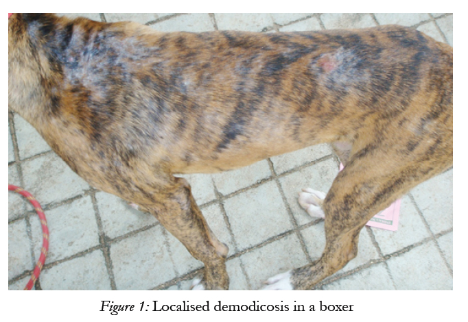
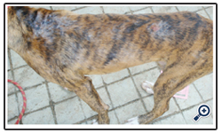
Figure 1: Localised demodicosis in a boxer.
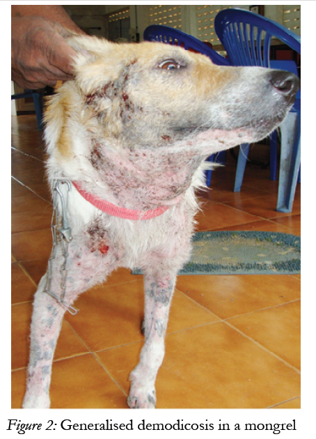
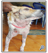
Figure 2: Generalised demodicosis in a mongrel.
Results and Discussion
Skin scrapings collected from all the 32 dogs with demodicosis revealed live mites of Demodex having long opisthosoma with pointed posterior end. Upon measurement of 320 adult (male and female) mites revealed total body length of 156-269 μm with a mean value of 211.81 ± 14.86 μm and width of 35-41 μm with mean width was 37.68±0.31 μm. Hence they were arbitrarily considered as D. canis (Figure:3). Tape impression smears taken from all the 32 dogs revealed, mites in 18 dogs with short, stumpy body with blunt opisthosomal posterior end. Morphometry examination was carried out on 320 adult (male and female) mites measured 96- 164 μm of length and 36-41 μm of width with a mean value of 137.15±22.84 μm of length and 38.28±0.19 μm of width. These mites were seen in tape impression smears and not observed in the hair pluck. Hence arbitrarily they were taken as D. cornei. D. cornei mites were diagnosed on tape impression smears only.
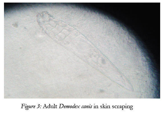
Figure 3: Adult Demodex canis in skin scraping.
Micrometry of both the mites was mentioned in the table-1. The mean total body length of the mites obtained from deep skin scrapings i.e. D. canis was almost agreeable with Chesney [4] (226.1±11.68μm), Sakulploy and Sangvaranond [8] (217.83 ± 30.06 microns), Gortel [9] (224 μm). The mean length of short mites obtained from the tape impression smears were 137.15 ± 12.84 μm of mean length. These findings were in accordance with Tamura et al. [10] who reported unidentified subspecies with a short opisthosoma and an obtuse end, short and wide body compared to Demodex canis with a body length of 139±21.6 μm. Similarly Sakulploy and Sangvaranond [8] and Lopezj et al. [11] reported the length of D. cornei 132.5 – 187.5 microns (mean 156.92 ± 11.12) and 120-155 μm (mean 139.3 ± 10.4) respectively. But, Saridomichelakis et al. [12], reported that the short tailed demodectic mite had a shorter body (145 to 200 μm; mean 165±19 μm). Chesney [4] reported a shorter and stubbier form of Demodex species with the size ranging from 90-148 μm (mean 122.6 μm SD 12.0 μm) in their studies.Though D. cornei was initially reported in four countries over three continents [13], the seemingly worldwide distribution of the parasite suggested that it is not uncommon and is merely overlooked or unrecognized. Measurements of width of the adult mites of D.canis were 35 – 41 μm (37.68±0.31), D. cornei 36- 41μm (38.28±0.19). These findings were similar to the Izdebska [14] who reported the range of width of D. canis (35-43 μm) and D. cornei (35-40 μm). Lengths of total body and opisthosoma of both types of the mites differed statistically significantl (P: 0.00) while gnathosoma and podosoma did not differ significantly (Table 1). Significant difference (P: 0.00) was also observed between the ratio of prosoma to opisthosoma in the two mites. There are no distinguishing features of history or clinical symptoms specific to D. cornei [2] and the symptoms may mimic classic Demodex infestation [15] also observed in the present study. Demodex cornei could be a mutant of D. canis or a new species [13]. The mites observed in deep skin scrapings and hair pluck were taken as D. canis based on their morphology i.e. pointed opisthosomal terminal end and size ranging from 250-300 μm. Though D. injai can also be found in deep skin scrapings, it would be much longer as its total body length was 334 - 368 μm [16]. In recent publications it was suggested that short-tailed Demodex mite may also be D. canis but may live on the surface of the epidermis. However, further genetical studies evaluating these mites (PCR test) will be needed to determine if these are indeed different species or just different forms of the same species [17]. If it were to be true, D. cornei should be found in every case of demodicosis. But in the present study, short stumpy Demodex mites were recorded in 18 out of 32 dogs i.e 56.25 % of cases and always associated with D. canis.
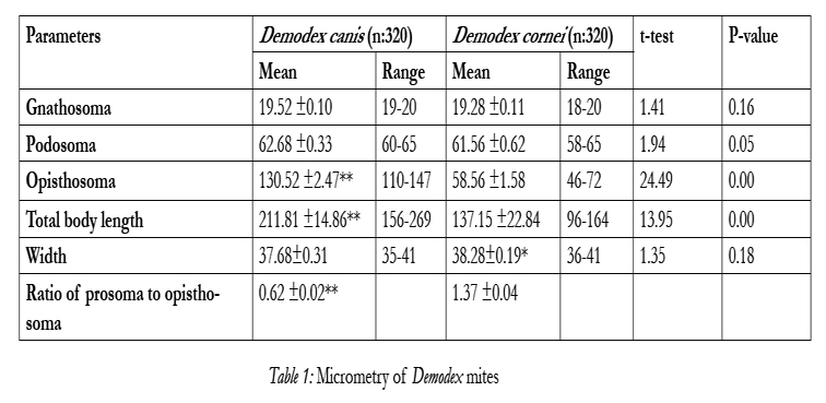

Table 1: Micrometry of Demodex mites.
In conclusion observation on the habitat of mites, its morphology and micrometry of opisthosoma, total body length and ratio of prosoma to opisthosoma will help to differentiation both the D.canis and D.cornei mites in dogs.
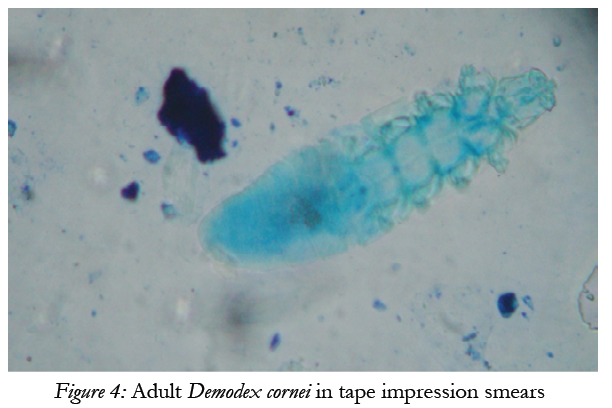
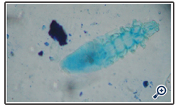
Figure 4: Adult Demodex cornei in tape impression smears
References
- Karakuram M C, Ural K, Cingi CC (2007) Evaluation of ivermectin tablets in the treatment of generalized demodicosis. Revue Medicine Veterinary 158(7): 380-383.
- Tater KC, Patterson AP (2008) Canine and Feline demodicosis.Veterinary Medicine. 444-461.
- Mueller RS, Meyer D, Emmanuel B, Louis CS (2009) Treatment of canine generaliged demodicosis with a ‘spot-on’ formulation containing 10% moxidectin and 2.5% imidacloprid (Advocate, Bayer Healthcare), Veterinary Dermatology, 20: 141-446.
- Chesney CJ (1999) Short form of Demodex species mite in the dog: occurrence and measurements. Journal of Small Animal Practice. 40: 58-61
- Paterson S (2008) Manual of skin diseases of the dog and cat, 2nd edition.104-109.
- Curtis CF (2001) Diagnostic techniques and sample collection. Clinical Techniques in Small Animal Practice 16 (4): 199-206.
- Rosenkrantz W (2008) Cutaneous cytology. A quick review of an indispensable test a supplement to Veterinary Medicine. 20-21.
- Sakulploy S, Sangvaranond A (2010) Canine demodicosis caused by Demodex canis and short opisthosomal Demodex cornei in Shi Tzu dog from Bangkok Metropolitan Thailand. Kasetsart Veterinarians. 20(1):27-35.
- Gortel K (2006) Update on canine demodicosis. Veterinary Clinical Small Animal Practice 36: 229-241
- Tamura Y, Kawamura Y, Inoue I, Ishino S (2001) Scanning electron microscopy description of a new species of Demodex canis spp. Veterinary Dermatology. 12, 275–278.
- Lopez R, Reyero D, Banson (2011) First report of canine demodicosis by short-bodied Demodex mite in Spain Rev. Inbero-Latinoam. Parasitology. 219-224.
- Saridomichelakis M, Koutinas A, Papadogiannakis E, Papazachariadou M, Liapi M, et al. (1999) Adult- onset demodicosis in two dogs due to Demodex canis and a short-tailed demodectic mite. Journal of Small Animal Practice. 40: 529-532.
- Carlotti DN (2010) Canine and feline demodicosis. 35th world SAVA, June 2-5, 2010.
- Izdebska JN (2010) Demodex sp. (Acari,Demodecidae) and Demodecosis in dogs: Characteristics, symptoms, occurrences. Bull Vet Inst Pulawy 54, 335-338.
- Schwassman M (2009) What’s new in dermatology (Proceedings) CVC Proceedings.
- Craig M (2003) BSAVA Manual of small animal dermatology. Second edition. Foster, A.P. and Foil, C.S.153.
- Kuznetsova E, Betteny S, Nikolaeva L, Majzoub M, Mueller R (2012) Influence of systemic antibiotics on the treatment of dogs with generalized demodicosis. Veterinary Parasitology. 148-155.


