Spectrum of Surgical Presentation of Eosinophilic Enteritis: A Case Series
Shetty SS1, Shetty CK2*
1 Medical Lecturer, Faculty of Medicine and Health Sciences, Department of Surgery, UCSI University, Kampus Kuala Terengganu, Terengganu, Malaysia.
2 Medical Lecturer, Faculty of Medicine and Health Sciences, Universiti Sultan Zainal Abidin (Unisza), Kampus Kota, Jalan sultan mahmud, Kuala
Terengganu, Terengganu, Malaysia.
*Corresponding Author
Dr Charan Kishor Shetty MBBS, MD,
Medical Lecturer,
Department of Forensic Medicine,
Faculty of Medicine,
Universiti Sultan Zainal Abidin (Unisza),
Kampus Kota, Kuala Terengganu, Terengganu,Malaysia 20400.
Tel: 609-6275546/609-6275740
Fax: 609-6275771/609-6275772
E-mail: shettykishor.k@rediff.com
Article Type: Case Report
Received: January 19, 2015; Accepted: March 10, 2015; Published: March 11, 2015
Citation: Shetty SS, Shetty CK (2015) Spectrum of Surgical Presentation of Eosinophilic Enteritis: A Case Series. Int J Surg Res 2(2) 8-12. doi: dx.doi.org/10.19070/2379-156X-150003
Copyright: Shetty CK© 2015. This is an open-access article distributed under the terms of the Creative Commons Attribution License, which permits unrestricted use, distribution and reproduction in any medium, provided the original author and source are credited.
Abstract
Eosinophilic enteritis is a rare disorder presenting mostly with diarrhea, malabsorption, abdominal pain, weight loss and hypersensitivity. Surgical manifestation of eosinophilic gastrointestinal disorders depends on the site and extent of involvement. In our case series of four patients two of them had ileocaecal masses with recurrent subacute intestinal obstruction with past history of intake of anti-tubercular drugs for 9 months. On histopathological examination both of them proved to have eosinophilic enterocolitis. Thus it is a clinical dilemma to differentiate between these two conditions. The other two patients presented as acute abdomen with perforation and intusseption. All four patients were treated surgically. Post operatively they recovered well with no symptoms on one year follow up. In Indian set up tuberculosis being rampant there may be under reporting or wrongly diagnosed cases of eosinophilic enteritis. Thus a strong clinical suspicion and awareness of this clinical entity is essential among surgical community.
2.Introduction
3.Case Presentation
3.1 Case 1
3.2 Case 2
3.3 Case 3
3.4 Case 4
4.Discussion
5.Conclusion
6.References
Keywords
Eosinophilic Colitis (EC); Eosinophilic Enteritis (EE); Obstruction; Perforation; Anti-Tubercular Drugs (AKT).
Introduction
Eosinophilic gastroenteritis is a rare disorder where affected patients present primarily with diarrhea, malabsorption, abdominal pain, weight loss and hypersensitivity. Surgical manifestation of eosinophilic gastrointestinal disorders depends on the site and extent of involvement and accordingly they can be divided in to eosinophilic esophagitis, eosinophilic enteritis, eosinophilic gastritis, and eosinophilic colitis. Eosinophilic gastrointestinal disorders can be primary as a part of a Hyper eosinophilic syndrome (HES) or Systemic disease (e.g. connective tissue disease, vasculitis, Celiac disease, inflammatory bowel disease), or secondary to infections (e.g. helminthic and fungi) and drugs (e.g. naproxen, clozapine, rifampicin, gold) [1].
Presentation tends to be dependent on which intestinal layer is most affected by the eosinophilic infiltration. Mucosa predominant disorder is associated with mucosal injury and patients present with malabsorption, diarrhoea, and protein-losing enteropathy. Transmural disease presents with colonic wall thickening and features of intestinal obstruction. Eosinophilic predominant ascites is a manifestation of serosal involvement. Eosinophilic colitis can present acutely with abdominal symptoms such as caecal volvulus causing intestinal obstruction, intussusception, and perforation [2]. Treatment of eosinophilic enterocolitis is antihistamines, mast cell stabilizers, glucocorticosteroids, and immunosuppressive agents along with surgery for acute surgical emergencies.
In Indian scenario where abdominal tuberculosis is widely prevalent, eosinophilic enterocolitis remain under reported. Eosinophilic colitis mimics abdominal tuberculosis with identical symptoms of fever, weight loss, abdominal pain, recurrent sub-acute intestinal obstruction and ascites. CT scan also has a similar picture of pulled up caecum, strictures, ascites especially in the transmural type of enterocolitis.
A 57-year old male with features of recurrent sub-acute intestinal obstruction from past one year. Patient was evaluated with ultrasonography which showed multiple dilated bowel loops with sluggish peristalsis. We suspected tuberculosis of abdomen as patient gave history of intake of anti-tubercular drugs for 9 months 1 year back. Patient contrast enhanced CT scan showed thickening in the ileocaecal region with mesenteric lymphadenopathy and dilated bowel loops. Patient had an elevated ESR and lymphocytosis. Patient had diagnostic laparoscopy which showed pulled up thickened shrunken caecum with ileal stricture. The patient underwent a right laparoscopic hemicolectomy (figure 1). Post operatively the patient recovered well. On histopathological examination of the specimen, eosinophilic infiltration was seen in all layers of intestine. Patient’s absolute eosinophilic count was 550/cumm.
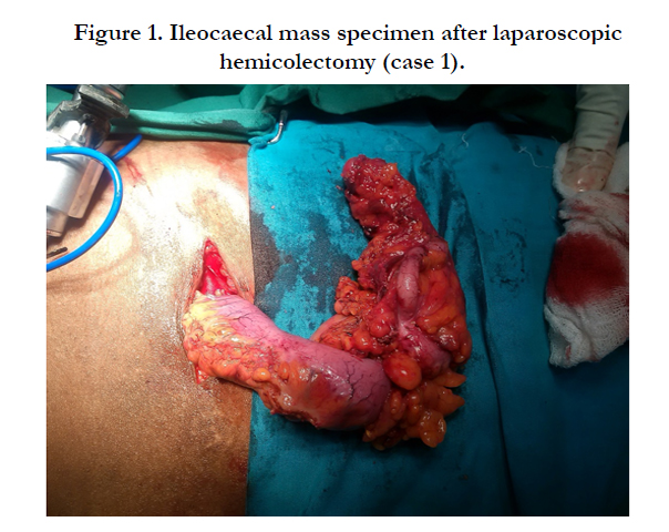
Figure 1. Ileocaecal mass specimen after laparoscopic hemicolectomy (case 1).
A 32-year old male patient arrived to emergency surgical department with acute abdominal pain. On examination the patient had tachycardia with blood pressure of 100/60mmHg and high colored urine. Abdominal examination revealed generalized tenderness, guarding and rigidity. Patients abdominal x-ray erect showed air under diaphragm. Patient underwent an exploratory laparotomy in view of features suggestive of perforative peritonitis. The patient had perforated ileum with two distal ileal strictures and a fibrosed caecum. Ileo transverse bypass was performed with closure of the ileal perforation after taking biopsy. (Figure 2). Post operatively after histopathological examination the patient developed eosinophilic enteritis. Patient was given a course of steroids and anti-helminthic drugs. However, his post-operative course was uneventful. We gave steroid in the second patient because we had not done any resective procedure in this patient. We only took multiple biopsies from perforated area and did an ileo transverse bypass procedure and left the affected bowel as patient presented in septic shock.
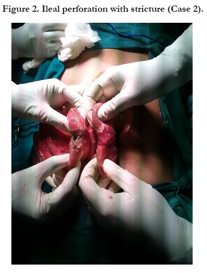
Figure 2. Ileal perforation with stricture (Case 2).
A 24-year old male came with history of obstipation, recurrent vomiting and abdominal distention since 1 day. On evaluation, the patient had tachycardia with low blood pressure. Patient’s abdominal x-ray showed multiple air fluid level and CT scan showed intestinal obstruction with target sign suggestive of intusseption. Patient was had exploratory laparotomy which showed dilated bowel loops with ileoileal intusseption at the level of distal ileum (figure 3). Resection anastomosis was performed. Specimen analysis showed eosinophilic enteritis. Patient had elevated absolute eosinophilic count. On one year follow up patient was symptom free.
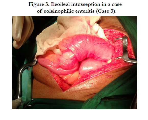
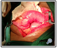
Figure 3. Ileoileal intusseption in a case of eoisinophilic enteritis (Case 3).
A 6- year old male with history of recurrent sub-acute intestinal obstruction since 3 months. Patient was evaluated with ultrasonography which showed multiple dilated bowel loops with sluggish peristalsis. Patients contrast enhanced CT scan showed thickening in the ileocaecal area with dilated bowel loops. Patient had an elevated ESR and lymphocytosis and also gave history of known case of tuberculosis with intake of anti-tubercular drugs 35 years back for a year. Patient had normal eosinophil count. Exploratory laparotomy and right hemi colectomy done in view of thickened pulled up illeoceacal junction and dilated proximal loops. Post operatively patient recovered well. On histopathological examination of specimen eosinophilic infiltration was seen in all layers of intestine. (Figure 4 and 5).

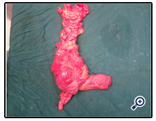
Figure 4. Resected specimen of ileum and caecum after right hemi colectomy (case 4).
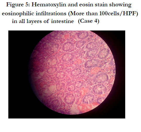
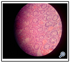
Figure 5: Hematoxylin and eosin stain showing eosinophilic infiltrations (More than 100cells/HPF) in all layers of intestine (Case 4)
Discussion
Eosinophilic enteritis (EE) was first described by Kaiser in 1937 [3]. In India, Venkataraman et al have reported seven cases of EE over a ten-year period [4]. Diagnosis is one of exclusion and the criteria put forward for the diagnosis are the presence of gastrointestinal (GIT) symptoms, infiltration of the GIT by eosinophils in one or more areas, absence of parasitic infestation and exclusion of eosinophilic involvement in organs other than the GIT [5]. The diagnosis of EE is made from the presence of gastrointestinal symptoms, peripheral eosinophilia, endoscopic and histological findings, and eosinophilic ascites, with no well-defined causes of eosinophilia on thorough evaluation. In the present case series only two patients had elevated peripheral eosinophil counts but all had eosinophilic infiltration of > 100 cells/ hpf on histopathological examination. Hence in a suspected case of EE a colonoscopic biopsy at multiple sites showing eosinophilic infiltration can be one of the diagnostic tools in confirming the disease.
In our case series of four patients two of them had ileocaecal mass with recurrent subacute intestinal obstruction with past history of intake of anti-tubercular drugs for 9 months. On histopathological examination both of them proved to have eosinophilic enterocolitis. Thus, it is a clinical dilemma to differentiate between these two conditions. The other two patients presented as acute abdomen with perforation and intusseption. All four patients were treated surgically. Post operatively they recovered well with no symptoms on one year follow up.
Various case reports have been reported where eosinophilic enteritis mimics tuberculosis and ulcerative colitis. In the present case series also out of four patients two of them had a history of abdominal tuberculosis in the past and treatment for the same was taken. Lange et al proposed rifampicin as a cause of eosinophilic colitis as its side effect [6]. In present case series whether patients were wrongly diagnosed cases of tuberculosis in actual case of eosinophilic enteritis or patient had eosinophilic enteritis as a side effect to consumption of rifampicin was an enigma.
Both these patients did not have a proven biopsy suggestive of tuberculosis before starting treatment. According to history they were on anti-tubercular drugs only based on clinical suspicion and CT picture. Drugs reported to cause colonic eosinophilia include nonsteroidal anti-inflammatories, tacrolimus, carbamazrpine, rifampicin, sulphasalazine, and naproxen. None of the patients in the case series had any h/o allergic rhinitis or atopy or food allergy. Thus, it is essential to conduct colonoscopic biopsy to differentiate between eosinophilic enterocolitis and tuberculosis. Biopsy has to be taken at multiple sites as the disease has tendency for skip lesions.
Eosinophilic enterocolitis also presents as a mass lesion which can be confused with neoplasm. In the third case, the patient had a lump on CT scan with target lesion which was suspected as malignancy causing a leading point for intusseption but was surprisingly proved as EE. Thus eosinophilic gastro intestinal disorders even though rare has to kept as differential diagnosis as it is a benign and an easily treatable entity.
Supportive treatment with pharmacotherapy, mainly oral glucocorticosteroids, is indicated for those with obstructive symptoms. Patients with mucosal layer involvement may benefit from antiinflammatory medications (e.g., oral glucocorticoids) and/or diet elimination therapy, particularly if they report a history of food intolerance or allergy. Drugs, such as montelukast, ketotifen, and mycophenolate mofetil, and alternative Chinese medicines have been advocated but are generally not successful. Recurrence is common even after surgical resection.
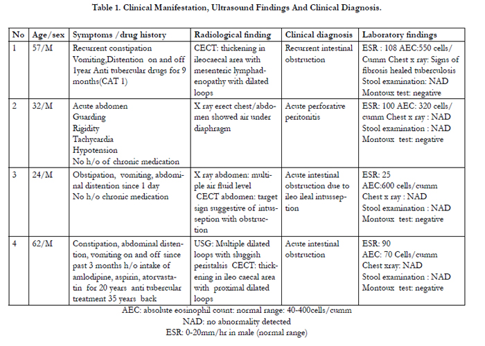

Table 1. Clinical Manifestation, Ultrasound Findings And Clinical Diagnosis.
AEC: absolute eosinophil count: normal range: 40-400cells/cumm
NAD: no abnormality detected
ESR: 0-20mm/hr in male (normal range)
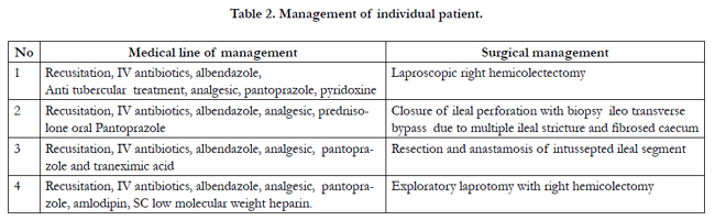
Table 2. Management of individual patient.
Conclusion
Eosinophilic enteritis even though a rare disorder has a varied spectrum of presentation which is easily misinterpreted as neoplasm, tuberculosis and inflammatory bowel disease. Since it can be easily treated medically and surgically it has to be diagnosed with certainty. In Indian set up, tuberculosis being rampant there may be under reporting or wrongly diagnosed cases of eosinophilic enteritis. Thus, a strong clinical suspicion and awareness of this clinical entity is essential among surgical fraternity.
References
- Abdulrahman A. Alfadda, Martin A. Storr, Eldon A. Shaffer (2010) Eosinophilic colitis: epidemiology, clinical features, and current management. Ther Adv Gastroenterol 4(5): 301-309.
- Ong GY, Hsu CC, Changchien CS, Lu SN, Huang SC (2002) Eosinophilic gastroenteritis involving the distal small intestine and proximal colon.Chang Gung Med J 25(1): 56-61.
- Kaijser R (1937) Zurkenntnis der allergisschen des verdauungskabals vonstandpunkt des chiruugan aus.Arch Klin Chir 36:188.
- Venkataraman S, Ramakrishna BS, Mathan M, Chacko A, Chandy G, et al. (1998) Eosinophilic gastroenteritis -an Indian experience. Indian J Gastroenterol. 17(4):148-149.
- Talley NJ, Shorter RG, Phillips SF, Zinsmeister AR (1990) Eosinophilic gastroenteritis: a clinicopathological study of patients with disease of the mucosa, muscle layer, and subserosal tissues. Gut 31(1): 54-58.
- Lange P, Oun H, Fuller S, Turney JH (1994) Eosinophilic colitis due to rifampicin. Lancet 344 (8932): 1296-1297.


