Suppression of Conjunctival Scarring by Chymase Inhibitor in a Canine Symblepharon Model
Tajiri K1, Sugiyama T1, 3*, Katsumura K4, Jin D2, Takai S2, Shimizu K5, Ikeda T1
1 Department of Ophthalmology, Osaka Medical College, Takatsuki-city, Osaka, Japan.
2 Department of Pharmacology, Osaka Medical College, Takatsuki-city, Osaka, Japan.
3 Nakano Eye Clinic of Kyoto Medical Co-operative, Kyoto, Japan.
4 Katsumura Eye Clinic, Osaka, Japan.
5 Department of Ophthalmology, Takatsuki Hospital, Takatsuki-city, Osaka, Japan.
*Corresponding Author
Tetsuya Sugiyama (MD, PhD),
Nakano Eye Clinic of Kyoto Medical Cooperative, 2,
Jurakumawari-higashimachi, Nakagyo-ku, Kyoto 604-8404, Japan.
Tel: +81-75-801-4151
E-mail: tsugiyama@kyo-con.or.jp
Received: September 07, 2016; Accepted: October 12, 2016; Published: October 15, 2016
Citation: Tajiri K, Sugiyama T, Katsumura K, Jin D, Takai S, et al., (2016) Suppression of Conjunctival Scarring by Chymase Inhibitor in a Canine Symblepharon Model. Int J Opthalmol Eye Res, S7:002, 6-12. doi: dx.doi.org/10.19070/2332-290X-SI07002
Copyright: Sugiyama T© 2016. This is an open-access article distributed under the terms of the Creative Commons Attribution License, which permits unrestricted use,distribution and reproduction in any medium, provided the original author and source are credited.
Abstract
Purpose: To investigate the suppressive effects of a chymase inhibitor (CI) on conjunctival scarring in a canine model of symblepharon induced by alkali burns.
Methods: Symblepharon models were made in beagle eyes. A cotton pad soaked in 1 N sodium hydroxide (NaOH) was placed in the conjunctival sac of each eye for 90 seconds, followed by washing the sac with 100 ml of physiological saline. Immediately post treatment, one eye drop (50 μl) including 1 mM-CI or the vehicle (CI-treated group and vehicle-treated group, respectively) was instilled in 5 eyes of 5 beagles once daily for 5 weeks. Fellow eyes were left untreated and served as the normal group. The severity of symblepharon was graded by macroscopic observation.
After the eyes were enucleated, conjunctival and scleral tissue specimens were histologically evaluated.
Results: Macroscopic observation revealed that symblepharon was induced in all NaOH-treated eyes, while symblepharon scores were significantly lower in the CI group than in the vehicle-treated group. Histological observation indicated a significant reduction in the adhered area in the CI-treated group compared to the vehicle-treated group. Immunohistochemical analysis demonstrated that vimentin-, α-smooth muscle actin-, chymase-, and angiotensin II-positive cells as well as mast cells increased in the vehicle-treated group compared to the normal group, whereas they were reduced in the CI-treated group.
Conclusions: Our macroscopic and histological findings indicated that a multiple application of CI eye drops suppressed conjunctival scarring in a canine symblepharon model, suggesting that the topical application of CI may be a promising therapy for symblepharon.
2.Introduction
3.Methods
3.1.Animals
3.2.Preliminary Experiment: Induction of a Canine Symblepharon Model
3.3.Experimental Protocol
3.4.Macroscopic Observation
3.5.Histology and Immunohistochemistry
3.6.Statistical Analysis
4.Results
4.1.Preliminary Experiment
4.2.Effects of CI in the Symblepharon Model
5.Discussion
6.Conclusion
7.Acknowledgements
8.References
Keywords
Chymase Inhibitor; Symblepharon; Scarring; Alkali Burn; Beagles.
Introduction
Stevens-Johnson syndrome, ocular cicatricial pemphigoid, and thermal or chemical injury are all serious disorders that are known to scar the ocular surface. Clinical problems that arise during the scarring period include visual impairment due to conjunctival epithelium on the cornea resulting from limbal stem-cell failure, severe dry eye due to destruction of the meibomian gland structure, obstruction of the lacrimal gland conduit, loss of conjunctival goblet cells, and problems related to grooming and function; e.g., skin-like keratinization of the ocular surface, trichiasis and entropion due to scar formation, eye-movement disorder due to symblepharon, and inability to open the eyelid [1-4].
To control inflammation in the acute phase, or to control scarring- associated change post surgical intervention (e.g., cultured epithelial transplantation [5, 6], amniotic membrane transplantation [7, 8], or plastic surgery treatment in the scarring phase [9]) local and systematic steroid therapy is often selected [1-3]. However, the use of steroids is known to produce systematic side effects such as diabetes or infection, as well as focal-related side effects (e.g., cataract or glaucoma). Thus, new medication options in the clinical setting to replace the use of steroids have been long-awaited [10].
In animal models of alkali injury, a kind of chemical injury, severe inflammation of the ocular surface, including corneal edema, erosion, ulcer, and conjunctival ischemia, occurs in the acute phase and prolongs into the chronic phase, thus resulting in conjunctival scarring-related change including symblepharon [11-18].
Chymase is a serine protease mostly derived from mast cells that are primarily present in heart and blood vessels [19]. Although there is some evidence that smooth muscle expresses chymase, only mast cells appear capable of accumulating chymase in secretory granules [20]. Chymase acts as angiotensin II production enzymes other than angiotensin converting enzyme (ACE) that are involved in tissue remodeling and production of the extracellular matrix [21]. Whether or not the ACE or chymase acts as an angiotensin II-producing enzyme depends on the species of animal, yet in humans, monkeys, dogs, and hamsters, chymase is known to play a significant role [22]. There have been numerous investigations regarding chymase and fibrosis in the heart [23, 24], lungs [25], liver [26], kidneys [27], and vessels [28, 29]. Moreover, mast cells and chymase play an important role in wound healing and keloid formation of skin through the transforming growth factor beta 1 (TGF-β1)/SMAD signaling pathway [30, 31].
Several studies have reported an involvement of chymase in allergic conjunctivitis. One of those reports suggested that chymase was released from mast cells post antigen challenge, followed by the induction of conjunctivitis symptoms through histamine release from mast cells [32]. Another in vitro study indicated that human mast-cell chymase caused conjunctival epithelial cell detachment by degrading fibronectin, thus leading to secondary apoptosis as the mechanism of conjunctival epithelial injury in vernal keratoconjunctivitis [33]. A relationship between chymase and conjunctival fibroblasts has also been reported [34]. The findings of that study indicated that chymase stimulated the proliferation of Tenon's capsule fibroblasts, and that chymase inhibitor (CI) had a suppressive effect on subconjunctival scarring in a canine conjunctival flap model. Another study reported the suppressive effect of the CI contained in gelatin hydrogel (GH) on the fibroblast proliferation in a canine filtration surgery model [35]. In addition, the involvement of chymase has been suggested in the mechanisms of mitomycin C action, used currently in the clinical setting as a conjunctival adhesion inhibitor, in a monkey trabeculectomy model [36].
In this present study, we induced a canine model of symblepharon by alkali burn and investigated whether a CI had the suppressive effects on conjunctival scarring in this model.
Twenty-six beagle dogs weighing 9-10 kg each were obtained from Japan SLC, Inc., Hamamatsu City, Shizuoka, Japan. Each day, the dogs were fed their regular diet of chow, had free access to tap water, and were housed in an air-conditioned room at a temperature of approximately 23°C and 60% humidity with a 12-hour light-dark cycle. This study was approved by the Institutional Animal Care and Use Committee of Osaka Medical College, and the experimental procedures used for all animals were conducted in accordance with the ARVO Statement for Use of Animals in Ophthalmic and Vision Research.
Under general anesthesia using 35 mg/kg of pentobarbital, a cotton pad (Asahi Eisei Zairyo Co., Ltd., Osaka, Japan) measuring 3 × 20 mm was soaked with 1N sodium hydroxide (NaOH) and then placed in the upper and lower conjunctival sacs of 1 eye for 15 seconds (n = 2), 60 seconds (n = 9), and 90 seconds (n = 5). The cotton pad was then removed (Fig. 1A) and the eye was washed with 100 ml of physiological saline. Five weeks later, macroscopic observation of the symblepharon of each treated eye was conducted.
A symblepharon model was induced by the same method as described above (i.e., a 90-second exposure according to the results of the preliminary experiment). A CI [Suc-Val-Pro-PheP(OPh)2, a gift from Prof. Józef Oleksyszyn, Wroclaw University of Technology, Wroclaw, Poland] was used for treatment [37]. The CI was adjusted as eye drops to be diluted to 1 mM with 0.01% dimethyl sulfoxide solution. The eyes were treated with a 1-drop (50 μl) instillation of 1 mM CI solution in 5 eyes of 5 dogs (CI-treated group) and the vehicle (0.01% dimethyl sulfoxide solution) in 5 eyes of 5 dogs (vehicle-treated group), respectively, once daily for 5-weeks post alkali burn. The fellow eyes without alkali burns (10 eyes of 10 dogs) were defined as the normal group.
Five-weeks later, the severity of symblepharon was evaluated by scoring as described below. The dogs were then killed by injecting a lethal dose of pentobarbital sodium, followed by the removal of the eyeballs with palpebral conjunctiva, bulbar conjunctiva, and lid to loaf for histological evaluation as described below.
The severity of symblepharon was scored as follows: Score 0, no symblepharon; Score 1, symblepharon exists. When moving the eyelid, neither the eyeball nor nictitating membrane moves; Score 2, symblepharon exists. There is strong adhesion, such that when moving the eyelid, the eyeball and nictitating membrane move together.
The eyeballs with palpebral conjunctiva, bulbar conjunctiva, and lid to loaf were fixed in Carnoy's solution (Muto Pure Chemicals Co., Ltd., Tokyo, Japan) and embedded in paraffin. Under targeting of the portion that caused the symblepharon, 5-μm-thick sections including palpebral and bulbar conjunctiva were then cut, mounted on silanized slides (Dako Japan Co. Ltd., Kyoto, Japan), and deparaffinized with xylene and a series of graded ethanol. First, hematoxylin and eosin (HE) and Azan-Mallory staining was conducted to measure the adhered areas of the palpebral conjunctiva to the bulbar conjunctiva in the vehicle-treated and CItreated groups. The procedure for measuring the adhered areas was follows: 1) The depths of the conjunctival sac were measured along the parallel line to the palpebral conjunctiva in the sections from normal eyes (as shown in Figure 2A) and the normal-depth value of the conjunctival sac was obtained by averaging them. 2) The adhered areas, surrounded by the eyelid, the sclera and the line obtained from the normal-depth value of the conjunctival sac, were measured in the vehicle-treated and CI-treated groups using Image J software (as shown in Figures. 2B and 2C).
Next, Toluidine blue staining was used to identify mast cells. In order to determine the distribution of chymase, immunohisto chemical staining was performed using anti-dog chymase antibody as previously described [38]. This antibody was a gift from Prof. George H. Caughey (University of California San Francisco, USA), raised by injection of α-chymase purified from dog mastocytoma cells into rabbits; this antibody had specificity for dog tissue [39]. Expression of angiotensin II was determined by rabbit anti-human angiotensin II polyclonal antibody (IgG Corporation, Nashville, TN, USA). To examine the cellular pattern of the subconjunctival tissue, monoclonal antibodies for mouse anti-human α-smooth muscle actin (α-SMA; M0851, Dako Denmark A/S, Glostrup, Denmark), a marker of myofibroblasts, and mouse anti-bovine vimentin (M0725, Dako Denmark A/S), a marker of vascular endothelial cells and fibroblasts, were used. The sections were incubated overnight at 4°C with each antibody, followed by reaction with appropriate reagents from a streptavidin-biotin peroxidase kit (Dako Denmark A/S) and 3-amino-9-ethylcarbazole for 5 to 10 minutes. The sections were then lightly counterstained with hematoxylin.
For the statistical analysis, each measurement was expressed as the mean ± standard deviation (SD). Symblepharon scores were analyzed by use of the Wilcoxon’s signed ranks test. Other parameters were evaluated by use of the paired t-test or the Tukey- Kramer test. Differences were considered statistically significant at a P-value of < 0.05.
Results
Severe conjunctival melting was observed in all eyes immediately after the exposure to 1 N NaOH (Figure 1A). With the macroscopic observation in the late phase, shortening of the conjunctival sac, adhesion between palpebral conjunctiva and bulbar conjunctiva, and blepharophimosis were observed. There were two types of adhesion observed later as follows: 1) diffuse adhesion in the conjunctival sac, which caused a shortening of the conjunctival sac, and 2) focal adhesion between the palpebral conjunctiva and the bulbar conjunctiva, or between the palpebral conjunctiva and nictitating membrane, which was observed as a bundle of adhesions. When diffuse and/or focal adhesions were observed (Figs. 1B and 1C), the case was determined as acquiring symblepharon.
In 5 (100%) of 5 eyes exposed for 90 seconds and in 3 (33%) of 9 eyes exposed for 60 seconds, symblepharon was observed within 5 weeks post treatment with NaOH (Figures. 1B and 1C). However, in both eyes treated for 15 seconds, the conjunctiva healed early and symblepharon did not occur (Figure 1D).
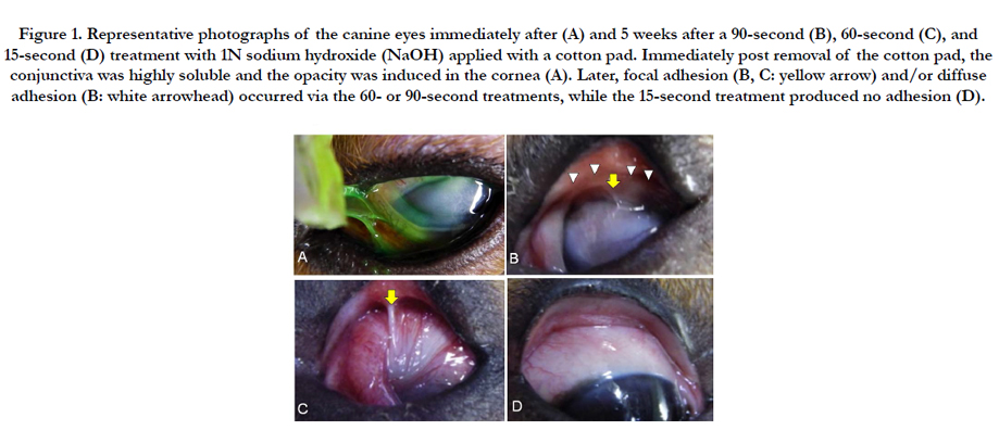
Figure 1. Representative photographs of the canine eyes immediately after (A) and 5 weeks after a 90-second (B), 60-second (C), and 15-second (D) treatment with 1N sodium hydroxide (NaOH) applied with a cotton pad. Immediately post removal of the cotton pad, the conjunctiva was highly soluble and the opacity was induced in the cornea (A). Later, focal adhesion (B, C: yellow arrow) and/or diffuse adhesion (B: white arrowhead) occurred via the 60- or 90-second treatments, while the 15-second treatment produced no adhesion (D).
In all of the CI-treated-group eyes, the severity of symblepharon was scored as 1 point, while the symblepharon score was 2 points in 4 of 5 eyes of the vehicle-treated eyes and 1 point in the remaining eye. The mean symblepharon scores were 1.8 (± 0.4) and 1.0 (± 0) in the vehicle-treated and CI-treated groups, respectively, thus illustrating that the score was significantly lower in the CItreated group than in the vehicle-treated group (P = 0.046, Wilcoxon’s signed ranks test).
In the vehicle-treated and CI-treated groups, adhesion occurred between the palpebral and bulbar conjunctivas. Before measuring the adhered areas, normal-depth value of the conjunctival sac was determined as 8.41 mm from the data of normal eyes. Then, measurement of the adhered areas revealed significant reduction of those areas in the CI-treated group compared to the vehicletreated group (Figure 2).
Vimentin- and α-SMA-positive cells were increased in the alkali burn eyes (vehicle-treated and CI-treated groups), yet were more suppressed in the CI-treated group than in the vehicle-treated group (Figure 3, Table 1).
Mast cells (Toluidine blue staining), angiotensin II-positive cells, and chymase-positive cells were examined (Figure 4, Table 1). In the vehicle-treated group, mast cells, angiotensin II-positive cells, and chymase-positive cells were increased in comparison to the normal group. In the CI-treated group, the numbers of all of those cells were suppressed.
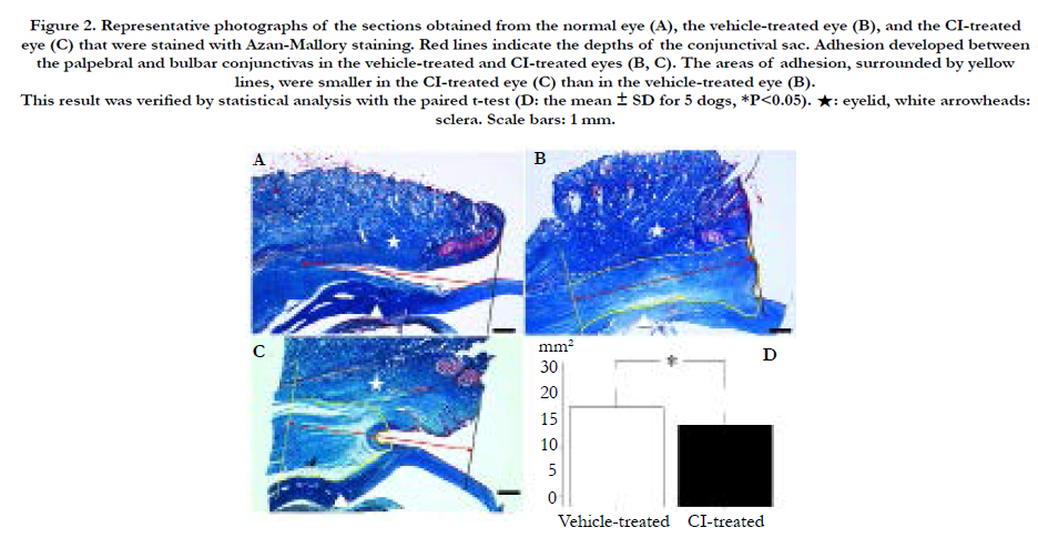
Figure 2. Representative photographs of the sections obtained from the normal eye (A), the vehicle-treated eye (B), and the CI-treated eye (C) that were stained with Azan-Mallory staining. Red lines indicate the depths of the conjunctival sac. Adhesion developed between the palpebral and bulbar conjunctivas in the vehicle-treated and CI-treated eyes (B, C). The areas of adhesion, surrounded by yellow lines, were smaller in the CI-treated eye (C) than in the vehicle-treated eye (B). This result was verified by statistical analysis with the paired t-test (D: the mean ± SD for 5 dogs, *P<0.05). ★: eyelid, white arrowheads: sclera. Scale bars: 1 mm.
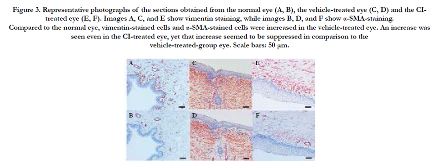
Figure 3. Representative photographs of the sections obtained from the normal eye (A, B), the vehicle-treated eye (C, D) and the CItreated eye (E, F). Images A, C, and E show vimentin staining, while images B, D, and F show α-SMA-staining. Compared to the normal eye, vimentin-stained cells and α-SMA-stained cells were increased in the vehicle-treated eye. An increase was seen even in the CI-treated eye, yet that increase seemed to be suppressed in comparison to the vehicle-treated-group eye. Scale bars: 50 μm.
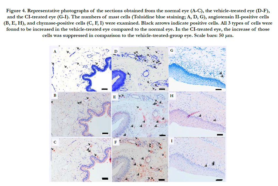
Figure 4. Representative photographs of the sections obtained from the normal eye (A-C), the vehicle-treated eye (D-F), and the CI-treated eye (G-I). The numbers of mast cells (Toluidine blue staining; A, D, G), angiotensin II-positive cells (B, E, H), and chymase-positive cells (C, F, I) were examined. Black arrows indicate positive cells. All 3 types of cells were found to be increased in the vehicle-treated eye compared to the normal eye. In the CI-treated eye, the increase of those cells was suppressed in comparison to the vehicle-treated-group eye. Scale bars: 50 μm.
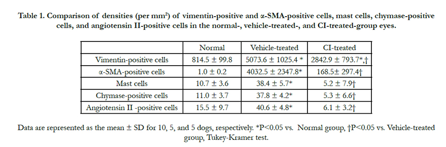

Table 1. Comparison of densities (per mm²) of vimentin-positive and α-SMA-positive cells, mast cells, chymase-positive cells, and angiotensin II-positive cells in the normal-, vehicle-treated-, and CI-treated-group eyes.
Discussion
The findings of the current study demonstrated for the first time that the topical application of CI inhibited conjunctival fibrosis post alkali burn in a canine symblepharon model. In addition, and to the best of our knowledge, this is the first report on multiple application of CI as eye drops in an animal model.
Dogs were used in this present study because unlike in rats and rabbits, the conversion of angiotensin I to angiotensin II in the vascular tissue of humans, monkeys, dogs and hamsters is partly dependent on chymase [40, 41]. In addition, the similarly of dogs to humans in regard to the fibrosis of the conjunctiva, such as symblepharon formation post ocular alkali injury, has previously been reported [42].
Our results suggested that a 90-second placement of 1 N NaOH in the conjunctival sac creates a stable canine symblepharon model in the chronic phase. To the best of our knowledge, although there have been many reports on animal models of alkali-burned cornea [11-18], there have been no reports of models of symblepharon induced by alkali injury. As NaOH is often used at 1 N in models of alkali-burned cornea, we opted to apply it at that concentration in this present study. According to the time-dependent effect of NaOH in the preliminary experiment, a 90-second placement of a NaOH-soaked cotton pad into the conjunctival sac was performed in the main experiment.
The concentration of the CI eye drops in the current study was determined based on our previous report [35]. In that study, the gelatin hydrogel (a 5 × 5 × 1.5-mm block) included the CI solution at 10 μM; the total amount of the CI in the block of gelatin hydrogel was therefore 375 pmol, 80% of which was gradually released over a 14-day period. On the other hand, 50 μl of the CI at 1 mM was applied once daily for 35 days in the present study. The total amount of the CI was 17500 pmol, approximately 50-times larger than the amount applied in the previous study. We considered that the effect of alkali burn on the ocular tissue was much more severe than that of filtration surgery, and thus decided to set the concentration at 1 mM.
Through the macroscopic observation, shortening of the conjunctival sac, adhesion between the palpebral conjunctiva and bulbar conjunctiva, and blepharophimosis were observed in the current symblepharon model. Whereas, the adhesion changes, as evaluated by the symblepharon score, were significantly suppressed in the CI-treated group compared with the vehicle-treated group. In other words, the strong adhesion which restricted ocular movement was not observed in the CI-treated group. The degrees of adhesion are presumably affected by the contraction of the tissue and the range of adhesion. Histological analysis also revealed that the adhered areas were significantly reduced in the CI-treated group. Previous studies have reported the preventive effect of the same CI as in the present study in animal models of adhesion formation [34, 35, 43, 44]. In addition, another CI was reported to attenuate extracellular matrix loss and tissue remodeling in a canine model of cardiovascular fibrosis [45].
In this present study, immunohistochemical analysis revealed that fibroblasts (vimentin-positive cells) were increased in the alkali injured
eyes in both the vehicle-treated group and CI-treated group compared to the normal group, yet the increase was significantly suppressed in the CI-treated group. Myofibroblasts (α-SMApositive cells) were also increased in the alkali injured eyes, specifically in the vehicle-treated group, although no significant increase was observed in the CI-treated group. It has been reported that the proliferation of fibroblasts promotes fibrosis and adhesion change between palpebral conjunctiva and bulbar conjunctiva and results in symblepharon formation [46]. However, the results of another study showed that the proliferation of myofibroblasts which produce actin filaments promotes shrinking of the tissue, thus resulting in a stronger adhesion [47]. The findings in this current study suggest that CI suppresses symblepharon formation through the inhibition of fibroblasts as well as myofibroblasts.
In this present study, mast cells, chymase-positive cells, and angiotensin II-positive cells were increased in the vehicle-treated group in comparison to the normal group. To date, there are few reports on mast cells and chymase in alkali-burn models, however, there have been many reports on their involvement in post-surgical adhesion as well as uveitis or allergic conjunctivitis [32-35, 44, 45, 48, 49]. In this current study, those cells were found to be rather reduced in the CI-treated group, probably due to the inhibitory effects of CI on the accumulation of mast cells.
It should be noted that this present study did include several limitations. First, the most suitable CI dosage (i.e., the eye-drop concentration and the number of instillations per day) is a question that requires further investigation. In addition, the effect of the CI should be compared with other conventional or probable drugs for the treatment of symblepharon, such as steroids. Furthermore, other pathogenic factors, except for chymase, might also be involved in the symblepharon model we used in the current study; tumor necrosis factor α, TGF-β1, Interleukin 1 and Interleukin-6 and so on. These other factors should also be studied in the future.
Conclusion
Our results, via both macroscopic and histological observation, suggest that CI eye drops suppress conjunctival scarring in a canine symblepharon model. The topical application of CI for the treatment of symblepharon is a therapy that appears to be worthy of further investigation.
Acknowledgements
The authors thank John Bush for reviewing the manuscript. Supported by Japanese Ministry of Education Grant 23792020 (KK).
References
- Sotozono C, Ueta M, Kinoshita S (2009) The management of severe ocular complications of Stevens-Johnson syndrome and toxic epidermal necrolysis. Arch Dermatol. 145(11): 1336-1337.
- Kirzhner M, Jakobiec FA (2011) Ocular cicatricial pemphigoid: a review of clinical features, immunopathology, differential diagnosis, and current management. Semin Ophthalmol. 26(4-5): 270-277.
- Fish R, Davidson RS (2010) Management of ocular thermal and chemical injuries, including amniotic membrane therapy. Curr Opin Ophthalmol. 21(4): 317-321.
- Cabalag MS, Wasiak J, Syed Q, Paul E, Hall AJ, et al., (2015) Cleland H. Early and late complications of ocular burn injuries. J Plast Reconstr Aesthet Surg. 68(3): 356-361.
- Tsubota K, Satake Y, Kaido M, Shinozaki N, Shimmura S, et al., (1999) Treatment of severe ocular-surface disorders with corneal epithelial stem-cell transplantation. N Engl J Med. 340(22): 1697-1703.
- Nishida K, Yamato M, Hayashida Y, Watanabe K, Yamamoto K, et al., (2004) Corneal reconstruction with tissue-engineered cell sheets composed of autologous oral mucosal epithelium. N Engl J Med. 351(12): 1187-1196.
- Nakamura T, Yoshitani M, Rigby H, Fullwood NJ, Ito W, et al., (2004) Sterilized, freeze-dried amniotic membrane: a useful substrate for ocular surface reconstruction. Invest Ophthalmol Vis Sci. 45(1): 93-99.
- Choi JA, Jin HJ, Jung S, Yang E, Choi JS, et al., (2009) Effects of amnioticmembrane suspension in human corneal wound healing In Vitro. Mol Vis. 15: 2230-2238.
- Pargament JM, Armenia J, Nerad JA (2015) Physical and chemical injuries to eyes and eyelids. Clin Dermatol. 33(2): 234-237.
- Onaran Z, Usta G, Koçak M, Ornek K, Büyükkoçak U (2011) Topicalophthalmic cyclosporine in the treatment of toxic epidermal necrolysis. Case Rep Med. 2011: 416842.
- Foster CS, Zelt RP, Mai-Phan T, Kenyon KR (1982) Immunosuppression and selective inflammatory cell depletion. Studies on a guinea pig model of cornealulceration after ocular alkali burning. Arch Ophthalmol 100(11): 1820-1824.
- Burns FR, Gray RD, Paterson CA (1990) Inhibition of alkali-induced cornealulceration and perforation by a thiol peptide. Invest Ophthalmol Vis Sci. 31(1): 107-114.
- Alio JL, Ayala MJ, Mulet ME, Artola A, Ruiz JM, et al., (1995) Antioxidant therapy in the treatment of experimental acute corneal inflammation. Ophthalmic Res. 27(3): 136-143.
- Ishizaki M, Shimoda M, Wakamatsu K, Ogro T, Yamanaka N, et al., (1997) Stromal fibroblasts are associated with collagen IV in scar tissues of alkaliburned and lacerated corneas. Curr Eye Res. 16(6): 339-348.
- Chung JH, Kang YG, Kim HJ (1998) Effect of 0.1% dexamethasone on epithelial healing in experimental corneal alkali wounds: morphological changes during the repair process. Graefes Arch Clin Exp Ophthalmol .236(7): 537-545.
- Kitano A, Okada Y, Yamanaka O, Shirai K, Mohan RR, et al., (2010) Therapeutic potential of Trichostatin A to control inflammatory and fibrogenic disorders of the ocular surface. Mol Vis. 16: 2964-2973.
- Ferrari G, Bignami F, Giacmini C, Franchini S, Rama P (2013) Safety and efficacy of topical infliximab in a mouse model of ocular surface scarring. Invest Ophthalmol Vis Sci. 54(3): 1680-1688.
- Espandar L, Caldwell D, Watson R, Blanco-Mezquita T, Zhang S, et al., (2014) Application of adipose-derived stem cells on scleral contact lens carrier in an animal model of severe acute alkaline burn. Eye Contact Lens.40(4): 243-247.
- Liao Y, Husain A (1995) The chymase-angiotensin system in humans: biochemistry, molecular biology and potential role in cardiovascular diseases. Can J Cardiol. 11: 13F-19F.
- Caughey GH (2007) Mast cell tryptases and chymases in inflammation and host defense. Immunological Reviews. 217: 141-154.
- Saito H (2005) Role of mast cell proteases in tissue remodeling. Chem Immunol Allergy. 87: 80-84.
- Takai S, Jin D, Miyazaki M (2012) Multiple mechanisms for the action of chymase inhibitors. J Pharmacol Sci. 118(3): 311-316.
- Oyamada S, Bianchi C, Takai S, Chu LM, Sellke FW (2011) Chymase inhibition reduces infarction and matrix metalloproteinase-9 activation and attenuates inflammation and fibrosis after acute myocardial ischemia/reperfusion. J Pharmacol Exp Ther. 339(1): 143-151.
- Takai S, Jin D, Sakaguchi M, Muramastu M, Miyazaki M et al., (2003) A novel chymase inhibitor, 4-[1-{[bis-(4-Methyl-phenyl)-methyl]-carbamoyl}- 3-(2-ethoxy-benzyl)-4-oxo-azeti dine-2-yloxy]-benzoic acid (BCEAB), suppressed cardiac fibrosis in cardiomyopathic hamsters. J Pharmacol Exp Ther.305(1): 17-23.
- Kosanovic D, Dahal BK, Wygrecka M, Reiss I, Günther A, et al., (2013) Mast cell chymase: an indispensable instrument in the pathological symphony of idiopathic pulmonary fibrosis? Histol Histopathol. 28(6): 691-699.
- Imai Y, Takai S, Jin D, Komeda K, Tashiro K, et al., (2014) Chymase inhibition attenuates lipopolysaccharide/ d-galactosamine-induced acute liver failure in hamsters. Pharmacology. 93(1-2): 47-56.
- Beghdadi W, Madjene LC, Claver J, Pejler G, Beaudoin L, et al., (2013) Mast cell chymase protects against renal fibrosis in murine unilateral ureteral obstruction. Kidney Int. 84(2): 317-326.
- Wypasek E, Natorska J, Grudzień G, Filip G, Sadowski J, et al., (2013) Mast cells in human stenotic aortic valves are associated with the severity of stenosis. Inflammation. 36(2): 449-456.
- Takai S, Shiota N, Sakaguchi, Muraguchi H, Matsumura E, et al., (1997) Characterization of chymase from human vascular tissues. Clin Chim Acta. 265(1): 13–20.
- Douaiher J, Succar J, Lancerotto L, Gurish MF, Orgill DP, et al., (2014) Development of mast cells and importance of their tryptase and chymase serine proteases in inflammation and wound healing. Adv Immunol. 122: 211-252.
- Dong X, Zhang C, Ma S, Wen (2014) Mast cell chymase in keloid induces profibrotic response via transforming growth factor-β1/Smad activation in keloid fibroblasts. Int J Clin Exp Pathol. 7(7): 3596-3607.
- Nabe T, Kijitani Y, Kitagawa Y, Sakano E, Ueno T, et al., (2013) Involvement of chymase in allergic conjunctivitis of guinea pigs. Exp Eye Res. 113: 74-79.
- Ebihara N, Takai S, Miyazaki M, Murakami A (2005) Mast cell chymase induces conjunctival epithelial cell apoptosis by a mechanism involving degradation of fibronectin. Curr Eye Res. 30(6): 429-435.
- Maruichi M, Takai S, Sugiyama T, Ueki M, Oku H, et al., (2004) Role of chymase on growth of cultured canine Tenon's capsule fibroblasts and scarring in a canine conjunctival flap model. Exp Eye Res. 79(1): 111-118.
- Kojima S, Sugiyama T, Takai S, Jin D, Shibata M, et al., (2011) Effects of gelatin hydrogel containing chymase inhibitor on scarring in a canine filtration surgery model. Invest Ophthalmol Vis Sci. 52(10): 7672-7680.
- Okada K, Sugiyama T, Takai S, Jin D, Ishida O, et al., (2009) Effects ofmitomycin C on the expression of chymase and mast cells in the conjunctival scar of a monkey trabeculectomy model. Mol Vis. 13: 2029-2036.
- Oleksyszyn J, Powers JC (1991) Irreversible inhibition of serine proteases by peptide derivatives of (alpha-aminoalkyl) phosphonate diphenyl esters.Biochemistry. 30(2): 485–493.
- Jin D, Ueda H, Takai S, Okamoto Y, Muramatsu M, et al., (2005) Effect of chymase inhibition on the arteriovenous fistula stenosis in dogs. J Am Soc Nephol. 16(4): 1024–1034.
- Fang KC, Wolters PJ, Steinhoff M, Bidgol A, Blount JL, Caughey GH (1999) Mast cell expression of gelatinases A and B is regulated by kit ligand and TGF-β. J Immunol. 162(9): 5528-5535.
- Miyazaki M, Takai S (2000) Role of chymase on vascular proliferation. J Renin Angiotensin Aldosterone Syst. 1(1): 23-26.
- Hollenberg NK (2000) Implications of species difference for clinical investigation: studies on the renin-angiotensin system. Hypertension. 35(1): 150- 154.
- Busse C, Hartley C, Kafarnik C, Pivetta M (2015) Ocular alkaline injury in four dogs - presentation, treatment, and follow-up - a case series. Vet Ophthalmol. 18(2): 127-134.
- Okamoto Y, Takai S, Miyazaki M (2002) Chymase inhibitor suppresses adhesion formation in a hamster experimental model. Eur J Pharmacol. 435(2- 3): 265-267.
- Soga Y, Takai S, Koyama T, Okamoto Y, Ikeda T, et al., (2004) Attenuation of adhesion formation after cardiac surgery with a chymase inhibitor in a hamster model. J Thorac Cardiovasc Surg. 127(1): 72-78.
- Pat B, Chen Y, Killingsworth C, Gladden JD, Shi K, et al., (2010) Chymase inhibition prevents fibronectin and myofibrillar loss and improves cardiomyocyte function and LV torsion angle in dogs with isolated mitral regurgitation.Circulation.122(15): 1488-1495.
- Roat MI, Ohji M, Hunt LE, Thoft RA (1993) Conjunctival epithelial cell hypermitosis and goblet cell hyperplasia in atopic keratoconjunctivitis. Am J Ophthalmol. 116(4): 456-463.
- Masur SK, Conors RJ Jr, Cheung JK, Antohi S (1999) Matrix adhesioncharacteristics of corneal myofibroblasts. Invest Ophthalmol Vis Sci. 40(5): 904-910.
- Li Q, Whitcup SM, Fujino Y, Nussenblatt RB, Chan CC (1993) The role of mast cells in endotoxin-induced uveitis. Invest Ophthalmol Vis Sci. 34(1): 256-259.
- Lee CH, Lang LS, Orr EL (1993) Changes in ocular mast cell numbers and histamine distribution during experimental autoimmune uveitis. Reg Immunol. 5(2): 106-113.


