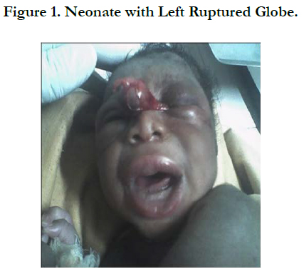
Globe Rupture in a Neonate Following Forceps Delivery
Théra JP1*, Théra B2
1 Department of Pediatric Ophthalmology, Institute of African Tropical Ophthalmology (IOTA), Bamako, BP, Africa.
2 General Practitioner, “Cabinet Médical Anna”, Bamako, Africa.
*Corresponding Author
Japhet Pobanou THERA,
Department of Pediatric Ophthalmology,
Institute of African Tropical Ophthalmology (IOTA),
Bamako, BP: 1560, Africa.
Tel: (00223) 76 43 88 10
E-mail: therajaphet@yahoo.fr
Received: September 30, 2016; Accepted: Decmber 22, 2016; Published: December 23, 2016
Citation: Théra JP, Théra B (2016) Globe Rupture in a Neonate Following Forceps Delivery. Int J Ophthalmol Eye Res. 4(11), 270-271. doi: dx.doi.org/10.19070/2332-290X-1600057
Copyright: Théra JP© 2016. This is an open-access article distributed under the terms of the Creative Commons Attribution License, which permits unrestricted use, distribution and reproduction in any medium, provided the original author and source are credited.
Abstract
Ocular injuries following forceps assisted delivery are rare in modern medicine. Forceps delivery can cause serious injuries to the neonate when used in an inappropriate way. The application of forceps by a physician who is not well qualified can end up with huge brain and ocular trauma. Commonly the injuries are secondary to the compression of the globe between the blade of the obstetric forceps and the orbit. We report a case of traumatic ruptured eyeball in a neonate following a forceps delivery.
2.Introduction
3.Materials and Methods
4.Result and Discussion
5.Conclusion
6.References
Keywords
Eyeball Rupture; Neonate; Forceps Delivery.
Introduction
Ocular injuries associated to birth trauma due to assisted vaginal delivery have been documented in the literature [1]. More than 700 types of forceps have been described [2].
Application and traction differ according to the type of instrument and require extensive training and knowledge of obstetric mechanics. Certain deliveries can be difficult and require careful evaluation informed by experience. If the fetus is not progressing after three pulls, this route of delivery can be abandoned [3]. Vision is one of the most valued and powerful senses. Intact binocular vision plays an important role in development, independence, quality of life, and personal safety. Ocular injury is a frequent and preventable cause of visual impairment [4]. In the case presented here, the blindness could have been prevented by performing caesarian section.
Materials and Methods
A male neonate weighting 4,125 grams was referred to our office for ocular trauma following forceps assisted delivery. He was the first born of a 16-year-old illiterate housewife. The pregnancy evolved normally. The indication of the forceps was a maternal exhaustion with a vertex presentation.
The forceps was used by an obstetrician. After the delivery, the patient was sent immediately to our office. After thorough exam using the hand held slit lamp, clinical findings were: ruptured left globe with prolapsed of ocular contents. The right eye was normal. The patient was prepared for exam under general anesthesia and to repair the injured eye. In the theatre, the left eye was prepped with 5% Povidone iodine and a sterile eye drape was placed. Exam under general anesthesia revealed a full thickness corneal laceration with traumatic evisceration. Thus we terminated the evisceration by removing the remaining cornea as well as the eye content. Interrupted sutures using 7.0 vicryl were used to close the sclera and the conjunctiva. Dexamethasone-Neomycin ointment was administered at the end of the procedure, then a sterile eye pad was applied. On hospital day one, the eye pad was renewed and the patient was discharged for a routine follow up.
Result and Discussion
A rupture is a full thickness injury of the tissue that makes up the external boundaries of the globe. Ocular structures that are vulnerable to rupture injury include the limbus, areas of sclera thinning just posterior to rectus muscle insertions, and previons surgical incision sites [4].
Forceps injury to the eye occurs during complicated forceps delivery. Inappropriate forceps application can cause serious injuries to the neonate like cuts, bruises and even death [5, 6]. In the current case, the forceps was used by an obstetrician with undoubtedly inappropriate application. Consequently the eyeball was ruptured with subsequent prolapsed of the left eye contents; this unilateral blindness compromises the future of the neonate.
If the fetus’s head needs to be rotated, there is now a tendency to use manual rotation, rotation with a vacuum extractor, or caesarean section in preference to kielland’s rotational forceps [7]. Ocular injuries following forceps delivery are not common worldwide; McAnena reported 11 cases of ophthalmic injuries secondary to forceps delivery; among them, two had oculomotor nerve palsy associated with intracranial hemorrhage both requiring surgical ptosis repair [8].
Forceps induced ocular lesions are among ocular injuries. Ocular injuries are the most common cause of acquired monocular blindness in children. In the case described here, the neonate was unilaterally blind.
Conclusion
Although ocular forceps related injuries are rare, they can damage the eye and its adnexa with a subsequent sight loss. All the practitioners who use obstetric forceps have to master the way they are used for a safe delivery. Newborns delivered by forceps should be referred to a pediatric ophthalmologist to rule out ocular trauma.
References
- Fasina O, Ugalahi MO, Olusanya BA (2015) Unilateral visual loss following assisted vaginal forceps delivery in a Nigerian neonate. Afr J Trauma. 4(2): 65-65.
- Okunwobi-Smith Y, Cooke I, Mackenzie IZ (2000) Decision to delivery intervals for assisted vaginal vertex delivery. Br J Obstetr Gynaecol. 107(4): 467-471.
- Feraud O (2008) Forceps: description, Obstetric Mecanics, indications and Contra-Indications. J Obstetr Gynecol. 37: S202-S209.
- Shane H, Omofolasade K, Millicent P (2009) Penetrating Eye Injury: A Case Study. Am J Clin Med. 6 (1): 42-49.
- Lesueur L, Arne JL, Mignon-Conte M, Malecaze F (1994) Structural and ultrastructural changes in the developmental process of premature infants’ and childrens’ corneas. Cornea 13: 331-338.
- O’Mahony F, Settatree R, Platt C, Johanson R (2005) Review of singleton fetal and neonatal death associated with cranial and cephalic delivery during a national intrapartum related confidential enquiry. BJOG. 112(5): 619-626.
- Roshi RP, Deirdre JM (2004) Forceps delivery in modern obstetric practice. BMJ. 328 (7451): 1302-1305.
- McAnena L, O’Keefe M, Kirwan C, Murphy J (2005) Forceps deliveryrelated ophthalmic injuries: A case series. J Pediatr Ophthalmol. Strabismus. 52(6): 355-359.
- Takvam JA, Midelfart A (1993) Survey of eye injuries in Norwegian children. Acta Ophthalmol. 71(4): 500-505.


