Evaluation of The Effectiveness of Using Calcium Sulfate Hemihydrate Graft for Socket Preservation after Teeth Extraction
Jihad Debes1, Mohamad Hassan Jaafo2, Mohey Aldeen Amam1, Kenan Saoud1, Safaa Shihabi3*
1 Department Oral & Maxillofacial Surgery, Faculty of Dentistry, University of Damascus, Syria.
2 Associate Professor in Department Oral & Maxillofacial Surgery, Faculty of Dentistry, University of Damascus, Syria.
3 Department of Pediatric Dentistry, Faculty of Dentistry, Damascus University, Syria.
*Corresponding Author
Safaa Shihabi,
Department of Pediatric Dentistry, Faculty of Dentistry, Damascus University, Syria.
E-mail: safaa2671991@gmail.com
Received: August 29, 2021; Accepted: October 22, 2021; Published: November 09, 2021
Citation: Jihad Debes, Mohamad Hassan Jaafo, Mohey Aldeen Amam, Kenan Saoud, Safaa Shihabi. Evaluation of The Effectiveness of Using Calcium Sulfate Hemihydrate Graft for Socket Preservation after Teeth Extraction. Int J Dentistry Oral Sci. 2021;8(10):4934-4938. doi: dx.doi.org/10.19070/2377-8075-21000997
Copyright: Safaa Shihabi�2021. This is an open-access article distributed under the terms of the Creative Commons Attribution License, which permits unrestricted use, distribution and reproduction in any medium, provided the original author and source are credited.
Abstract
Background: The protection of the socket absorption followed by teeth extraction is one of the most essential matters, as it
facilitates the next implantation process and gives better functional and cosmetic results. Therefore, many methods were used
to preserve the socket absorption, including bone grafts, which is the most available and less expensive material.
Aim of Study: to evaluate the effectiveness of calcium sulfate in reducing bone absorption
and preventing alveolitis after teeth extraction.
Materials & Methods: 24 teeth were extracted; 12 sockets were immediately grafted with Calcium Sulfate after extraction
however, 12 sockets were left to heal spontaneously without adding any material. In both groups, the horizontal bone absorption
of the crestal bone was evaluated 6 months after the extraction using CBCT technique. The occurrence of alveolitis was
evaluated clinically.
Results: Radiographic results after 6 months showed that the average of horizontal bone absorption in the study sample was
(0,575� 0.24) mm, and the absorption rate was (8.5%) while in the control group (without Bone graft) the average of horizontal
bone absorption was (1,483� 0.65) and the absorption rate was (22.7%). The incidence of alveolitis in the study group
was (0%), while it was 41.7% in the control group.
Conclusion: The application of calcium sulfate grafts after extractions reduces dimension changes in the alveolar bone at
the horizontal level, but doesn't prevent its occurrence. On the other hand, it can prevent the occurrence of post-extraction
Alveolitis.
2.Introduction
3.Materials and Methods
3.Results
4.Discussion
5.Conclusion
5.References
Keywords
Bone Grafts; Bone Resorption; Calcium Sulfate; CBCT; Socket Preservation.
Introduction
The alveolar socket can be defined as the bony tissue that surrounds
the whole erupted tooth, and it forms in conjunction with
the development and eruption of the teeth.[1]
After teeth extraction, a modeling and remodeling process occurred,
this process leads to many changes in the width and
height of the alveolar bone, the resorption of the buccal wall is
higher than the palatal and lingual wall. [2]
After extraction, the absorption emerges at 2 stages. In the first
stage the bundle bone is absorbed quickly and replaced by a premature
bony tissue that is soon replaced by a lamellar bone that
submerges the alveolar socket within 180 days. In the second
stage, the outer surface of the alveolar bone is remodeled through
the interaction between the osteoclast and osteoblast which leads
to decrease the dimension of vertical and horizontal alveolar
bone.[3]
Bone restoration is more efficient during the 5th and 10th week
after extraction, but still occurs until the fourth month.[4]
Many techniques were used to preserve the socket such as: Tissue
engineering approaches, Bio-Col or Resorbable Hemostatic
Plug Technique, Open Barrier Techniques, and Hyaluronic Acid
in alveolar ridge preservation, [5] Immediate dental implantation,
[6] and dental bone grafts (Autogenous bone grafts, Allografts, Synthetic and Alloplastic grafts, Xenografts).[7, 8]
Alveolitis is considered as One of the most common postoperative
complications followed by the extraction of permanent teeth.
Recent studies demonstrated that alveolitis occurs on 1-3 days up
to 1 week in 95-100% of reported cases. There are numerous reasons
that cause alveolitis including: traumatic extraction, roots or
bone fragments remaining in the wound, Physical dislodgement
of the clot, excessive irrigation or curettage of the alveolus after
extraction. [9]
The methods were suggested to prevent alveolitis were: systemic
and topical antibiotic�s, using chlorhexidine 0.12%, using sanitized
gloves, steroids. However, Treating alveolitis is misleading because
it is caused by various reasons. Some ways to treat alveolitis are:
prescribing antibiotics, systemic sedatives/pain killers, using ALVOGYL
which is the most common way of treating alveolitis.[10]
Calcium Sulfate is known as Plaster of Paris. It is an active biocompatible
compound which absorbed within 30-60 days. [8] Calcium
sulfate has many uses such as: correction of bone defects,
preservation of alveolar sockets, Sinus Augmentation, Guided
Tissue Regeneration. [11]
Cotzee et al pointed out that when calcium sulfate is used in
direct contact with bone or periosteum the bone regeneration
process was accelerated. A study by Gabriele et al also showed
full absorption of calcium sulfate and substitution with new bone
after sinus lift operation.[12]
Sicher and Weinman reported that the presence of calcium salts
was the primary motif that caused the differentiation of cells to
osteoblasts.[13]
Several studies have shown the easiness and reliability of using
Cone beam computed tomography (CBCT) in measurements of
bone dimension.[14]
Other studies reported the accuracy and reliability of the measurements
taken by CBCT scan.[15]
Materials And Methods
Study Design
A Split-Mouth Randomized Control Trial was accomplished in
Oral and Maxillofacial Surgery at Damascus University. The study
sample consisted of 24 alveolar sockets that are electively chosen
from 12 patients how have two teeth need to be extracted in the
upper jaw side.
Any patient doesn�t suffer from any systemic diseases and has
no contraindications for applying local anesthesia or minor oral
surgery, his age was between 20-50 years, doesn't have any personal
habits that could badly effect post extraction healing such
as smoking or drinking alcohol, and has a good oral hygiene was
included in this study.
Samples were divided into 2 groups
1st Group (Control Group): consists of 12 empty alveolar sockets
(post extraction) of 12 patients. the extraction was done without
adding any grafts and the healing occurred spontaneously.
2nd Group (Experimental Group): consists of 12 empty alveolar
sockets (post extraction) from the same patients in the 1st group,
but the sockets was filled with calcium sulfate without covering it
with any membrane.
The clinical Procedure: the X-ray and the medical history of the
patients were done, the patients were enrolled in the experiment
after taking their consent. The operational procedure is done as
follows:
� After sanitizing the mouth with chlorhexidine 0.12 %, a local
anesthetic (buccal and palatal) was done by lidocaine 2% with epinephrine
1/80.000.
� The periodontal ligament of the tooth was cut using blade # 15.
� The teeth were extracted by small elevators and suitable forceps
where the surgical trauma of the soft and hard tissues was as small
as possible.
� Confirmation of the absence of any bony fragments in the alveolar
socket was done by bone excavators then the cleaning the
of alveolar cavity was done by a physiological serum in the control
group.
� In the experimental group the bone graft (calcium sulfate) DentoGen
from ORTHOGEN� was applied applied in the alveolar
socket according to manufacturer's instructions.as follows: the
socket was dried and the graft was mixed (powder and liquid),
(Waiting 2-4 minutes is required to make sure the graft is dry then
applying the moisturized gauze with the liquid enclosed with the
graft to achieve the perfect hardness according to the recommendations
of the manufacturing company.
A CBCT 3D X-ray was done in the next day after the surgery (T1)
and another one after 6 months (T2) using the same device with
specific standards and settings. The photos were compared by
using a single methodology where 2 levels were determined. The
first one in the axial section that passes through 2 fixed points
[ANS (anterior nasal spine) and PNS (posterior nasal spine)]. The
second level is in the coronal section that passes tangentially with
the lower edge of hard palate. (Figure 1)
The width of the alveolar socket crest was measured in both levels
immediately AND after 6 months of extraction in both control
and experimental groups as shown below in the figures 2 and 3.
Results
The research sample was consisted of 24 teeth extracted from 12
patients. 12 Alveolar sockets were grafted with calcium sulfate and
the others were left to heal without any. Then the bone absorption
of the horizontal crestal bone was evaluated after 6 months�
post extraction in both the control and the experimental groups
using CBCT. In addition, a clinical evaluation was performed to
the alveolar bone in both groups to detect if the alveolitis was
occurred.
Kolmogorov-Smirnov test was used to study the equality of the
natural distribution of the measurements studied in the sample
research. Based on the results of the Kolmogorov-Smirnov test
the type of the probability tests to be used in the current research
will be determined.
The radial mean for the measurement of the bone width post
extraction in the experimental group was 6.733 � 1.14, however;
it was 6.525 � 1.08 in the control group with p -value 0.651 (no
statistical differences)
The radial mean for the measurement of the bone width after 6
months in the control group was 6.158 � 1.10; however; it was
5.042 � 1.18 in the experimental group with a p � value 0.026 (less
than 0.05) indicating a statistical significance.
In order to study the differences in the width measurement changes
in both immediate post extraction and after 6 months in both
of the study groups, T student test was used for the independent
samples, and the results are shown in table (1)
According to the results in table (1), the value of the T-student
test - for independent samples to study the differences in the
width change immediately post extraction and after 6 months between
the experimental and the control group reached (4.467),
with p-value (0.000) less than the significance level (0.05) which
indicates a statistical significance in the amount of width change.
This statistical difference is favorable for the experimental group
because the mean of change of the width is (-0.575) less than the
mean of change for the control group (-1.483).
In order to study the changes in the alveolitis between the control
and the experimental group Chi- Square Test was used and the
result is shown in the following tables.
It is evident through table 3 that the Chi-Square value in the study
the difference in alveolitis between the experimental and control
groups is (6.316) with P- value (0.012) less than 0.05 indicating
a significant difference in the alveolitis in the two study groups.
This statistical difference is favorable for the experimental group
as there were no cases of alveolitis compared to 5 cases of alveolitis
in the control group.


Figure 1. Reference points for measurements.


Bar graph (1) Shows the difference between the two groups of the sample research in the amount of width change in both levels.


Bar Graph (2). Shows the reoccurrence of alveolitis in the control and study groups.

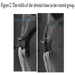
Figure 2. The width of the alveolar bone in the control group.


Figure 3. The width of the alveolar socket crest in the experimental group.


Table 1. T student test results showing the differences between the two samples regarding the amount change in the control and the experimental group.

Table 2. Percentage of the recurrence of alveolitis in the Control group and Experimental group.

Table 3. Chi Square Test result to study the difference in alveolitis between the experimental and control groups.
Discussion
The aim of our study was to evaluate the effectivness of Calcium
Sulfate graft in the prevention of alveolitis and decreasing horizontal
bone absorption 6 months after the extraction. Where the
research samples were divided into two groups, a control group
where the alveolar socket is left to heal spontaneously after extraction
without adding any substance, and an experimental group
where Calcium Sulfate graft is inserted in the cavity post extraction
without any covering membrane or any other substance and
studying the changes after 6 months.
Shilpa Budhiraja and his colleagues noted that using calcium sulfate
as a membrane would mimic the use of a collagen membrane
in getting clinical benefits, hence; it can be used as an economical
substitute instead the collagen membrane. He also noted that it
has an absorbing and porous nature and it omits the need for
suturing after being applied. [16]
After 6 months, radial results have shown that the average amount
of horizontal absorption (mm) in the experimental group was
0.575 � 0.24 with an absorption rate 8.5% whereas in the control
group (no graft) it was 1.483 � 0.65 with an absorption rate 22.7
%. As for the level of alveolitis the percentage of alveolitis occurrence
in the experimental group was 0% whereas in the control
group it was 41.7%.
These results can be attributed to the reasons related to the characteristics
of calcium sulfate.
Chia Wei Cheah pointed out that due to its high porous structure
it will not interfere with the development of new blood vessels
during the healing process, in addition; it acts as a membrane to
prevent epithelial down growth into the socket. [17]
Our study was consistent with Cheah�s study (2014) in that using
calcium sulfate or calcium sulfate + platelet rich plasma gave effective
results in the preservation of the alveolar socket.
We disagreed with Cheah�s study that indicated that the absorption
of the horizontal bone between the post extraction phase
and after 4 months was 18.6% in the group that was grafted with
calcium sulfate only whereas it was 9.2% in the group grafted with
calcium sulfate + platelet rich plasma. This difference could be
explained due to two reasons. The first related to the procedure
(Minimal flap reflection was performed on buccal and palatal or
lingual surfaces to expose bone margin of the tooth to be extracted)
unlike our procedure where flapless extraction was done.
The second is the difference in the follow up timing where in the
Cheah study it was 4 months and ours was 6 months. [17]
We agreed with Reza Amirzagrar�s study indicating that calcium
sulfate grafts gave good results in socket preservation and that
there is no difference between it and Freeze Dried Bone Allograft (FDBA). [18]
Our study was consistent with Aimetti�s study and his colleagues
that indicated that grafting the alveolar socket with calcium sulfate
reduced the absorption of the horizontal bone. [19]
We agreed with Samira M. Toloue�s study and her colleagues who
indicated that the efficiency of calcium sulfate grafts matched that
of FBDA in preserving the dimension of the bone dimensions
after extraction. [20]
We disagreed with Barone�s study regarding the preservation of
the alveolar socket using a Xenograft as opposed to natural healing.
His study showed an average horizontal drop in the width of
the alveolar socket in the control groups of 4.3 � 0.8 mm and 2.5
� 1.2 mm in the experimental group after a 6 months follow up
period which are values that exceed the values of our study. This
is due to the differences in the evaluation methods that were clinical
and histological evaluation. Also his study included teeth with
multi roots and use of a different kind of graft. [21]
Conclusions
According to the results of our study we can conclude that Calcium
Sulfate is feasible and safe to use in grafting operations,
its characteristics allowed us to dispense the use of membranes
avoiding its complications. Calcium sulfate can decreasing horizontal
bone absorption after dental extraction and it was effective
in reducing the occurrence of Alveolitis.
Suggestions and Recommendations
Performing broader and longitudinal studies regarding the use of
calcium sulfate grafts in preserving the alveolar socket as well as
monitoring and evaluating implants in alveolar bones grafted using
similar grafts on the long term.
Performing studies later on to find other ways to decrease the
absorption of the used bone grafts and monitoring the cases long
term.
It is recommended to use the calcium sulfate according to the
instructions of the manufacturing company to get the best results.
Perform histological studies to identify the rate of natural bone
formation followed by the application of the bone graft.
References
-
[1]. Ara�jo MG, Silva CO, Misawa M, Sukekava F. Alveolar socket healing: what
can we learn? Periodontology 2000. 2015;68(1):122-34.Pubmed PMID:
25867983.
[2]. Discepoli N, Vignoletti F, Laino L, De Sanctis M, Mu�oz F, Sanz M. Early healing of the alveolar process after tooth extraction: an experimental study in the beagle dog. Journal of clinical periodontology. 2013;40(6):638-44. Pubmed PMID: 23534915.
[3]. Jamjoom A, Cohen RE. Grafts for Ridge Preservation. Journal of functional biomaterials. 2015;6(3):833-48. Pubmed PMID: 26262646.
[4]. Chen ST, Wilson TG, Jr., Hammerle CH. Immediate or early placement of implants following tooth extraction: review of biologic basis, clinical procedures, and outcomes. The International journal of oral & maxillofacial implants. 2004;19 Suppl:12-25. Pubmed PMID: 15635942./
[5]. Praphul VP SKS. Novel Approaches for Alveolar Ridge Preservation. Int J Rec Innov Med Clin Res. 2020;2(2):32-41.
[6]. Araujo MG, Wennstr�m JL, Lindhe J. Modeling of the buccal and lingual bone walls of fresh extraction sites following implant installation. Clinical oral implants research. 2006;17(6):606-14. Pubmed PMID: 17092217.
[7]. Irinakis T, Tabesh M. Preserving the socket dimensions with bone grafting in single sites: an esthetic surgical approach when planning delayed implant placement. Journal of Oral Implantology. 2007;33(3):156-63. Pubmed PMID: 17674682.
[8]. Kumar P, Vinitha B, Fathima G. Bone grafts in dentistry. Journal of pharmacy & bioallied sciences. 2013;5(Suppl 1):S125. Pubmed PMID: 23946565.
[9]. Blum I. Contemporary views on dry socket (alveolar osteitis): a clinical appraisal of standardization, aetiopathogenesis and management: a critical review. International journal of oral and maxillofacial surgery. 2002;31(3):309- 17. Pubmed PMID: 12190139.
[10]. Kolokythas A, Olech E, Miloro M. Alveolar osteitis: a comprehensive review of concepts and controversies. International journal of dentistry. 2010;2010. Pubmed PMID: 20652078.
[11]. Thomas MV, Puleo DA. Calcium sulfate: Properties and clinical applications. Journal of Biomedical Materials Research Part B: Applied Biomaterials: An Official Journal of The Society for Biomaterials, The Japanese Society for Biomaterials, and The Australian Society for Biomaterials and the Korean Society for Biomaterials. 2009;88(2):597-610. Pubmed PMID: 19025981.
[12]. Pecora GE, De Leonardis D, Della Rocca C, Cornelini R, Cortesini C. Short-term healing following the use of calcium sulfate as a grafting material for sinus augmentation: a clinical report. International Journal of Oral and Maxillofacial Implants. 1998;13(6):866-74. Pubmed PMID: 9857600.
[13]. Shaffer CD, App GR. The use of plaster of paris in treating infrabony periodontal defects in humans. Journal of periodontology. 1971;42(11):685-90. Pubmed PMID: 5288563.
[14]. Ludlow JB, Gubler M, Cevidanes L, Mol A. Precision of cephalometric landmark identification: cone-beam computed tomography vs conventional cephalometric views. American Journal of Orthodontics and Dentofacial Orthopedics. 2009;136(3):312. e1-. e10. Pubmed PMID: 19732656.
[15]. Damstra J, Fourie Z, Slater JJH, Ren Y. Accuracy of linear measurements from cone-beam computed tomography-derived surface models of different voxel sizes. American Journal of Orthodontics and Dentofacial Orthopedics. 2010;137(1):16. e1-. e6. Pubmed PMID: 20122425.
[16]. Budhiraja S, Bhavsar N, Kumar S, Desai K, Duseja S. Evaluation of calcium sulphate barrier to collagen membrane in intrabony defects. Journal of periodontal & implant science. 2012;42(6):237. Pubmed PMID: 23346468.
[17]. Cheah CW, Vaithilingam RD, Siar CH, Swaminathan D, Hornbuckle GC. Histologic, histomorphometric, and cone-beam computerized tomography analyses of calcium sulfate and platelet-rich plasma in socket preservation: A pilot study. Implant dentistry. 2014;23(5):593-601.Pubmed PMID: 25192162.
[18]. Amirzargar R, Shirani G, Mahmoudhashemi H, Khoshzaban A, Hasheminejad M. A comparative study of ridge preservation using calcium sulfate and collagen membrane with and without freeze-dried bone allograft following tooth extraction. Journal of Osseointegration. 2018;10(1):11-6.
[19]. Aimetti M, Romano F, Griga FB, Godio L. Clinical and histologic healing of human extraction sockets filled with calcium sulfate. International Journal of Oral & Maxillofacial Implants. 2009 Oct 1;24(5).
[20]. Toloue SM, Chesnoiu-Matei I, Blanchard SB. A clinical and histomorphometric study of calcium sulfate compared with freeze-dried bone allograft for alveolar ridge preservation. Journal of periodontology. 2012;83(7):847-55.
[21]. Barone A, Aldini NN, Fini M, Giardino R, Calvo Guirado JL, Covani U. Xenograft versus extraction alone for ridge preservation after tooth removal: a clinical and histomorphometric study. Journal of periodontology. 2008 Aug;79(8):1370-7.


