Microimplant-Assisted Rapid Palatal Expansion (MARPE) - A Comprehensive Review
Tamanna Hoque1*, Dilip Srinivasan2, Sangeetha Morekonda Gnaneswar3, Sushil Chakravarthi4, Krishnaraj Rajaram5, Ravi Kannan6
1 Post-Graduate Student, Department of Orthodontics and Dentofacial Orthopaedics, SRM Dental College, Ramapuram, Chennai, India.
2 Department of Orthodontics and Dentofacial Orthopaedics, SRM Dental College, Ramapuram, Chennai, India.
3 Senior lecturer, Department of Orthodontics and Dentofacial Orthopaedics, SRM Dental College, Ramapuram, Chennai, India.
4 Reader, Department of Orthodontics and Dentofacial Orthopaedics, SRM Dental College, Ramapuram, Chennai, India.
5 Professor, Department of Orthodontics and Dentofacial Orthopaedics, SRM Dental College, Ramapuram, Chennai, India.
6 Professor & head, Department of Orthodontics and Dentofacial Orthopaedics, and Dean, SRM Dental College, Ramapuram, Chennai, India.
*Corresponding Author
Tamanna Hoque,
Post-Graduate Student, Department of Orthodontics and Dentofacial Orthopaedics, SRM Dental College, Ramapuram, Chennai, India.
Tel: 919051646823
Fax: 044 - 2249 0526
E-mail: tamannadenthealth@gmail.com
Received: April 28, 2021; Accepted: October 20, 2021; Published: October 26, 2021
Citation:Tamanna Hoque, Dilip Srinivasan, Sangeetha Morekonda Gnaneswar, Sushil Chakravarthi, Krishnaraj Rajaram, Ravi Kannan. Microimplant-Assisted Rapid Palatal Expansion (MARPE) - A Comprehensive Review. Int J Dentistry Oral Sci. 2021;8(10):4848-4852. doi: dx.doi.org/10.19070/2377-8075-21000980
Copyright: Tamanna Hoque�2021. This is an open-access article distributed under the terms of the Creative Commons Attribution License, which permits unrestricted use, distribution and reproduction in any medium, provided the original author and source are credited.
Abstract
Maxillary transverse deficiency routinely requires expansion of the palate. In growing patients, well-documented expansion modalities are slow maxillary expansion (SME) and rapid palatal expansion (RME). However, in mature patients due to the complexity of interdigitation of midpalatal suture and decreased elasticity of bone, palatal expansion is challenging. Patients are frequently suggested to go for a more invasive procedure, like the Surgically Assisted Rapid Palatal Expansion(SARPE) expansion. More recently, with the emergence of implants, researchers havevalidated that it is possible to expand the maxilla in adult patients without carrying out osteotomies.This comprehensive review provides fundamental information, different designs,recent updates, surgical guides, clinical significance and limitations ofMicroimplant-Assisted Rapid Palatal Expansion(MARPE), which has become a generic term for the maxillary expansion appliance which transmits expansion forces to basal bones by a miniscrew anchorage system.MARPE represents a valid alternative to surgery in treating mature patients with a transverse maxillary deficiency with greater stability, safety, and fewer side effects.
2.Introduction
3.Materials and Methods
3.Results
4.Discussion
5.Conclusion
5.References
Keywords
Maxillary Expansion; Microimplant-Assisted Rapid Palatal Expansion (MARPE); Maxillary Skeletal Expander
(MSE); Maxillary Transverse Deficiency; Rapid Maxillary Expansion.
Introduction
A genomic biomarker is a measurement of the expression, function
aMaxillary transverse deficiency is one of the most pervasive
problems in the craniofacial region prevalent in all age groups,
from deciduous to permanent dentition [1]. It has been reported
that 9.4% of the entire population and nearly 30% of the adult
orthodontic patients have a maxillary transverse deficiency [1].
However, some reported that the prevalence of maxillary transverse
deficiency ranges from 8% to 23% in mixed and deciduous
dentitions and less than 10% in adults [2]. Maxillary transverse
deficiency has multifactorial etiology and some of the most prevalent
factors are narrow palatal dimensions, inheritance, ectopic
eruption, impaired maxillary transverse growth associated with
a palatal cleft and breathing disordersand soft tissue imbalance
like prolonged digit sucking, lower tongue position [3]. When the
maxilla and mandible fail to properly orient in the transverse dimension,
odontogenesis continues its processand teeth eruption
inabnormal positions leading to malocclusion [4, 5]. If maxilla
mandibular transverse discrepancies are not treated in an appropriate
time, they can aggravate and metamorphose into more
complex malocclusion, hindering facial growth and development
[6]. Maxillary transverse deficiency impacts the occlusion not only
in the transverse plane but also in the vertical and sagittal planes
leading to intricate situations, such as posterior unilateral or bilateral
crossbites, crowding, scissor bite, non-carious cervical wear,
adverse periodontal stress, low masticatory ability, functional shift
of the mandible, faulty buccolingual tipping of posterior teeth,
asymmetric mandibular position in growing patients, joint disorders and muscle function disharmony. However, the grave consequence
of maxillary transverse deficiency is the narrowing of the
nasal cavity, which increases nasal air resistance and might become
an etiologic factor of Obstructive Sleep Apnea Syndrome (OSAS)
[6, 7]. In Class III malocclusions nearly half of the patients have
maxillary skeletal retrusion, which contributes to transverse discrepancies
between the maxilla and mandible [8]. Dental crowding
and posterior crossbite are two easily recognizable clinical
features of transverse deficiency, while exaggerated buccal flaring
of the maxillary dentition and deep Curve of Wilson in the lower
dentition can mask the maxillary transverse constriction [7].
Traditionally, to correct transverse maxillary deficiencies RME
(Rapid Maxillary Expansion) and SME (Slow Maxillary Expansion)
appliances have been effectively used for years, despite certain
negative side effects like undesirable tooth tipping, limited
skeletal movement, root resorption, bone dehiscence, thinning
of the buccal cortical bone and relapse. For Adults due to the
complexity of interdigitation of midpalatal suture and decreased
elasticity of bonealternative methods like Surgically assisted RPE
(SARPE) which increases expansion possibilities, long term stability
and success, with reduced side effects, have been developed.
Despite its benefits, the procedure has its impediment of surgical
morbidity, high cost, periodontal complications [6]. With the
advent of orthodontic mini-implants, the possibilities for pure
orthopedic movement in the expansion of maxilla with RME
are explored around the world. This novel system, called Microimplant-
Assisted Rapid Palatal Expansion (MARPE), transmits
expansion forces to basal bones by a miniscrew anchorage system.
Theterm MARPE became a generic term, although designs
and activation protocols differed greatlywith different appliance
models. Due to different expansion force vectors and magnitude,
different dimensions of the implants, widely varying in anchor
location and the relative position of jackscrew to the skeletal anchor,
different designs yield varying results, often in contradiction
to each other [9]. Some MARPEs are tooth-bone-anchored or
hybrid and others are purely bone-borne [10].
Review Result
The treatment envelope of maxillary transverse deficiency has
been broadened to treat adult patients without surgery with
MARPE [11] (Fig.1).


Figure 1.


Figure 2.


Figure 3.


Figure 4.

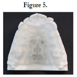
Figure 5.


Table 1.
Discussion
The midpalatal suture changes with age
Mid palatal sutural studies by Melsen [12] Isaacson et al [13] have
revealed a relationship between the increased interdigitation of
the midpalatal suture with the age of the subjects in hindering
maxillary separation. They also emphasized that the maximum
resistance is not due to the midpalatal suture but by the surrounding
maxillary articulation.Bishara and Staley [14] suggest that the
resistance to mid-palatal suture openingwas noticed at the sphenoid
and zygomatic bones, particularly at the superior parts of the
pterygoid plates of the sphenoid bone, and anterior part of the
zygomatic bone. Wertz [15] reported that the fulcrum of maxillary
separation tends to be displaced more inferiorly, nearer to the
activating force with an increase in age. The fulcrum may be high
near to frontomaxillary suture in children, whereas in adolescents
the fulcrum is much lower. These variances in age-dependent effects
may be due to the increased resistance in circum-maxillary
sutures during maxillary separation.
Fernanda Angelieri et al [16] studied the Cone-beam computed
tomography images of 140 subjects. They divided the Mid Palatal
Suture into five stages of maturation and defined them as:
1. Stage A- straight high-density sutural line, with no or little interdigitation.
2. Stage B- the scalloped appearance of the high-density sutural
line.
3. Stage C- two parallel, scalloped, high-density lines that were
close to each other, separated in some areas by small low-density
spaces.
4. Stage D- fusion completed in the palatine bone, with maturation
progressing from posterior to anterior.
5. Stage E- the fusion of the midpalatal suture has occurred in
the maxilla.
The study concluded that at stage C, a less skeletal response would
be expected than at stages A and B with the conventional RME
approach. For patients at stages D and E, surgically assisted RME
would be necessary.
Conversely, Wehrbein et al [17] emphasized that the term �suture
fusion� should be avoided in terms of radiologic terminology as
they found that a radiologically invisible mid-palatal suture is not
the histological equivalent of a fused or closed suture after analyzing
the palatal suture status of young adults ranging from 18 to
38 years of age.
Different designs of bone-borne palatal expanders using
micro-implants [1]
Type 1: miniscrews placed lateral to midpalatal suture
Type 2: miniscrews placed at the palatal slope
Type 3: miniscrews as in type 1 but with additional conventional
Hyrax arms
MARPE Design
Lee et al [18] used a Hyrax screw with an orthodontic miniscrew
also called a hybrid expander (Fig.2), two miniscrews placed anteriorly
in the palatal rugae and two posteriorly in the parasagittal
area. The activation protocol followed was one-quarter of a turn
(0.2 mm) once a day, with a total activation period of 40 days and
a 3-month retention period thereby successfully treated severe
transverse discrepancy 20-years-old patient.
Cunha et al [19] suggested that the position of posterior miniscrews
in MARPE may have a crucial role in providing adequate
stress distribution, favoring the complete disjunction of the midpalatal
suture with type I palatal split pattern (Fig.3).
Lim et al [20] suggested that the expansion effects of MARPE
were not limited to the maxilla only but also extended to the circummaxillary
structures and that the maxillary halves showed
buccal rotation, with the rotational center located near the frontonasal
suture. MacGinnis et al [2] used the finite element method
(FEM) to demonstrate that by changing the location of the expansion
screw, the stress distribution on the craniofacial complex
is altered. Likewise, placing the jackscrew closer to the center of
resistance, a more horizontal translation of the maxilla takes place
with less resultant buccal tipping. They also concluded that MARPE
propagates less stress to the buttresses and adjacent locations
in the maxillary complex compared to the conventional RPE.
MSE (Maxillary Skeletal Expander) developed by Dr. Won
moon[9]is a unique lineage of MARPE. It causes expansion of
the entire midface, agitating all peri-maxillary structures. When
MSE is applied in combination with FM (face mask), almost negligible
vertical side effects are detected, the existing anteroposterior
dental compensation can be reversed, the maxilla advances
efficiently in large magnitude, and resulted in some skeletal protraction
even in mature patients. This combination simulates
distraction-like movement, which forms a promising basis for
non-surgical orthopedic treatment modality for Class III adult patients.
The unique position of miniscrew in MSE (Fig.4) in the superior
and posterior aspect of the palate with four long implants
engaging the palatal bone bicortically gives a significant advantage
in overcoming the resistance from zygomatic buttress bones and
pterygopalatine sutures, possibly leading to a more parallel expansion
in contrast to many other designs of MARPE.
Carlson et al [21] suggested that the size of the jackscrew must be
chosen based on the maximum screw size that would adequately
fit in the palatal vault, concurrently allowing close adaptation of
the appliance to the tissue surface between the maxillary first molars.
This position exerts lateral forces against the pterygomaxillary
buttress of the bone, which is a major resistance factor in
maxillary expansion. The expansion rate (Table 1) was selected
based on the protocol developed by Dr. Won Moon through clinical
experience with the MARPE appliance [21].
Clement. A and Krishnaswamy N. R.[22] concluded that MSE
used in young adults produced 61% of the degree of expansion at
skeletal level, 20%alveolar, and 19% dental expansion. Cantarella
D et al [23] evaluated midfacial skeletal changes in the coronal
plane in late adolescent patients treated with a bone-anchored
maxillary expander using CBCT and found significant lateral
displacement of the zygomaticomaxillary complex and outward
rotation of zygomatic bone along with the maxilla with a common
center of rotation located near the superior aspect of the
frontozygomatic suture which ultimately leads to negligible dental
tipping of the molars. Cantarella D et al [24] study, revealed that
the opening of the mid-palatal suture in the anterior region was
4.8mm and at the posterior nasal spine was about 4.3mm and the
percentage of the mid-palatal split in the PNS was 90% that of
ANS, showing near the parallel opening.
Selection of mini-implants and site of placement
Nojima et al [25] suggested the following steps to select the length
of miniscrews to be used in the MARPE: 1. Procurement of dental casts, 2. Selection of DICOM visualization software and maxilla
orientation in CBCT images. 3. Measurement of bone thickness
on the coronal section of CBCT images. 4. Evaluation of
expander miniscrews fixation rings. 5.Selection of miniscrew. The
total length of the miniscrew (MI) is represented by the variables:
bone thickness (o), adding 1.0 to 2.0 mm which is necessary for
the miniscrew tip to surpass the cortical plate of the nasal fossa,
soft tissue thickness (m), fixation ring thickness (a), distance from
the ring to the palatal surface (d). The equation employed to calculate
the total miniscrew length is described, with the value in millimeters,
as MI= o + m + a + d + (1 or 2). Lee et al [26] suggested
the use of bicortical (cortical bone of palate and nasal floor) miniimplant
anchorage over monocortical anchorage to enhance miniimplant
stability, mitigate mini-implant deformation and fracture,
more parallel expansion in the coronal plane, and increased expansion
during bone-borne palatal expansion. Peri-implant stress
was preeminent in the monocortical anchorage model compared
with both bicortical anchorage models. Wilmes B et al [27] found
that the area immediately posterior to the palatal rugae, and the
paramedian area referred to as the "T-Zone", is amore suitable
region for insertion of palatal mini-implants due to the available
bone volume and bone is much thinner in posterior and lateral
areas. Lombardo et al [28] FEM study demonstrated that a miniscrew
of diameter 2 mm and length 11 mm inserted into the
palate can withstand loads between 240 and 480gf (gram force),
without causing a fracture to the bone, even in the absence of
osseointegration.
Surgical guide for MARPE
A surgical guide is an essential tool for correctly placing implants,
which aims to achieve a perfect interrelation of digital planning
and actual placement. It allows the three-dimensional orientation
of the expander close to the palate and guides perforations of
mini-implants, which is required to establish anchorage in areas
with adequate bone, assuring the system stability and a successful
outcome.Bruno L Minervino et al [29] suggested two fundamental
aspects concerning planning for the placement of MARPE.
Firstly, suture evaluation by CBCT to assess the possibility of
expansion secondly three-dimensional positioning of both expander
and mini-implants to assure insertion in an area with bone
support. Intraoral scanning of the maxilla is required followed
by superimposed to the computed tomography, using points at
the teeth region as a reference which allows determination of the
correct position between intraoral scanning and the tomography.
After this initial merging, the third digital file, namely the stereography
of the MARPE expander, is also merged. Finally, the expander
and four mini-implants are positioned using the software.
Miniscrew Assisted Palatal Appliance (MAPA) system protocol
� Maino G et al [30] introduced a new high-precision 3D Method
of the palatal miniscrew placement technique to prevent damage
to the anatomical structures. This template can ensure not only
that mini-implants are placed at the correct depth inthe maxillary
bone but also that multiple implants are parallelly placed. The
use of CBCT is strictly recommended in all cases of impacted
canines, laterally displaced lateral incisors, narrow maxilla, or anatomic
abnormalities that may affect the correct insertion of the
mini-implants. MAPA (Fig. 5) is designed to recreate the angle of
insertion and prevent the mini-implants from penetrating beyond
the required depth. Therefore, the 3D technological processes
assure efficient, accurate, and predictable orthodontic planning,
since they standardize the technique and reduce the risks.
Clinical Significance
Advantages Of Marpe
� MSE appliances transmit expansion force into the palatine bone
and produced a more parallel-type and more consistent suture
opening upon maxillary expansion. Widening of surrounding
craniofacial structures including the zygoma and the nasal bone
[2].
� Larger transverse skeletal expansion while lessening dental side
effects such as dental tipping, vertical alveolar bone loss, and alveolar
bending [1, 2].
� MARPE allows better vertical control, therefore, is also beneficial
in young dolichofacial patients [2].
� MARPE surpasses conventional RME by a significantly decreasing
excessive load on the buccal periodontal ligament of teeth to
which they are anchored [1].
� Bone-anchored maxillary expansion is superior to the conventional
RPE for OSA (obstructive sleep apnea) patients. For a postadolescent
OSA patient with Class II hyperdivergent pattern and
maxillary constriction, MARPE can be useful. MSE appliances reduce
upper airway resistance and increase intranasal capacity [31].
� BAME (bone-anchored maxillary expansion) allows full bonded
orthodontic therapy at the same time as the expansion. This could
shorten the total treatment time.
� A combination of MSE and Face mask can be a successful nonsurgical
orthopedic treatment modality for Class III adult patients
[24].
� MARPE results in greater stability, reduced relapse [29].
� Choi et al [32] and Park et al. [33] reported a success rate for
MARPE as 86.96% and 84.2% respectively.
Limitations Of MARPE
� The most frequent complication is the inflammation and hyperplasia
of the mucosa around the mini-implant [6].
� In the tooth-bone-anchored design of MARPE appliance, a significant
amount of dental tipping was reported in few studies due
to the thickness of the connecting arms which is soldered to the
molar bands [34].
� Unilateral expansion is not feasible in basic MARPE design,
modifications are required [35].
� Reduced or absent bone thickness, contraindicates MARPE
placement [29].
� Appliances present restricted to use with extreme maxillary atresia
or palatal asymmetry [25].
� Systemic conditions like type II diabetes and habits like smoking
should be carefully assessed and might contra-indicate the therapy
[6].
Conclusion
MARPE represents a valid alternative to surgery in treating patients
with a transverse maxillary deficiency with greater stability,
safety, and fewer side effects.
References
-
[1]. Di Luzio C, Bellisario A, Squillace F, Favale M, Caputo M. Miniscrew-Assisted
Rapid Palatal Expander (Marpe): A Efficient Alternative Treatment of
axillary Transverse Deficiency.
[2]. MacGinnis M, Chu H, Youssef G, Wu KW, Machado AW, Moon W. The effects of micro-implant assisted rapid palatal expansion (MARPE) on the nasomaxillary complex--a finite element method (FEM) analysis. Prog Orthod. 2014 Aug 29;15(1):52. Pubmed PMID: 25242527.
[3]. Southard TE, Marshall SD, Allareddy V, Shin K. Adult transverse diagnosis and treatment: A case-based review. InSeminars in Orthodontics 2019 Mar 1 (Vol. 25, No. 1, pp. 69-108). WB Saunders.
[4]. Nanda R, Snodell SF, Bollu P. Transverse growth of maxilla and mandible. InSeminars in Orthodontics 2012 Jun 1 (Vol. 18, No. 2, pp. 100-117). WB Saunders.
[5]. MULETT V�SQUEZ JA, CLAVIJO ESCOBAR AF, FUENTES LOYO IS, S�NCHEZ CANO PA. Correlation between transverse maxillary discrepancy and the inclination of first permanent molars. a pilot study. Revista Facultad de Odontolog�a Universidad de Antioquia. 2017 Jun;28(2):354-73.
[6]. Brunetto DP, Sant'Anna EF, Machado AW, Moon W. Non-surgical treatment of transverse deficiency in adults using Microimplant-assisted Rapid Palatal Expansion (MARPE). Dental Press J Orthod. 2017 Feb;22(1):110- 125. Pubmed PMID: 28444019.
[7]. N. R. Krishnaswamy, �APOS Trends in Orthodontics Expansion in the absence of crossbite � rationale and protocol,� APOS Trends Orthod., vol. 9, no. 3, pp. 126�137, 2019.
[8]. Koo YJ, Choi SH, Keum BT, Yu HS, Hwang CJ, Melsen B, et al. Maxillomandibular arch width differences at estimated centers of resistance: Comparison between normal occlusion and skeletal Class III malocclusion. Korean J Orthod. 2017 May;47(3):167-175. Pubmed PMID: 28523243.
[9]. Moon W. Class III treatment by combining facemask (FM) and maxillary skeletal expander (MSE). InSeminars in Orthodontics 2018 Mar 1 (Vol. 24, No. 1, pp. 95-107). WB Saunders.
[10]. Oh H, Park J, Lagravere-Vich MO. Comparison of traditional RPE with two types of micro-implant assisted RPE: CBCT study. InSeminars in Orthodontics 2019 Mar 1 (Vol. 25, No. 1, pp. 60-68). WB Saunders.
[11]. J. Robert L. Vanarsdall, Jr., Ignacio Blasi and and A. G. Secchi, �Periodontal- Orthodontic Interrelationships,� in Orthodontics: Current Principles and Techniques Edition: 6 thChapter: 22, 2017, pp. 621�668.
[12]. Melsen B. Palatal growth studied on human autopsy material. A histologic microradiographic study. Am J Orthod. 1975 Jul;68(1):42-54. Pubmed PMID: 1056143.
[13]. ZIMRING JF, ISAACSON RJ. FORCES PRODUCED BY RAPID MAXILLARY EXPANSION. 3. FORCES PRESENT DURING RETENTION. Angle Orthod. 1965 Jul;35:178-86. Pubmed PMID: 14331018.
[14]. Bishara SE, Staley RN. Maxillary expansion: clinical implications. Am J Orthod Dentofacial Orthop. 1987 Jan;91(1):3-14. Pubmed PMID: 3541577.
[15]. Wertz RA. Skeletal and dental changes accompanying rapid midpalatal suture opening. Am J Orthod. 1970 Jul;58(1):41-66. Pubmed PMID: 5269181.
[16]. Angelieri F, Cevidanes LH, Franchi L, Gon�alves JR, Benavides E, McNamara JA Jr. Midpalatal suture maturation: classification method for individual assessment before rapid maxillary expansion. Am J Orthod Dentofacial Orthop. 2013 Nov;144(5):759-69. Pubmed PMID: 24182592.
[17]. Wehrbein H, Yildizhan F. The mid-palatal suture in young adults. A radiological- histological investigation. Eur J Orthod. 2001 Apr;23(2):105-14. Pubmed PMID: 11398548.
[18]. Lee KJ, Park YC, Park JY, Hwang WS. Miniscrew-assisted nonsurgical palatal expansion before orthognathic surgery for a patient with severe mandibular prognathism. Am J Orthod Dentofacial Orthop. 2010 Jun;137(6):830-9. Pubmed PMID: 20685540.
[19]. Cunha ACD, Lee H, Nojima LI, Nojima MDCG, Lee KJ. Miniscrew-assisted rapid palatal expansion for managing arch perimeter in an adult patient. Dental Press J Orthod. 2017 May-Jun;22(3):97-108. Pubmed PMID: 28746493.
[20]. Lim HM, Park YC, Lee KJ, Kim KH, Choi YJ. Stability of dental, alveolar, and skeletal changes after miniscrew-assisted rapid palatal expansion. Korean J Orthod. 2017 Sep;47(5):313-322. Pubmed PMID: 28861393.
[21]. Carlson C, Sung J, McComb RW, Machado AW, Moon W. Microimplantassisted rapid palatal expansion appliance to orthopedically correct transverse maxillary deficiency in an adult. Am J Orthod Dentofacial Orthop. 2016 May;149(5):716-28. Pubmed PMID: 27131254.
[22]. Clement E, Krishnaswamy N. Skeletal and dentoalveolar changes after skeletal anchorage-assisted rapid palatal expansion in young adults: a cone beam computed tomography study. APOS Trends in Orthodontics. 2017 May 1;7(3):113-.
[23]. Cantarella D, Dominguez-Mompell R, Moschik C, Mallya SM, Pan HC, Alkahtani MR, et al. Midfacial changes in the coronal plane induced by microimplant-supported skeletal expander, studied with cone-beam computed tomography images. Am J Orthod Dentofacial Orthop. 2018 Sep;154(3):337-345. Pubmed PMID: 30173836.
[24]. Cantarella D, Dominguez-Mompell R, Mallya SM, Moschik C, Pan HC, Miller J, et al. Changes in the midpalatal and pterygopalatine sutures induced by micro-implant-supported skeletal expander, analyzed with a novel 3D method based on CBCT imaging. Prog Orthod. 2017 Nov 1;18(1):34. Pubmed PMID: 29090368.
[25]. Nojima LI, Nojima MDCG, Cunha ACD, Guss NO, Sant'Anna EF. Miniimplant selection protocol applied to MARPE. Dental Press J Orthod. 2018 Sep-Oct;23(5):93-101. Pubmed PMID: 30427498.
[26]. Lee RJ, Moon W, Hong C. Effects of monocortical and bicortical miniimplant anchorage on bone-borne palatal expansion using finite element analysis. Am J Orthod Dentofacial Orthop. 2017 May;151(5):887-897. Pubmed PMID: 28457266.
[27]. Wilmes B, Ludwig B, Vasudavan S, Nienkemper M, Drescher D. The TZone: Median vs. Paramedian Insertion of Palatal Mini-Implants. J Clin Orthod. 2016 Sep;50(9):543-551. Pubmed PMID: 27809213.
[28]. Lombardo L, Gracco A, Zampini F, Stefanoni F, Mollica F. Optimal palatal configuration for miniscrew applications. Angle Orthod. 2010 Jan;80(1):145-52. Pubmed PMID: 19852654.
[29]. Minervino BL, Barriviera M, Curado MM, Gandini LG. MARPE Guide: A Case Report. J Contemp Dent Pract. 2019 Sep 1;20(9):1102-1107. Pubmed PMID: 31797837.
[30]. Maino G, Paoletto E, Lombardo L, Siciliani G. MAPA: a new high-precision 3D method of palatal miniscrew placement. European Journal of Clinical Orthodontics. 2015;3(2):41-7.
[31]. S. Kim, Orthodontics in Obstructive Sleep Apnea Patients. 2020. [32]. Choi SH, Shi KK, Cha JY, Park YC, Lee KJ. Nonsurgical miniscrew-assisted rapid maxillary expansion results in acceptable stability in young adults. Angle Orthod. 2016 Sep;86(5):713-20. Pubmed PMID: 26938955.
[33]. Park JJ, Park YC, Lee KJ, Cha JY, Tahk JH, Choi YJ. Skeletal and dentoalveolar changes after miniscrew-assisted rapid palatal expansion in young adults: A cone-beam computed tomography study. Korean J Orthod. 2017 Mar;47(2):77-86. Pubmed PMID: 28337417.
[34]. Winsauer H, Walter A, Scherfler M, Ploder O. What are the limits of microimplant- assisted palatal expanders? Am J Orthod Dentofacial Orthop. 2017 Jan;151(1):3-4. Pubmed PMID: 28024780.
[35]. Dzingle J, Mehta S, Chen PJ, Yadav S. Correction of Unilateral Posterior Crossbite with U-MARPE. Turk J Orthod. 2020 Jul 20;33(3):192-196. Pubmed PMID: 32974066.


