Effectiveness of Ultrasonic Activation of Endosequense BC Sealer and the Type of Gutta-Percha Pointon the Root Canal Filling Quality (An In-Vitro Study)
Adnan alafandi1, Samar Akil2, Haya Ajalyakeen3, Abdulmalek Adi4, Muaaz Alkhouli5*
1 Department of Endodontics, Faculty of Dentistry, Damascus University, Syria.
2 Professor at Department of Endodontics, Faculty of Dentistry, Damascus University, Syria.
3 Department of oral Histology and Pathology, Faculty of Dentistry, Damascus University, Syria.
4 Department of Pediatric Dentistry, Faculty of Dentistry, Damascus University, Syria.
5 Department of Pediatric Dentistry, Faculty of Dentistry, Damascus University, Syria.
*Corresponding Author
Muaaz Alkhouli MSc, DDS,
Department of Pediatric Dentistry, Faculty of Dentistry, Damascus University, Syria.
Tel: 00963966133383
E-mail: muaaz.alkhouli@outlook.com
Received: November 14, 2020; Accepted: December 01, 2020; Published: December 05, 2020
Citation:Adnan alafandi, Samar Akil, Haya Ajalyakeen, Abdulmalek Adi, Muaaz Alkhouli. Effectiveness of Ultrasonic Activation of Endosequense BC Sealer and the Type of Gutta-Percha Pointon the Root Canal Filling Quality (An In-Vitro Study). Int J Dentistry Oral Sci. 2020;7(12):1160-1163. doi: dx.doi.org/10.19070/2377-8075-20000230
Copyright: Muaaz Alkhouli©2020. This is an open-access article distributed under the terms of the Creative Commons Attribution License, which permits unrestricted use, distribution and reproduction in any medium, provided the original author and source are credited.
Abstract
Objectives: This study aims to evaluate the effectiveness of ultrasonic activation and BC gutta-percha points in the obturation
of root canals.
Materials and Methods: The study sample consisted of 40 single rooted lower premolars extracted for orthodontic reasons.
Root canal treatment was done after decoronation and equalization the length of the roots. The sample was divided randomly
into four groups (n = 10) depending on whether or not ultrasonic activation used and the type of gutta-perhapoints: group 1:
ultrasonic activation used for EndoSequence BC sealer are used with traditional gutta-percha (UAGP), group 2: ultrasonic activation
used for EndoSequence BC sealer are used with BC gutta-percha (UABC), group 3: ultrasonic activation was not used for
BC sealer with traditional gutta-percha (NAGP). group 4: ultrasonic activation was not used with BC gutta-percha (NABC). After
filling the teeth, a microleakage test with 2% methylene blue was performed for all samples. Then, all samples were studied on a
20X stereomicroscope after clearing them. The data was subjected to statistical analysis using one-way ANOVA Test with a p value
(p<0.05) and the confidence level (95%).
Results: There were statistically significant differences in the average amount of microleakage (mm) between the filling groups
with ultrasonic activation and the non-activation groups. While using endosequence BC points showed no significant differences.
Conclusion: Ultrasonic activation of EndoSequence BC sealer improves root filling quality and reduces microleakege percentage.
While using endosequence BC points showed no reduction in the microleakage level.
2.Introduction
3.Materials and Methods
4.Results
5.Discussion
6.Conclusions
7.Refereces
Keywords
Ultrasonic Activation; Root Filling Quality; Endosequence BC Sealer; Endosequence BC Points.
Introduction
The success of root canal treatment mainly depends on the three
dimensional obturation of the root canal system, which prevents
bacteria from penetrating into periapical tissues [1]. Bioceramic
sealers have been introduced recently due to their characteristics,
they are biocompatible, nontoxic, have no shrinking, and chemically
stable within the biological environment [2]. BC sealers are
set to use with cold obturation technique, such as Single coneobturation
technique. Although BC sealers with single cone have
shown good results, they have many disadvantages such as voids,
gaps formation and bad adaptation with root canal walls [3].
Ultrasonic activation is used to enhance irrigants cleaning [4]. It
is introduced recently to improve root filling quality [5]. Endosequence
BC Points (Brasseler USA) are a new guttapercha impregnated
and coated with BC nanoparticles [6]. According to the
manufacturer, the bioceramic particles in EndoSequence Sealer
attach to the bioceramic particles in the BC sealer to modulate a
gap-free filling.
In this research, we studied the effect of ultrasonic activation
andthe advantages of using Endosequence BC points on the apical
microleakage.
The study sample consisted of 40 single rooted lower premolars
extracted for orthodontic reasons, with fully formed apices.
The sample was divided randomly into four groups (n = 10) depending
on whether or not ultrasonic activation used and the type
of gutta-percha points.
Group 1: ultrasonic activation used for Endosequence BC sealer
with traditional gutta-percha points (Sure-endo; Korea) (UAGP)
Group 2: ultrasonic activation used for Endosequence BC sealer
with Endosequence BC gutta-percha points (Brasseler; USA)
(UABC)
Group 3: Endosequence BC sealer with traditional gutta-percha
(NAGP) without ultrasonic activation.
Group 4: Endosequence BC sealer with BC gutta-percha (NABC)
without ultrasonic activation.
Roots were cleaned and stored in distilled water. Crowns were
decoronated using a thin diamond bur (FG 167, HoricoDental;
Germany) to achieve standard length of 15 mm. The working
length was established by subtracting 0.5 mm from the length.
Root canal shaping were performed with SC2 (size 25, taper 0.4)
RevoS Niti rotary instrument (MicroMega; France). Root canals
were irrigated with 1 ml 5.25% NaOCl between instruments
changes. A final irrigationwas applied using 5 ml 5.25% NaOCl
for 1 min and 5 ml EDTA solution (MetaBiomed, Korea) for another
1 min and then 3 ml of distilled water as a final rinse.
Endosequence BC sealer (Brassler; USA) is a premixed tube form.
Sealer was injected into the coronal one third of the canals using
the tip of the syringe. Then, the sealer was inserted into the canals
using 25 k-file (Mani, Japan), 1 mm shorter than the working
length. This was done for the all four groups.
After that, ultrasonic activation of the sealer was performed for
20 seconds 2 mm short of the working length in the first and
second groups. Ultrasonic activation was performed using 0.2ultrasonic
tip (Ultra x eighteeth; Changzhou) at power level of 2
(45 KHZ).
The Master cone of both types of gutta-percha (25/04) wascoated
with a thin layer of the sealer and inserted slowly into canals
for all the groups in the same way.
After radiographic confirmation of complete filling of canals,
excess gutta-percha was removed with heated instrument. Then
coronal orifices were sealed with GIC (Fuji plus) restorative material.
All the specimens were then stored in a humidifier with 100%
humidity and temperature maintained at 37°C for 1 week. After
that, samples were dried and coated with nail varnish (leaving a
margin of 1 mm from the apex).
A Dye Penetration test with 2% methylene blue was performed
for all samples for 24 hours. After that, samples were washed with
water for 15 minutes. Samples were decalcified with nitric acid for
3 days, and dehydrated with alcohol for 16 hours, and cleared with
methyl salicylate for 4 hours (figure 1).
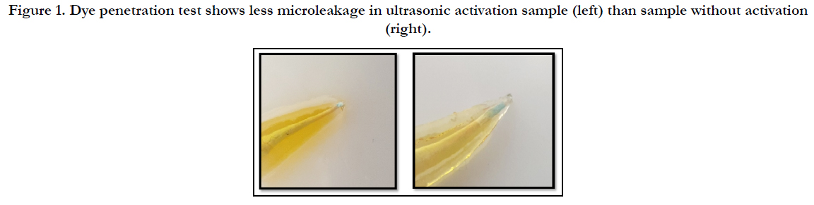
Figure 1. Dye penetration test shows less microleakage in ultrasonic activation sample (left) than sample without activation (right).
All samples were studied on a 20X stereomicroscope by two blinded investigators.
Data were subjected to statistical analysis using one-way ANOVA
Test with a P value (p<0.05) and the confidence level (95%).
Moreover,we used Bonferroni test for multiple comparisons between
the study groups.
Results
There was a significant difference between the microleakage of
the apical third of the groups (P = 0.000). The use of ultrasonic
activation with Endosequence BC sealer resulted in lower apical
microleakage than with no activation (P< 0.05). While using Endosequence
BC points showed no significant differences.
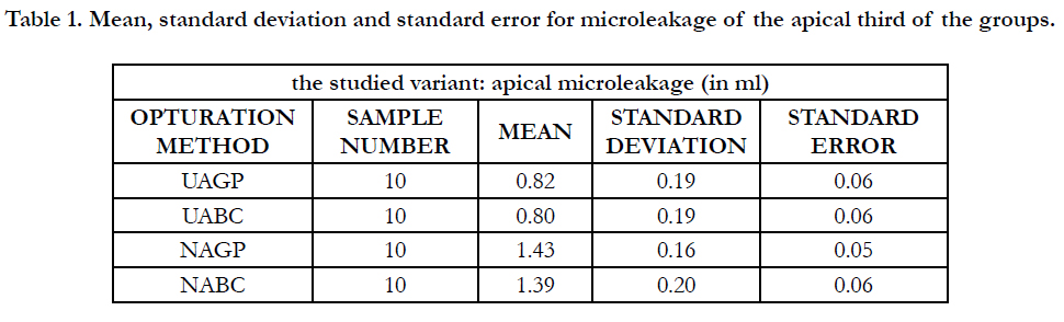

Table 1. Mean, standard deviation and standard error for microleakage of the apical third of the groups.
one-way ANOVA Test (table 2) was conducted and showed positive differentiation rates between at least two groups with P-value less than 0.05. and for further investigation boneferroni test (table 3) was conducted afterwards to make multiple comparisons between the groups. The results showed positive differentiation between ultrasonic activation and non-activation groups. While no significant differentiation was found between endosequence BC gutta-percha and the traditional gutta-percha.
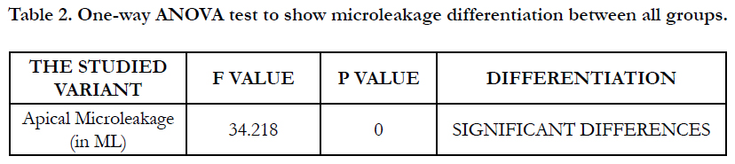
Table 2. One-way ANOVA test to show microleakage differentiation between all groups.
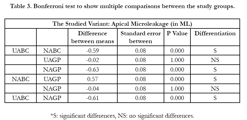
Table 3. Bonferroni test to show multiple comparisons between the study groups.
Discussion
This study was conducted on extracted lower premolers with
a single root, single rounded canal, free of internal or external
absorbtion in order to obtain a good canal obturation [7]. The
same preparation technique was used in all canals with repeated
irrigation with 5.25% NaOCl, and with 17% EDTA solution to
remove the smear layer which make a better sealer penetration in
the dentinal tubules [8]. A single paper point cone was used to dry
each canal in order to make a better sealing between BC sealer and
canals walls [9].
Single cone oburation technique has been introduced to reduce
working time and obturation mistakes (10), as well as sealer quantity
in the filling material. Even though, it showed higher number
of voids and gaps in the filling material (11); which has led to
suggest the use of ultrasonic activation of the sealer during root
canal obturtion(12)(13).In this study, ultrasonic activation did not
show higher exceed of sealer material over the apex; this can be
ascribed to the distance (2mm) between the ultrasonic tip and
the apex, beside the low frequency of ultrasonic activation device
(14).
Apical third of the canal is the most important third because it
has more lateral canals than the other two thirds, and these lateral
canalsare connected with periapical tissues. However, they are a
home for bacteria which derive from pulpal or periapical tissues
[15]. Those bacteria are the direct reason for periapical lesions.
Dye Penetration Technique is one of the most commonmethods
which are used to evaluate root filling quality and sealer sealability.
This technique is simple, cheap and do notneed any complicatedequipment
[16].
Cohen's kappa test was used to calculate intra-examiner reproducibility
and inter-examiner reliability for the assessment of test
variables. The kappa for intra-examiner agreement and inter-examiner
reliability was 0.90.
Results showed that samples with ultrasonic activation had a lower
apical microleakage which can refer to the increase in tubular
penetration for the ultrasonic activation groups [13]. Ultrasonic
activation also reduces the number of voids and gaps [17] and can
result in better filling for the lateral canals [12].
Arslan in 2016, has found better peneteration of sealer into lateral
canals with the ultrasonic activation use compared to sonic activation
and non-activation groups [5].
Wiesse et al., [14] studied the effect of ultrasonic activation of
root canal sealer on the push-out bond strength and interfacial
adaptation to root canal dentine. Those studies are in agreement
with our results, as they conclude that ultrasonic activation resulted in higher bond strength and better interfacial adaptation of
sealers to canal walls.
The type of gutta-percha cone did not make a significant difference
in microleakage between the groups. These findings may
refer to incapability of the nano-particles in Endosequence BC
points to make better bonding with sealer or canal walls. The
mentioned result can be attributed to the method used in this
study, whichexamined the microleakage between sealer and canal
walls and the less between sealer and cone material.
Yanpiset et al, examined the bacterial leakage around BC guttapercha
points with BC sealer compared to traditional gutta-percha
with BC sealer using single cone obturation technique. Results of
this study agreed with our study as they obtained a comparable
scores among the two gutta-percha types [18].
Al Haddad in 2018 [19] compared the apical sealing of BC point
with traditional one by applying push-out test on their specimens
and showed resembling results between the two types. These findings
are also in the line with the present study in spite of the different
testing method.
Limitation of our study is the small sample size.
Conclusions
Under the conditions of this study, the use of ultrasonic activation
of EndoSequence BC sealer improves root filling quality and
reduces microleakege percentage. While using endosequence BC
points showed no reduction in the microleakage level.
References
- Naseri M, Kangarlou A, Khavid A, Goodini M. Evaluation of the quality of four root canal obturation techniques using micro-computed tomography. Iran Endod J. 2013;8(3):89.PubmedPMID: 23922567.
- Loushine BA, Bryan TE, Looney SW, Gillen BM, Loushine RJ, Weller RN, et al. Setting properties and cytotoxicity evaluation of a premixed bioceramic root canal sealer. J Endod. 2011;37(5):673–7. Pubmed PMID: 21496669.
- Whitworth J. Methods of filling root canals: principles and practices. Endod Top. 2005;12(1):2–24.
- Galler KM, Grubmüller V, Schlichting R, Widbiller M, Eidt A, Schuller C, et al. Penetration depth of irrigants into root dentine after sonic, ultrasonic and photoacoustic activation. Int Endod J. 2019;52(8):1210–7. Pubmed PMID: 30828819.
- Arslan H, Abbas A, Karatas E. Influence of ultrasonic and sonic activation of epoxy-amine resin-based sealer on penetration of sealer into lateral canals. Clin Oral Investig. 2016;20(8):2161-4. Pubmed PMID: 26818582.
- Gervini MJ, Pacheco-Yanes J, Gonçalves LS, Lopes HP, Vieira VTL, Siqueira Jr JF, et al. Fracture resistance of roots filled with either a bioceramic or an epoxy resin-based sealer. ENDO-ENDODONTIC Pract TODAY. 2018;12(2):119–23.
- Nilsson E, Bonte E, Bayet F, Lasfargues J-J. Management of internal root resorption on permanent teeth. Int J Dent. 2013;2013. Pubmed PMID: 24348560.
- Vemuri S, Kolanu SK, Varri S, Pabbati RK, Penumaka R, Bolla N. Effect of different final irrigating solutions on smear layer removal in apical third of root canal: A scanning electron microscope study. J Conserv Dent JCD. 2016;19(1):87. Pubmed PMID: 26957801.
- Taşdemir T, Er K, Çelik D, Tahan E, Serper A, Ceyhanli KT, et al. Bond strength of calcium silicate-based sealers to dentine dried with different techniques. Med Princ Pract. 2014;23(4):373–6.Pubmed PMID: 24903084.
- Pereira AC, Nishiyama CK, de Castro Pinto L. Single-cone obturation technique: a literature review. RSBO Rev Sul-Brasileira Odontol. 2012;9(4):442–7.
- Taşdemir T, Er K, Yildirim T, Buruk K, Çelik D, Cora S, et al. Comparison of the sealing ability of three filling techniques in canals shaped with two different rotary systems: a bacterial leakage study. Oral Surgery, Oral Med Oral Pathol Oral Radiol Endodontology. 2009;108(3):e129–34.Pubmed PMID: 19716483.
- Sungur DD, Moinzadeh A-T, Wesselink PR, Tarhan SÇ, Özok AR. Sealing efficacy of a single-cone root filling after post space preparation. Clin Oral Investig. 2016;20(5):1071–7. Pubmed PMID: 26411973.
- Chadgal S, Farooq R, Purra AR, Ahangar FA, Amin K, Lone OH. Ultrasonic activation of a bioceramic sealer and its dentinal tubule penetration: An in vitro study. Ann Int Med Dent Res. 2018;4(2):51-4.
- Wiesse PEB, Silva‐Sousa YT, Pereira RD, Estrela C, Domingues LM, Pécora JD, et al. Effect of ultrasonic and sonic activation of root canal sealers on the push‐out bond strength and interfacial adaptation to root canal dentine. Int Endod J. 2018;51(1):102-11. Pubmed PMID: 28543092.
- Solomon C, Chalfin H, Kellert M, Weseley P. The endodontic-periodontal lesion: a rational approach to treatment. J Am Dent Assoc. 1995;126(4):473– 9. Pubmed PMID: 7722108.
- AI‐Ghamdi A, Wennberg A. Testing of sealing ability of endodontic filling materials. Dent Traumatol. 1994;10(6):249–55.Pubmed PMID: 7867611.
- Kim JA, Hwang YC, Rosa V, Yu MK, Lee KW, Min KS. Root Canal Filling Quality of a Premixed Calcium Silicate Endodontic Sealer Applied Using Gutta-percha Cone-mediated Ultrasonic Activation. J Endod [Internet]. 2018;44(1):133–8. Pubmed PMID: 29102078.
- Yanpiset K, Banomyong D, Chotvorrarak K, Srisatjaluk RL. Bacterial leakage and micro-computed tomography evaluation in round-shaped canals obturated with bioceramic cone and sealer using matched single cone technique. Restor Dent Endod. 2018;43(3). Pubmed PMID: 30135849.
- Al-Haddad AY, Kutty MG, Che Ab Aziz ZA. Al-Haddad, A. Y., Kutty, M. G. en Che Ab Aziz, Z. A. (2018) “Push-out bond strength of experimental apatite calcium phosphate based coated gutta-percha”, International journal of biomaterials. Hindawi, 2018.Push-out bond strength of experimental apatite c. Int J Biomater. 2018;2018.


