Canine Impaction Among Orthodontic Patients - A Retrospective Study
Nilesh Suresh1, Naveen Kumar M2*
1 Department of Orthodontics and Dentofacial Orthopaedics, Saveetha Dental College and Hospital, Saveetha Institute of Medical and Technical Sciences, Saveetha University, 162, Poonamallee High Road ,Chennai-600077, Tamil Nadu, India.
2 Senior Lecturer, Saveetha Dental College and Hospital, Saveetha Institute of Medical and Technical Sciences, Saveetha University, 162, Poonamallee High Road ,Chennai-600077, Tamil Nadu, India.
*Corresponding Author
Naveen Kumar M,
Senior Lecturer, Department of Public Health Dentistry, Saveetha Dental College and Hospitals, Saveetha Institute of Medical and Technical Sciences, Saveetha University, Chennai,
600077, India.
Tel: +91 9791217568
E-mail: drnaveen2012@gmail.com
Received: November 10, 2020; Accepted: December 15, 2020; Published: December 18, 2020
Citation: Nilesh Suresh, Naveen Kumar M. Canine Impaction Among Orthodontic Patients - A Retrospective Study. Int J Dentistry Oral Sci. 2020;7(12):1283-1288. doi: dx.doi.org/10.19070/2377-8075-20000254
Copyright: Naveen Kumar M©2020. This is an open-access article distributed under the terms of the Creative Commons Attribution License, which permits unrestricted use, distribution and reproduction in any medium, provided the original author and source are credited.
Abstract
Treatment of Canine impactions is one of the most complex procedures in orthodontic treatment. The aim of this study was to present detailed information regarding the impacted maxillary and mandibular canines and their patterns in the oral cavity the prevalence of various canine anomalies, like ectopic canine, transmigration, transposition and agenesis of permanent canines among South Indian population. A total of 500 patients OPG’s were thoroughly evaluated and therefore the prevalence of various canine anomalies like impacted maxillary and mandibular canine, transmigration, transposition, agenesis and ectopic canine eruptions were evaluated. The canine angulation, vertical position in reference to the occlusal surface of adjacent tooth’s and therefore the overlapping of adjacent teeth’s crown by impacted canine was evaluated by tracings. Out of 500 subjects, 11 patients had impacted canines. The prevalence of canine impaction was 2.21%, with maxillary canine impaction of 1.53%, mandibular canine impaction of 0.68%, canine agenesis 0.06%, transmigration 0.12%, canine transposition 0.18% and the ectopic canine was 5.5%. There is no gender difference in canine impaction. The prevalence of canine impaction is 2.21%.
2.Introduction
3.Material and Methods
4.Statistical Analysis
5.Results
6.Discussion
7.Conclusion
8.Refereces
Keywords
Agenesis; Ectopic Canine; Impacted Canine; Transmigration; Transposition.
Introduction
The different sorts of canine anomalies like ectopic canine eruption,
canine transmigration, canine transposition, agenesis, impaction,
usually occur because of the disturbances during development
and eruption. Since the canines are the longest teeth within
the oral cavity and therefore the shape, position of the canines
contribute to the guidance of the teeth into the intercuspal position,
the canine teeth should be evaluated thoroughly in order to
deliver the best treatment to the patients.
The impaction of tooth have been studied by many authors and
various terminologies have been given in the literature to define
impaction including delayed eruption, primary retention, submerged
teeth, impacted teeth etc [1]. According to Abron et al,
impaction can be defined as a deceleration of the normal eruption
process of the tooth1 and according to Lindauer et al, it can
be defined as a impaction if it was not erupted after completion
of the root development or if the eruption of the contralateral
tooth was there for at least 6 months with completion of root
formation.
The ectopic eruption is a condition where because of deficiency
of growth in the jaw or segment of jaw, a primary tooth assumes
a path of eruption that intercepts its premature loss and produces
a consequent malposition of the adult permanent teeth. Tooth
transposition, is also a special type of ectopic eruption. It can
be defined as a condition where the position of two teeth is interchanged
or a condition where a tooth develops in the place
of another tooth [2]. It can be divided into two types, complete
and incomplete transposition. Complete transposition is when the
crown and root surface of the teeth is completely transposed in
different positions. In incomplete transposition, only the crown is
displaced in another tooth position but the root remains in their
normal positions [3].
The transmigration refers to a condition where a tooth crosses
a midline. Previously, transmigration term was used where the
whole impacted canine had migrated and crossed the midline of
the mandible [4]. But according to Javid, transmigration can be
defined as a condition where one half or more of impacted canine
crosses the midline [5, 6]. According to various studies the
prevalence of transmigration is suggested to be 0.1 to 0.34% in
different populations [7-9].
Overall, the incidence of impacted maxillary canine is suggested
to be 0.9–2.2% [10, 11]. But the incidence for mandibular canine
impaction is at least 20 times lower than that of maxillary canine
impaction [12]. However, the transmigration of canine, canine
agenesis and canine transposition are even rarer anomalies.
The aim of our study was to determine the prevalence of impacted
canines and its pattern among orthodontic patients.
Materials And Methodology
This is a retrospective clinical study. The subjects for this study
were selected from the patients who had come to the department
of Orthodontics. All the patients who came to the department
of Orthodontics were thoroughly examined and checked for any
missing permanent canine, retained primary canine and other canine
anomalies. A total number of 500 patients were evaluated
for this study.
Patients were advised for OPG x-ray for confirmation of the
clinical examination. Different canine anomalies were determined
from the Orthopantomogram. The method given by Lindauer et
al was used to consider canine as impacted.
The tracings were made on acetate paper. The impacted canine,
central incisor, lateral incisor on the impacted side were traced by
lead pencil.
The impacted canines were evaluated for level, angulation and
overlapping in relation to adjacent tooth. The angulation of the
impacted canine was evaluated by tracing the long axis of the impacted
canine in reference to mid-sagittal plane. The angulations
were classified into mesioangular, vertical, distoangular and horizontal.
Since there were no exact criteria to classify according to the degree
of angulations between the long axis of the impacted canine
and the mid-sagittal plane, we performed a survey to decide the
exact criteria; 10 senior resident orthodontists were asked to classify
different angulations between mid-sagittal plane and long axis
of impacted canine ranging from 5° to more than 75°. After the
survey the following angulation classification was used.
When the long axis of the impacted canine was directed towards
the mid sagittal plane and therefore the angle is made near the
coronal area of the impacted canine with a range of angle between
15–70 degree. Distoangular: when the long axis of the impacted
canine is far away from the mid-sagittal plane and forming
the angle above the apical region of impacted canine. Vertical:
when the long axis of the impacted canine is almost parallel with the mid-sagittal plane and if the angle was between 0–15 degree.
Horizontal: when the long axis of the impacted canine meets the
mid sagittal plane at an angle of about more than 70 degrees.
Level A, The impacted canine crown is touching the cervical line
of the adjacent teeth. Level B, The impacted canine crown is positioned
between the adjacent teeth cervical line and the adjacent
teeth root apex. Level C, The impacted canines crown is positioned
below the root apex of the adjacent teeth.
To determine the overlap of the adjacent incisor root by the impacted
canine the following classification was used in this study
[13, 14]. Grade 1, no overlapping of the adjacent teeth; Grade 2,
overlapping of adjacent roots less than half width; Grade 3, overlapping
of greater than half root width, but not the whole root;
Grade 4, overlapping of complete root width or greater than that
(Figure .1).
For this study, complete transposition was considered when the
crown and root surface of teeth was completely transposed in the
different positions.
Javid’s definition for transmigration was used for this study according
to which a canine was considered transmigrated when the
one half of impacted canine or more than that of the impacted
canine crosses the midline 4. To further classify the transmigrant
canines, the classification given by the Mupparapu was used [15].
The classification is as follows: Type 1, canine positioned mesioangularly
across the midline, labial or lingual to the anterior teeth.
Type 2, canine horizontally impacted near the inferior border of
the mandible inferior to the apices of the incisor teeth. Type 3,
canine erupting on the contra lateral side.
Type 4, canine horizontally impacted near the inferior border of
the mandible below the apices of posterior teeth. Type 5, canine
positioned vertically in the midline with the long axis of the tooth
crossing the midline.
Results And Discussion
Out of 500 subjects, 23 (twenty three) patients had at least one
impacted maxillary or mandibular canine. Among the eleven subjects
the total number of impacted canine teeth found was 35
and one missing permanent canine. The distribution of different
patterns of 35 canine anomalies were as follows: transmigration,
3 teeth and only in the mandibular arch; canine transposition, 5
teeth (1 complete, 2 incomplete), 26 impacted canine (16 in the
maxillary arch and 10 in the mandibular arch). The present study,
the prevalence of overall (both maxillary and mandibular) canine
impaction found was 2.21%, only maxillary canine impaction was
1.53% and mandibular canine impaction 0.68%, canine agenesis
0.06%, canine transmigration 0.12% and only within the mandibular
arch, canine transposition was 0.18% and only unilateral. The
ectopic canine was found in 5.5% of patients. More than 95%
of ectopic canines were present in the maxillary arch. Almost all
the patients that took part in the study were not conscious of the
condition but only two patients had complained of bulging of the
soft tissues, because the tooth was erupting in the upper buccal
mucosa.
Figures 2, 3 and 4 depicts the patterns of impacted canines, which include the angulation, level of impaction and grade (overlapping
of adjacent teeth). In the angulation category the mesioangular
angulation was the most common finding, followed by vertical,
then horizontal. In this study none of the impacted canine
showed distoangular angulation. In the presentation of vertical
heights (Level) of impacted canine, the level B was the most
prevalent and level A and level C showed almost equal frequency.
Grade 1 and grade 2 again showed almost equal incidence and
most prevalent in the grade’s category, followed by grade 4, while
grade 3 was the least finding.
It has been observed that canine impaction is more in males
(52.2%) than females (42.8%) [Figure.5]. It has also been observed
that the prevalence of canine impaction was more in the
maxilla (62.9%) than mandible (37.1%) [Figure.6].
The association between gender and angular measurement of impacted
canine showed no statistical significance (p value= 0.410)
with Pearson’s chi square value of 1.782 [Figure. 7].
The association between gender and level of impacted canine
showed no significance. (p value= 0.57) with Pearson’s chi square
value of 1.124 [Figure.8].
The association between gender and grade of impacted canine
showed no significance. (p value= 0.984) with Pearson’s chi square
value of 0.158 [Figure.9].
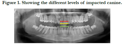
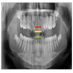
Figure 1. Showing the different levels of impacted canine.
Figure 2. Bar chart representing the frequency distribution of angular measurements of impacted canines. The X-axis represents the various types of angular measurements of impacted canines. The Y-axis represents the number of impacted canines . Mesioangular impacted canine (red colour) was the most common compared to vertical and horizontal impaction.
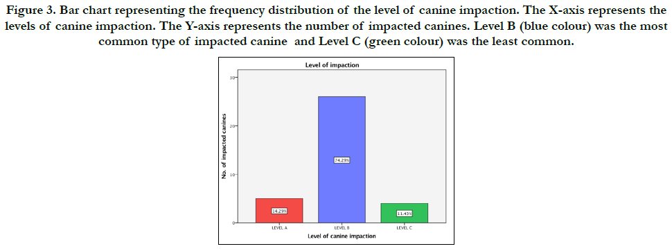
Figure 3. Bar chart representing the frequency distribution of the level of canine impaction. The X-axis represents the levels of canine impaction. The Y-axis represents the number of impacted canines. Level B (blue colour) was the most common type of impacted canine and Level C (green colour) was the least common.
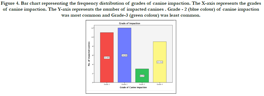
Figure 4. Bar chart representing the frequency distribution of grades of canine impaction. The X-axis represents the grades of canine impaction. The Y-axis represents the number of impacted canines . Grade - 2 (blue colour) of canine impaction was most common and Grade-3 (green colour) was least common.
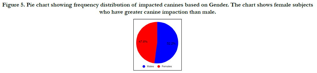
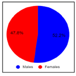
Figure 5. Pie chart showing frequency distribution of impacted canines based on Gender. The chart shows female subjects who have greater canine impaction than male.
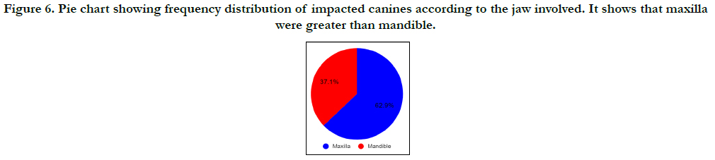
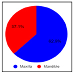
Figure 6. Pie chart showing frequency distribution of impacted canines according to the jaw involved. It shows that maxilla were greater than mandible.
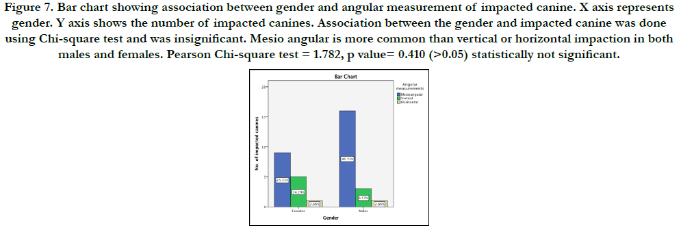
Figure 7. Bar chart showing association between gender and angular measurement of impacted canine. X axis represents gender. Y axis shows the number of impacted canines. Association between the gender and impacted canine was done using Chi-square test and was insignificant. Mesio angular is more common than vertical or horizontal impaction in both males and females. Pearson Chi-square test = 1.782, p value= 0.410 (>0.05) statistically not significant.
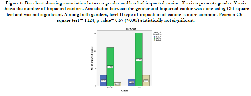
Figure 8. Bar chart showing association between gender and level of impacted canine. X axis represents gender. Y axis shows the number of impacted canines. Association between the gender and impacted canine was done using Chi-square test and was not significant. Among both genders, level B type of impaction of canine is more common. Pearson Chisquare test = 1.124, p value= 0.57 (>0.05) statistically not significant.
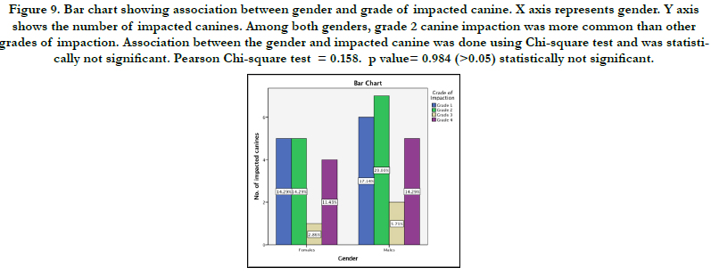
Figure 9. Bar chart showing association between gender and grade of impacted canine. X axis represents gender. Y axis shows the number of impacted canines. Among both genders, grade 2 canine impaction was more common than other grades of impaction. Association between the gender and impacted canine was done using Chi-square test and was statistically not significant. Pearson Chi-square test = 0.158. p value= 0.984 (>0.05) statistically not significant.
In the present study the prevalence of impacted canines among the central Indian population was estimated to be 2.21%. The prevalence of impacted maxillary canine was 1.53%, which is lower than the study by Chu et al. where they did find the prevalence of 2.1% in Caucasian and Chinese populations.
The prevalence of impacted mandibular canine in this study was found to be 0.68%, which is higher than the study done by Rohrer A [12, 15] where they have found the ratio of maxillary and mandibular impacted canine 20:1 ratio (2.06% and (0.1%), Grover and Lorton [16] reported 0.22%, Chu et al reported 0.07% among 7486 patients. In other studies by Aydin et al. [17] among Turkish population, the incidence reported was higher than the present study 0.44% which was studied among 4500 patients.
According to Takahama and Aiyama [18] the unilateral impaction was the most common finding, and according to Harzer the side mostly affected was the left one. Other studies had different views, the higher incidence side being the right side [19, 20]. In our study the most common impaction found was the unilateral canine impaction, which was observed in 14 subjects and the most common side affected was the right side in both genders, similar to the studies by Takahama and Aiyama [18], while Bass [21] found that the bilateral impaction was the most common finding. But in our study only 8 (eight) subjects out of [23] (twenty three) were found with bilateral canine impaction.
When it comes to the distribution of the prevalence of impacted canine according to the gender then the majority of studies found the higher prevalence to be among the females [9]. But equal occurrence of impacted canine in both the genders was reported by some studies [22]. In the present study we have also found almost equal prevalence among male and female subjects.
The tooth transposition occurs most frequently on the left side then the right side, in the maxillary arch, unilateral then bilateral and in females. Various studies finding the most common transposition occurrence to be between the canine and first premolar [23, 24] and less frequent with the lateral incisor [23]. In this study one complete transposition and two incomplete canine transpositions were observed. The complete canine transposition occurred between the canine and the lateral incisor and primary canine was also retained. In the other two cases no retained deciduous canine and also the lateral incisor was in normal shape. The prevalence found was 0.18%, the side involved in all three cases was the right side. This study does not agree with other studies which are in favor of the left side to be the most common affected side by canine transposition.
The present study found the prevalence of canine transmigration within the mandibular arch 0.12% and in the maxillary arch none. This study result shows less incidence compared to the study done by Sharma G, Nagpal A [23, 25], where they did the study among 3000 panoramic radiographs of north Indian population. The study by Aktan et al. [7] among Turkish subpopulation also shows a higher prevalence of 0.34% among 5000 subjects. [26- 40].
Conclusion
From the limitation of the study the prevalence of canine impaction among Orthodontic patients was greater in females compared
to male and higher in maxillary arch then mandibular arch.
Among gender mesio angular is common among male and level
B impaction is higher among female subjects. Grade 2 impaction
is common among both gender. Knowledge of canine teeth development
and eruption is necessary for the dentist to diagnose
the incidence of impaction at an early age in order to reduce the
malocclusion probability.
Acknowledgement
I would like to record my deep sense of gratitude to my research
supervisor Dr. Naveen Kumar M, Senior lecturer, Department
of Orthodontics, Saveetha Dental College and Hospitals, Chennai
for his inspiring guidance and encouragement with my work
during all stages. There was an equal contribution from all the
authors.
References
- Aslan BI, Üçüncü N. Clinical consideration and management of impacted maxillary canine teeth. Emerging Trends in Oral Health Sciences and Dentistry. 2015 Mar 11:465-501.
- Pindborg JJ. Pathology of the dental hard tissues. Saunders; 1970:442.
- Shapira Y, Kuftinec MM. Maxillary tooth transpositions: characteristic features and accompanying dental anomalies. Am J Orthod Dentofacial Orthop. 2001 Feb;119(2):127-34.Pubmed PMID: 11174558.
- Tarsitano JJ, Wooten JW, Burditt JT. Transmigration of nonerupted mandibular canines: report of cases. J. Am. Dent. Assoc. 1971 Jun 1;82(6):1395-7.
- Javid BR. Transmigration of impacted mandibular cuspids. Int J Oral Surg. 1985;14: 547–9.
- Pippi R, Kaitsas R. Mandibular canine transmigration: aethio‐pathogenetic aspects and six new reported cases. Oral Surg. 2008 May;1(2):78-83.
- Kamiloglu B, Kelahmet U. Prevalence of impacted and transmigrated canine teeth in a Cypriote orthodontic population in the Northern Cyprus area. BMC Res Notes. 2014 Jun 7;7:346.Pubmed PMID: 24906489.
- Aktan AM, Kara S, Akgünlü F, Malkoç S. The incidence of canine transmigration and tooth impaction in a Turkish subpopulation. Eur J Orthod. 2010 Oct;32(5):575-81.Pubmed PMID: 20237077.
- Gündüz K, Celenk P. The incidence of impacted transmigrant canines: a retrospective study. Oral Radiol. 2010 Dec;26(2):77-81.
- DACHI SF, HOWELL FV. A survey of 3,874 routine full-mouth radiographs. I. A study of retained roots and teeth. Oral Surg Oral Med Oral Pathol. 1961 Aug;14:1165-9.Pubmed PMID: 13719264.
- Thilander B, Myrberg N. The prevalence of malocclusion in Swedish schoolchildren. Scand J Dent Res. 1973;81(1):12-20.Pubmed PMID: 4510864.
- Röhrer A. Displaced and impacted canines A radiographic research [Internet]. Vol. 15, International Journal of Orthodontia, Oral Surgery and Radiography. 1929. p. 1003–20.
- . Stivaros N, Mandall NA. Radiographic factors affecting the management of impacted upper permanent canines. J Orthod. 2000 Jun;27(2):169-73. Pubmed PMID: 10867073.
- Power SM, Short MB. An investigation into the response of palatally displaced canines to the removal of deciduous canines and an assessment of factors contributing to favourable eruption. Br J Orthod. 1993 Aug;20(3):215- 23.Pubmed PMID: 8399054.
- Mupparapu M. Patterns of intra-osseous transmigration and ectopic eruption of mandibular canines: review of literature and report of nine additional cases. Dentomaxillofac Radiol. 2002 Nov;31(6):355-60.Pubmed PMID: 12424633.
- Grover PS, Lorton L. The incidence of unerupted permanent teeth and related clinical cases. Oral Surg Oral Med Oral Pathol. 1985 Apr;59(4):420-5. Pubmed PMID: 3858781.
- Aydin U, Yilmaz HH, Yildirim D. Incidence of canine impaction and transmigration in a patient population. Dentomaxillofac Radiol. 2004 May;33(3):164-9.Pubmed PMID: 15371316.
- Takahama Y, Aiyama Y. Maxillary canine impaction as a possible microform of cleft lip and palate. Eur J Orthod. 1982 Nov;4(4):275-7.Pubmed PMID: 6959819.
- Stahl F, Grabowski R. Maxillary canine displacement and genetically determined predisposition to disturbed development of the dentition. J Orofac Orthop. 2003 May;64(3):167-77.Pubmed PMID: 12835889.
- Sacerdoti R, Baccetti T. Dentoskeletal features associated with unilateral or bilateral palatal displacement of maxillary canines. Angle Orthod. 2004 Dec;74(6):725-32.
- Observations of the misplaced upper canine tooth [Internet]. Vol. 54, American Journal of Orthodontics. 1968. p. 550.
- Brin I, Becker A, Shalhav M. Position of the maxillary permanent canine in relation to anomalous or missing lateral incisors: a population study. Eur J Orthod. 1986 Feb 1;8(1):12-6.
- Joshi MR, Bhatt NA. Canine transposition [Internet]. Vol. 31, Oral Surgery, Oral Medicine, Oral Pathology. 1971. p. 49–54.
- Shapira Y. Transposition of Canines [Internet]. Vol. 100, The Journal of the American Dental Association. 1980. p. 710–2.
- Kumar S, Jayaswal P, Pentapati KC, Valiathan A, Kotak N. Investigation of the transmigrated canine in an orthodontic patient population. J. Orthod. 2012 Jun;39(2):89-94.
- Sivamurthy G, Sundari S. Stress distribution patterns at mini-implant site during retraction and intrusion--a three-dimensional finite element study. Prog Orthod. 2016;17:4.Pubmed PMID: 26780464.
- Samantha C, Sundari S, Chandrasekhar S, Sivamurty G, Dinesh S. Comparative Evaluation of Two Bis-GMA Based Orthodontic Bonding Adhesives - A Randomized Clinical Trial. J Clin Diagn Res. 2017 Apr;11(4):ZC40- ZC44.Pubmed PMID: 28571259.
- Krishnan S, Pandian S, Kumar S A. Effect of bisphosphonates on orthodontic tooth movement-an update. J Clin Diagn Res. 2015 Apr;9(4):ZE01-5. Pubmed PMID: 26023659.
- Vikram NR, Prabhakar R, Kumar SA, Karthikeyan MK, Saravanan R. Ball Headed Mini Implant. J Clin Diagn Res. 2017 Jan;11(1):ZL02-3.
- Kamisetty SK, Verma JK, Arun, Sundari S, Chandrasekhar S, Kumar A. SBS vs Inhouse Recycling Methods-An Invitro Evaluation. J Clin Diagn Res. 2015 Sep;9(9):ZC04-8.Pubmed PMID: 26501002.
- Viswanath A, Ramamurthy J, Dinesh SP, Srinivas A. Obstructive sleep apnea: awakening the hidden truth. Niger J Clin Pract. 2015 Jan-Feb;18(1):1-7. Pubmed PMID: 25511335.
- Felicita AS. Quantification of intrusive/retraction force and moment generated during en-masse retraction of maxillary anterior teeth using mini-implants: A conceptual approach. Dental Press J Orthod. 2017 Sep- Oct;22(5):47-55.Pubmed PMID: 29160344.
- Rubika J, Sumathi Felicita A, Sivambiga V. Gonial angle as an indicator for the prediction of growth pattern. World J. Dent. 2015;6(3):161-3.
- Jain RK, Kumar SP, Manjula WS. Comparison of intrusion effects on maxillary incisors among mini implant anchorage, j-hook headgear and utility arch. J Clin Diagn Res. 2014 Jul;8(7):ZC21-4.Pubmed PMID: 25177631.
- Pandian KS, Krishnan S, Kumar SA. Angular photogrammetric analysis of the soft-tissue facial profile of Indian adults. Indian J Dent Res. 2018 Mar- Apr;29(2):137-143.Pubmed PMID: 29652003.
- Ramesh Kumar KR, Shanta Sundari KK, Venkatesan A, Chandrasekar S. Depth of resin penetration into enamel with 3 types of enamel conditioning methods: a confocal microscopic study. Am J Orthod Dentofacial Orthop. 2011 Oct;140(4):479-85.Pubmed PMID: 21967934.
- Felicita AS. Orthodontic management of a dilacerated central incisor and partially impacted canine with unilateral extraction - A case report. Saudi Dent J. 2017 Oct;29(4):185-193.Pubmed PMID: 29033530.
- Felicita AS, Chandrasekar S, Shanthasundari KK. Determination of craniofacial relation among the subethnic Indian population: a modified approach - (Sagittal relation). Indian J Dent Res. 2012 May-Jun;23(3):305-12. Pubmed PMID: 23059564.
- Dinesh SP, Arun AV, Sundari KK, Samantha C, Ambika K. An indigenously designed apparatus for measuring orthodontic force. J Clin Diagn Res. 2013 Nov;7(11):2623-6.Pubmed PMID: 24392423.
- Felicita AS. Orthodontic extrusion of Ellis Class VIII fracture of maxillary lateral incisor - The sling shot method. Saudi Dent J. 2018 Jul;30(3):265- 269.Pubmed PMID: 29942113.


