To Evaluate the Efficacy of New Brush Covered Irrigation Needle in Removing Root Canal Debris In Vitro : A Scanning Electron Microscopy Study
Piyush Oswal1, Santosh S. Martande2*, Suresh Shenvi3
1 Department of Conservative Dentistry and Endodontics, Dr. D Y Patil Vidyapeeth, Pimpri, Pune, 400 018, Maharashtra, India.
2 Department of Periodontology, Dr. D Y Patil Vidyapeeth, Pimpri, Pune, 400 018, Maharashtra, India.
3 Department of Conservative Dentistry and Endodontics, KLE VK Institute of Dental Sciences, Belgaum, KAHER, Karnataka, India.
*Corresponding Author
Dr. Santosh S. Martande MDS,
Department of Periodontology, Dr. D Y Patil Vidyapeeth, Pimpri, Pune, 400 018, Maharashtra, India.
Tel: 9890353072
E-mail: santoshmartande@gmail.com
Received: November 18, 2020; Accepted: December 15, 2020; Published: December 16, 2020
Citation:Piyush Oswal, Santosh S. Martande, Suresh Shenvi. To Evaluate the Efficacy of New Brush Covered Irrigation Needle in Removing Root Canal Debris In Vitro: A Scanning Electron Microscopy Study Int J Dentistry Oral Sci. 2020;7(12):1211-1215. doi: dx.doi.org/10.19070/2377-8075-20000239
Copyright: Santosh S. Martande©2020. This is an open-access article distributed under the terms of the Creative Commons Attribution License, which permits unrestricted use, distribution and reproduction in any medium, provided the original author and source are credited.
Abstract
Objective: The main purpose of the study was to compare the efficacy of NaviTip and NaviTip FX in debriding the walls of the
canals using a Scanning Electron Microscope (SEM). The secondary purpose of the study was to evaluate if use of 17 % EDTA
along with 5.25% NaOCl had an additional effect in root canal debridement.
Material and Methods: Access cavity preparations were performed in 40 extracted incisor teeth. All the teeth were randomly
allotted to any of 4 groups consisting of 10 teeth- Group 1: Irrigation with 1ml of 5.25% NaOCl with NaviTip needle after each
instrumentation; Group 2: Irrigation with 1ml of 5.25% NaOCl using NaviTip FX with a manual left and right rotary motion
combined with up and down motions, and a brushing action on dentin walls after each instrumentation; Group 3: Irrigation with
1ml of 5.25% NaOCl followed by 1ml of 17%EDTA with NaviTip needle after each instrumentation; Group 4: Irrigation with
1ml of 5.25% NaOCl followed by 1ml of 17%EDTA using NaviTip FX with a manual left and right rotary motion combined with
up and down motion, and a brushing action on dentin walls after each instrumentation. Later each selected sample was further
split horizontally into three halves as coronal (10mm from apex), middle (5mm from apex) and apical (1mm from apex) thirds and
treated with sputter-coated withgold using fine-coat ion sputter JFC-1100. Samples were analyzedfinally by SEM analysis.
Result: All the groups showed significantly lesser smear layer at 10mm when compared to 5mm and 1mm. Further, it signifies that
NaviTip FX (Group 4) when used along with 5.25% NaOCL and 17% EDTA removed more debris as compared to Group 1 and
2 at 10mmand 5mm; and removed more debris at 1mm compared to Group 3.
Conclusion: Navi Tip could be replaced by Navi Tip FX for the purpose of cleaning and removal of smear layer. EDTA, which
is considered as one of the best irrigants for removal dentinal debris may be ineffective in some narrower canals and could be
adjuncted with manual irrigation devices such as Navi Tip FX to improve its action.
2.Introduction
3.Material and Methods
4.Statistical Analysis
5.Results
6.Discussion
7.Conclusion
8.Refereces
Keywords
Root Canal Debridement; Root Canal Irrigation; 5.25% NaOCl; 17% EDTA; Scanning Electron Microscopy.
Introduction
The core of endodontic instrumentation lies in the cleaning,
shaping and canal preparation. It is contemplated as an important
step as it removes the vital and necrotic pulp which contains
the microflora that can cause failure of the endodontic treatment
[1, 2]. Furthermore, the debris that contains the chips of dentin,
other organic and inorganic content block the canals for proper
flow of sealer leading to a poor obturation. Several studies have
been conducted and regardthat the debris should be completely
removed for a successful endodontic treatment. For this purpose,
the canals are thoroughly instrumented with both hand and rotary
instruments in an effort to remove the debris totally [3, 4].
Literature has multiple evidences which state that mechanical
instrumentation alone cannot remove the debris totally, hence,
chemical methods have also been advocated for successful removal
of debris and disinfection of the canals [5]. Even if the
debris has been successfully removed, the instrumented canal
walls leave a 1 to 2 μthin layer called as smear layer which clogs
the dentinal tubules, thus preventing the flow of sealer [6]. The
use of rotary instruments also leaves certain areas such as isthmuses,
cul-de-sacs and canal fins inaccessible 7. 1 to 6 % NaOCl
and 17% EDTA are considered to be the best intracanal irrigants
that are used as an adjunct to mechanical debridement to remove the smear layer. Most importantly, the irrigant should come into
contact with the walls of the canals for which irrigation devices
are used. The manual irrigation devices are cost effective and aid
in irrigating the canals as effectively as powered irrigation devices.
NaviTip and NaviTip FX are the two manual irrigation devices
that are used to deliver intracanal irrigants and act upon canal
walls to remove the smear layer. The NaviTip is available in various
lengths varying from 17 to 27 mm in 29 and 30 gauges. It
consists of delivery cannula which is rigid from base to the middle
third beyond which is flexible in the apical third. This design
facilitates for easy movement of the tip through curved canals.
The latter variant i.e. NaviTip FX consists of same design but is
flocked with an irregularly grit surface that helps for mechanical
debridement while irrigating the walls of the canals.
Several studies have been performed using NaviTip and NaviTip
FX individually and have proven to be efficacious. These devices
have gained importance as they deliver intracanal irrigant whilst
mechanically removing the smear layer or debris. According to
our knowledge, very meagre evidence is available comparing
the efficacy of these two devices. So, the main purpose of the
study was to compare the efficacy of NaviTip and NaviTip FX
in debriding the walls of the canals using a Scanning Electron
Microscope (SEM). The secondary purpose of the study was to
evaluate if use of 17 % EDTA along with 5.25% NaOClhad an
additional effect in root canal debridement.
Material and Methodsn
The study was performed as an invitro study using scanning electron
microscopy. Forty anterior incisors were collected from Department
of Oral and Maxillofacial Surgery and were stored in
the running water until the teeth were subjected to microscopy.
Healthy, non-carious, incisors which did not show any signs of
pathologic root resorption, fractures and double roots in mesiodistal
radiographs were chosen into the study.
Access cavity preparations were performed to the pulpchambers
and a #15 K-type file was inserted into the canaluntil the tip
was just visible at the apical foramen. The length of the file was
measuredand 1mm was subtracted from this length to establish
working length after which sticky wax was used to seal the tooth
apex. The initial canal enlargement was done using Gates Glidden
drills size 2, 3 and 4. Instrumentation was performed using
crown down technique with k-files size #15, 20, 25, 30, 35 and 40
to theworking length. In between the instrumentation the canals
were irrigated using one of the following protocols explained below.
All the teeth were divided and randomly allotted to any of the
following 4 groups each consisting of 10 teeth- Group 1: Irrigation
was done with 1ml of 5.25% NaOCl with NaviTip needle
aftereach instrumentation; Group 2: Irrigation was done with 1ml
of 5.25% NaOCl using NaviTip FX with amanual left and right
rotary motion combined with up and down motions, and a brushing
action on dentin walls after each instrumentation; Group 3:
Irrigation was done with 1ml of 5.25% NaOCl followed by 1ml
of 17%EDTA with NaviTip needle after each instrumentation;
Group 4: Irrigation was done with 1ml of 5.25% NaOCl followed
by 1ml of 17%EDTA using NaviTip FX with a manual left and right rotary motioncombined with up and down motion, and a
brushing action on dentin wallsafter each instrumentation. After
canal preparation all the canals were dried with paper points and
vertical grooves were placed bucco-lingually using carborundum
discs. The teeth were separated using chisel-malletand that half of
the tooth with a more visible apex was used. Each selected sample
was further split horizontally into three halves as coronal (10mm
from apex), middle (5mm from apex) and apical (1mm from apex)
thirds with markings using a sharp instrument. The samples were
treated with sputter-coated withgold using fine-coat ion sputter
JFC-1100 (Fine coat ion sputter JFC-1100, JEOL Ltd., Tokyo, Japan),
and then evaluated using the SEM (JeolJSM-6360 LV, JEOL
Ltd.). and Paque’s protocol was used for standardisation of the
microscopy. Debris on the canal wall was evaluated using the following scoring
system:
Score 1: Clean root canal, only few small debris particles.
Score 2: Few small isles of debris covering less than 25% of the roocanal wall.
Score 3: Many accumulations of debris covering more than 25% buless than 50% of the root canal wall.
Score 4: More than 50% of the root canal wall covered by debris.
Statistical Analysis
The results were statisticallyanalysed using theKruskal Walls and
Mann-Whitney U-test at significance levelp< 0.05 using IBM
SPSS software version 21.
Resultss
The inter group comparisons were done using the Kruskal Walls
test while pairwise comparisons were done using Mann-Whitney
U test. Figures 1, 2, 3 and 4 depict the Scanning Electron Microscopy
images of the canal walls for the amount of smear layer
removed. All the groups showed significantly lesser smear layer
at 10mm when compared to 5mm and 1mm. Further, it signifies
that NaviTip FX (Group 4) when used along with 5.25% NaOCLand
17% EDTA removed more debris as compared to Group
1 and 2 at 10mmand 5mm; and removed more debris at 1mm
compared to Group 3.
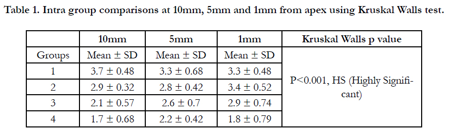

Table 1. Intra group comparisons at 10mm, 5mm and 1mm from apex using Kruskal Walls test.
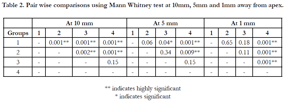
Table 2. Pair wise comparisons using Mann Whitney test at 10mm, 5mm and 1mm away from apex.
The pair wise inter group comparisons of Mann Whitney test have showed that at 10mm away from apex, Groups 2, 3 and 4 were cleaner than Group 1; Groups3 and 4 were cleaner that Group 2 and showed statistical significance while there was no difference between Group 3 and 4. At 5mm away from root apex, Groups 3 and 4 were cleaner than Group 1 while only Group 4 was cleaner than Group 2 and showed statistical significance. The remaining pairwise comparisons remained statistically insignificant. At 1mm away from apex, only Group 4 had significantly cleaner canals than all other groups while the remaining pairwise comparisons remained insignificant.
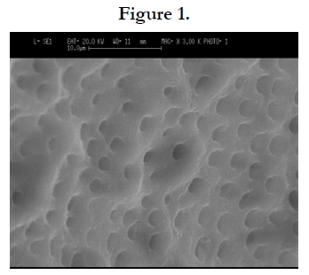
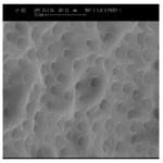
Figure 1.
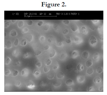
Figure 2.
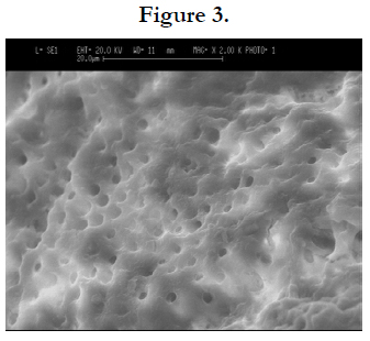
Figure 3.
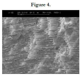
Figure 4.
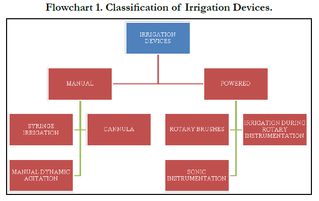
Flowchart 1. Classification of Irrigation Devices.
Discussion
This in vitro study using a scanning electron microscope was performed
primarily to evaluate the difference in the working efficiency
between NaviTip and NaviTip FX to remove the smear
layer from the canal walls of the root. 40 teeth without any pathologic
changes have been selected for the study. All the teeth were endodontically treated using the crown down technique. To simulate
the clinical condition, the apices of the teeth were sealed
using sticky wax. Later, they were divided into three sections as
coronal third (10mm from apex), middle third (5mm from apex)
and apical third (1mm from apex). The examiners were blinded
regarding the nature of study during the microscopic examination
to standardise the procedure and to avoid examination bias.
Results of the study have shown that all the four groups removed
the smear layer better in the coronal third when compared to middle
third and apical third but there were differences in the amount
of smear layer removed when compared with the other groups.
Group 4 that was instrumented with NaviTip FX and treated with
17% EDTA showed superior results than the other four groups at
all the three levels. This provides insight into the better capabilities
of the NaviTipFX in removing the smear layer. Solaiman et al
have conducted a scanning electron microscopic study on thirty
single rooted teeth using Navi Tip FX and 5% NaOCl against a
control group which did not have bristles on the irrigation device
[8]. NaviTip FX showed superior results in terms of removal
of smear layer in the coronal third of the canals8. Similarly, several
studies have been performed individually using NaviTip and
NaviTip FX and both the instruments have performed well and
were able to significantly remove the smear layer. In this present
study, Navi Tip FX has performed superiorly than Navi Tip that
is evident from the results i.e. table 2. The apical third of the root
showed canals walls with less smear layer than the other three
groups. Although 17% EDTA helps in lubricating the canal walls
and helps in easy smear layer removal that is evident from the
pairwise comparison between Group 3 and 4 at 10mm and 5mm
away from apex, its action could have been be nullified at the apical
third of the root where the NaviTip FX performed well even
if 17% EDTA was not applied. This is evident from the pairwise
comparisons between Group 4 and other three groups at the apical third (1mm away from the apex).
The superiority of NaviTip FX over NaviTip lies in the design
and the method in which the instrument is used. NaviTip FX has
an irregularly grit surface that helps in removal of smear layer and
prevents clogging of the apical third with dentinal debris. The
up and down motion combined with brush like strokes prevents
from creating any ledges that might lead to failure of the endodontic
treatment. Ethylene Diamine Tetra Acetic Acid (EDTA) is
a chelating agent that forms disodium chelate salts with calcium.
This property helps in removal of smear layer and several studies
have proven its efficacy [9-11]. The probable reason for EDTA
not performing equivalent to the test group is because of the narrower
canals that might have caused clogging in the apical third
of the root and this could be overcome using the Navi Tip FX.
According to our knowledge, very few studies have been conducted
comparing the Navi Tip and Navi Tip FX and this study
could shed some light on the superiority of the Navi Tip FX. The
limitations of this study could be considered as the future prospective
where multirooted, curved and longer canals should be
irrigated with this new device to test its effectiveness for regular
clinical use.
Conclusion
Within the limitations of present study, it can be concluded that
the Navi Tip could be replaced by Navi Tip FX for the purpose
of cleaning and removal of smear layer. EDTA, which is considered
as one of the best irrigants for removal dentinal debris may
be ineffective in some narrower canals and could be adjuncted
with manual irrigation devices such as Navi Tip FX to improve
its action.
References
- Wong R. Conventional endodontic failure and retreatment. Dent. Clin. N. Am. 2004 Jan;48(1):265-89.
- Basmadjian-Charles CL, Farge P, Bourgeois DM, Lebrun T. Factors influencing the long-term results of endodontic treatment: a review of the literature. Int Dent J. 2002 Apr;52(2):81-6.Pubmed PMID: 12013255.
- Khedmat S, Shokouhinejad N. Comparison of the efficacy of three chelating agents in smear layer removal. J Endod. 2008 May;34(5):599-602.Pubmed PMID: 18436043.
- Kuah HG, Lui JN, Tseng PS, Chen NN. The effect of EDTA with and without ultrasonics on removal of the smear layer. J Endod. 2009 Mar 1;35(3):393-6.
- Ferreira RB, Alfredo E, Porto de Arruda M, Silva Sousa YT, Sousa-Neto MD. Histological analysis of the cleaning capacity of nickel-titanium rotary instrumentation with ultrasonic irrigation in root canals. Aust Endod J. 2004 Aug;30(2):56-8.Pubmed PMID: 15378973.
- Garip Y, Sazak H, Gunday M, Hatipoglu S. Evaluation of smear layer removal after use of a canal brush: an SEM study. Oral Surg Oral Med Oral Pathol Oral Radiol Endod. 2010 Aug 1;110(2):e62-6.
- Walton RE. Histologic evaluation of different methods of enlarging the pulp canal space. J Endod. 1976 Oct;2(10):304-11.Pubmed PMID: 1068207.
- Al-Hadlaq SM, Al-Turaiki SA, Al-Sulami U, Saad AY. Efficacy of a new brush-covered irrigation needle in removing root canal debris: a scanning electron microscopic study. J Endod. 2006 Dec;32(12):1181-4.Pubmed PMID: 17174678.
- Wu L, Mu Y, Deng X, Zhang S, Zhou D. Comparison of the effect of four decalcifying agents combined with 60°C 3% sodium hypochlorite on smear layer removal. J Endod. 2012 Mar;38(3):381-4.Pubmed PMID: 22341079.
- Prado M, Gusman H, Gomes BP, Simão RA. Scanning electron microscopic investigation of the effectiveness of phosphoric acid in smear layer removal when compared with EDTA and citric acid. J Endod. 2011 Feb;37(2):255- 8.Pubmed PMID: 21238813.
- Dai L, Khechen K, Khan S, Gillen B, Loushine BA, Wimmer CE, Gutmann JL, Pashley D, Tay FR. The effect of QMix, an experimental antibacterial root canal irrigant, on removal of canal wall smear layer and debris. J Endod. 2011 Jan 1;37(1):80-4.


