Assessment of Prevalence And Gender Predilection Of Canine Impaction In Chennai Population
Joshini Shanmugam1, Aravind Kumar S2*, Suresh V3
1 Saveetha Dental College and Hospitals, Saveetha Institute of Medical and Technical Sciences, Saveetha University, Chennai, India.
2 Professor, Department of Orthodontics, Saveetha Dental College and Hospitals, Saveetha Institute of Medical and Technical Sciences, Saveetha University, Chennai, 600077, India.
3 Reader, Department of Prosthodontics, Saveetha Dental College and Hospitals, Saveetha Institute of Medical and Technical Sciences, Saveetha University, Chennai, 600077, India.
*Corresponding Author
Aravind Kumar S,
Professor, Department of Orthodontics, Saveetha Dental College and Hospitals, Saveetha Institute of Medical and Technical Sciences, Saveetha University, Chennai, 600077, India.
Tel: 9841299939
E-mail: aravindkumar@saveetha.com
Received: October 07, 2020; Accepted: November 22, 2020; Published: November 25, 2020
Citation:Joshini Shanmugam, Aravind Kumar S, Suresh V. Assessment of Prevalence And Gender Predilection Of Canine Impaction In Chennai Population. Int J Dentistry Oral Sci. 2020;7(11):1058-1062. doi: dx.doi.org/10.19070/2377-8075-20000209
Copyright: Aravind Kumar S© 2020. This is an open-access article distributed under the terms of the Creative Commons Attribution License, which permits unrestricted use, distribution and reproduction in any medium, provided the original author and source are credited.
Abstract
Tooth impaction is a pathological situation in which a tooth cannot or will not erupt into its normal functioning position. The
permanent canines are the foundation and pillar of an aesthetic smile and functional occlusion. Although most impacted teeth are
asymptomatic, some can cause complications such as pain, infection cysts, tumors, resorption of the adjacent teeth, jaw fractures,
malpositioning of the mandibular anterior teeth and marginal bone resorption near the adjacent teeth. Hence, it is important to
see the prevalence of canine impaction in the selected population so that awareness can be created among the public to report to
a dentist as early as possible. The aim of this study was to assess the prevalence of canine impaction in the Chennai population.
A retrospective cross sectional study was conducted using thes case records of patients who visited the outpatient department in
Saveetha Dental College from June 2019 to March 2020. The selection was done by non probability sampling. Data was collected
and then subjected to statistical analysis. 2069 patients who fulfilled the inclusion criteria were included in the study. Microsoft
excel 2016(microsoft office 10) data spreadsheet was used and later exported to the statistical package for social science for windows(
version 20.0 SPSS, Chicago III USA). The data was analysed through chi square. Out of 2069 patients, 5.12% reported
with canine impaction. Gender predilection shows that the canine impaction was slightly more prevalent in males(2.95%) than in
females(2.17%) (p<0.05) and maxillary canines being most commonly impacted (73.5%). Within the limitations of this study, it
was concluded that canine impaction is moderately prevalent in the Chennai population. The findings of the current study can be
used to create awareness among common people and dentists so that canine impacted cases can be reported at an early age and
treated without any complications.
2.Introduction
3.Materials and Method
4.Results and Discussion
5.Conclusion
6.Acknowledgement
7.References
Keywords
Canine; Eruption; Impaction; Prevalence.
Introduction
The different types of canine anomalies like ectopic canine eruption,
canine transmigration, canine transposition, agenesis, impaction,
usually occur due to the disturbances during development
and eruption. Since the canines are the longest teeth in the oral
cavity and the shape, position of the canines contribute to the
guidance of the teeth into the intercuspal position, the canine
teeth should be evaluated thoroughly in order to deliver the best
treatment to the patients.
Impacted teeth are those with a delayed time of eruption or that
are partially erupted [1-3]. The eruption of permanent maxillary
canine represents a complex series of events and is mostly genetically
based [4-6]. Failure of the eruption of permanent maxillary
canine is a common dental problem. Problems such as compromised
tooth movement, esthetics and functional outcome are
posed by impacted teeth [7-9].
Although most of the impacted teeth are asymptomatic, some
can cause complications such as pain, infection cysts, tumors,
resorption of adjacent teeth, jaw fractures, malpositioning of
anterior teeth and marginal bone resorption near adjacent teeth
[10-12]. A complex synchronised forward and lateral growth of
maxillary bone contributes to a successful development of maxillary
permanent canine.
As the eruption process is complex , it is inevitable that problems
may arise, leading to complications including tooth retardation or
failure of eruption. Diagnosis of canine impaction can be done
based on clinical and radiographic findings. Kettle recommended
that palpation of the buccal surface of the alveolar process
just distal to the lateral incisor may be helpful in the diagnosis
of canine impaction [12]. A bulge will indicate the presence of a
normally developing canine. A panoramic radiography is of great
clinical significance, to establish the correct surgical procedure11.
In addition to analytics studies, our team has been working on
various comparative studies [13-15]; and also recent advancements
[16-19] that are being considered as a breakthrough in orthodontics.
Various reviews [20-23] and clinical trials [24-26] also
have been conducted in order to create new views and effective
treatment options in future. The aim of the present study is to
determine the prevalence and gender predilection of impacted
canines in the Chennai population.
This study is a university setting. Study conducted in Saveetha
dental College., predominantly. Patients who reported to Saveetha
Dental College, Chennai were included for the study. Approval was
obtained from the institutional ethical committee [IEC]. Ethical
approval number- SDC/SIHEC/2020/DIASDATA/0619-0320.
Two examiners were involved in the study.
The study is a retrospective study. Data was collected from June
1, 2019 to March 31, 2020. Totally 2069 casesheets were reviewed.
The study population included patients who reported to the outpatient
department for dental treatment needs. Cross verification
of data for error was done by the presence of an additional reviewer.
Simple random sampling was done to minimise the sampling
bias.
Data of 2069 patients undergoing dental treatment was taken
from the hospital database. Repeated or incomplete patient records
were excluded. All the 2069 patient records were selected
and reviewed. Each patient’s case history was reviewed for any canine impaction. Also their OPG(if any) and clinical photographs
were obtained and studied. Data verification was done based on
the age, gender, presence of canine impaction. Data was entered
in the excel sheet in a methodical manner and was imported to
SPSS. Incomplete or uncensored data was excluded from the
study.
Data was recorded in Microsoft excel 2016 and then exported to
IBM SPSS 2.0 Software for data analysis. Independent variables
include - age, gender and dependent variables include presence
of canine impaction, arch. Descriptive and inferential statistics
was used. Inferential test includes the chi-square test which was
employed with a level of significance set at p<0.05.
Results and Discussion
Out of 2069 patients, 5.1% reported with canine impaction[figure
1]. Gender predilection shows that the canine impaction was
slightly more prevalent in males(2.95%) than in females(2.17%)
(p<0.05)[figure 2]. In regard to association of canine impaction
with arch shows that canine impaction was most prevalent in upper
arch(3.77%; n=80) followed by canine impaction in lower
arch(1.11%; n=23) and least occurrence of canine impaction in
both arches at the same time (0.24%; n=5)[figure 3] where p>0.05
is not statistically significant.
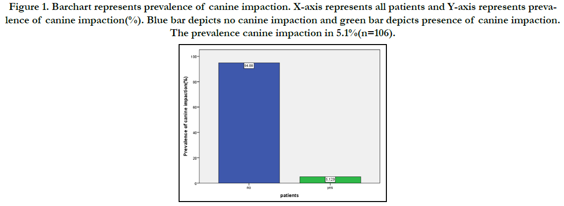
Figure 1. Pie Chart diagram showing gender wise distribution of maxillofacial traumas. There is a male predominance with 85.7% than females with 14.29%.
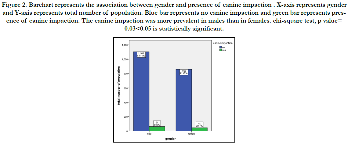
Figure 2. Bar graph depicting association between site of fracture and method of treatment where Green colour represents Mandibular fracture, X-Axis represents the Treatment done and Y-Axis represents the site of fracture. Mandible is the commonly involved Site and ORIF is the preferred treatment modality. Chi square value- 51.595 p=0.01 and hence it is statistically significant.
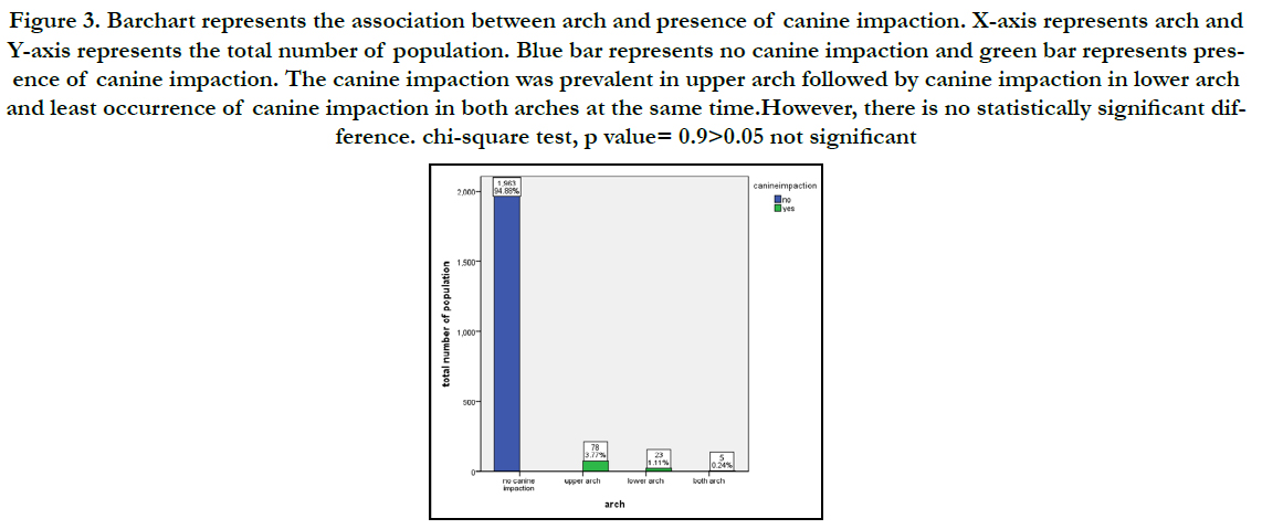
Figure 3. Barchart represents the association between arch and presence of canine impaction. X-axis represents arch and Y-axis represents the total number of population. Blue bar represents no canine impaction and green bar represents presence of canine impaction. The canine impaction was prevalent in upper arch followed by canine impaction in lower arch and least occurrence of canine impaction in both arches at the same time.However, there is no statistically significant difference. chi-square test, p value= 0.9>0.05 not significant.
The data for this retrospective study was based on residents of Chennai who reported to Saveetha Dental Hospital for treatment of impacted canine. Currently there are no existing studies investigating the prevalence of canine impaction in Chennai. Since all the data available was included without a sorting process, no bias was accepted in selection of patients. Knowledge of dental anomalies in patients is fundamental for treatment planning [27]. According to Stecker et al. [28], dental practitioners who are aware of ethnic differences in the occurrence of dental anomalies will be more aware in finding them in patients during routine examinations, and may be predictive of normal patterns of tooth development and/or eruption, allowing for prompt clinical intervention to avoid complicating pathology. The Canine impaction is one of the anomalies that should be considered by clinicians in detail. Hence, this study was conducted in order to create awareness on the severity of canine impaction among the public, so that they can report to a dentist at an early stage for treatment of impacted canines; as well as among dentists for better diagnosis. This study helps a dentist to understand the severity of impacted canines in chennai.
Although the investigated subjects may not represent the whole Chennai population, there was no significant variation in the prevalence and distribution of impacted canines. Comparison of the results of the present study with various populations was done. From the analysis, it was seen that 5.1% of canine impaction was prevalent in this study. This finding is close to a study done in the Puerto rican population [29] where prevalence of canine impaction was 3.2% and two other studies done in Riyadh which showed prevalence of 3.41% [30] and 3.37% [31] respectively. However, the prevalence found in this study, was relatively low when compared to the prevalence of impacted canines reported in other populations such as the 8.8% rate reported in Greek population1 and 6.04% rate in Mexican population [32]. The Japanese have shown to have the lowest frequency as reported in the literature, where the anomaly occurred in only 0.27% of the study population. Similar to these findings, study of a large series of full mouth dental radiographs in the USA revealed a figure of 0.92% [33]. While Brinet al.[34] in their study of an Israeli population, found a level of 1.5%. The different results from these studies may arise from racial differences and differences in the methodology of the study.
Taking into account the source of the analyzed data, which were derived from our Department, the large age range of the examined sample and the limited exclusion criteria, one might consider that the results of this study are not representative of the general population. However, the primary aim of this study was to investigate the frequency of impacted teeth in patients who attend our Department. A study done by SanthoshPatil et al.[35] had examined the patient from 8years to 72 years of age. The present study examined the similar range of age group from 18-50 years. In the present study, the maximum number of patients with impacted canines reported at the age 18-30 years when compared to the age group 31-50 years. Hence it was understood that 18-30 years age group were more aware of impacted canines and immediately reported to a dentist whereas the other age groups 31-50 years and showed that they were not much aware of canine impactions and reported much late to the dental clinic for treatment of impacted canines.
In association of canine impaction with arch, the present study reported that maximum number of patients reported with canine impactions in upper arch(3.77%; n=80) followed by canine impaction lower arch(1.11%; n=23) and least number of patients reported with canine impactions in both arches at the same time(0.24%; n=5). This finding correlates with Roher [36] where it was observed that impacted canines were 20 times more frequent in the maxilla than in the mandible. The rarity of impacted canines in the mandible was confirmed in this study. Indeed, most of the impacted canines were located in the maxilla, which had also been established as the predominant location by others [37]. Impacted canines of the mandible, of which there were only six cases in our study, are very rare in the general population. Grover and Lorton examined 5,000 radiographs and found 142 examples of impacted canines in the maxilla (2.84%) and only 11 in the mandible (0.22%) [38]. In one study that included 1.000 Turkish patients, the incidence of maxillary canine impaction was 2.9%, while the incidence of impacted mandibular canines was 0.3% [39]. In another study, Shah et al., detected only eight impacted canines in the mandible among 7.886 patients (0.10%) [40].
In a chinese population [39], the prevalence of canine impaction was high in the lower arch. However, this differs from the findings of the present study as high prevalence was seen in upper arch (2.67%).
The significance obtained from the statistical tests done to observe the relationship between impaction and gender shows that canine impaction is more prevalent in males than females in the present study. This finding does not correlate with Jacobs [40] who states that prevalence of canine impaction is higher in females than male. Most of the studies report that there is no difference between impacted canines and gender [35].
This study has certain limitations that may have affected the results. For example, the data collection period could have been longer, different population could have been taken for comparison. The sample size was small and the sample population was only representative of the patient pool at saveetha dental college and hospitals. Wider population groups should be studied in chennai. However, some authors still believe that the prevalence rates of canine impaction may reflect the prevalence rates of these anomalies in the general population.
The current research shows that despite its drawbacks, canine impaction is moderately prevalent in the Chennai population. This suggests the need to spread awareness on diagnosis and treatment modalities of exposure of impacted canines among dentists and public.
Conclusion
The prevalence of canine impaction is moderate in the Chennai
population (4.32%). The present study findings have shown
that the 18-30 years age group was aware of impacted canines
when compared to older age groups and prevalence of canine
impaction was prevalent in males than in females. The eruption
of canine plays a vital role in facial appearance, dental aesthetics,
arch development and functional occlusion. Thus, the early diagnosis
of canine impaction at an early stage is crucial to carry out
a proper and successful orthodontic treatment.
Acknowledgement
This research was supported by Saveetha Dental College and
Hospital. We thank the department of Orthodontics, Saveetha
dental college for providing insight and expertise that greatly assisted this research.
References
- Fardi A, Kondylidou-Sidira A, Bachour Z, Parisis N, Tsirlis A. Incidence of impacted and supernumerary teeth-a radiographic study in a North Greek population. Med Oral Patol Oral Cir Bucal. 2011 Jan 1;16(1):e56-61.Pubmed PMID: 20711166.
- Becker A, Sharabi S, Chaushu S. Maxillary tooth size variation in dentitions with palatal canine displacement. Eur J Orthod. 2002 Jun;24(3):313-8.Pubmed PMID: 12143095.
- . Jacoby H. The etiology of maxillary canine impactions. Am. J. Orthod 1983 Aug 1;84(2):125-32.
- Becker A. Impacted Teeth and Resorption of the Roots of Adjacent Teeth. Orthodontic Treatment of Impacted Teeth. 2013 Mar 30:173-210.
- Ericson S, Kurol J. Radiographic examination of ectopically erupting maxillary canines. Am J OrthodDentofacialOrthop. 1987 Jun;91(6):483-92.Pubmed PMID: 3473928.
- Ericson S, Kurol J. Resorption of maxillary lateral incisors caused by ectopic eruption of the canines. A clinical and radiographic analysis of predisposing factors. Am J OrthodDentofacialOrthop. 1988 Dec;94(6):503-13.Pubmed PMID: 3195514.
- Tsukiboshi M, Andreasen JO. Autotransplantation of teeth [Internet]. Quintessence Pub. Co.; 2001. 192 p.
- Hupp JR. Principles of Management of Impacted Teeth [Internet]. Contemporary Oral and Maxillofacial Surgery. 2014; 143–67. Available from: http://dx.doi.org/10.1016/b978-0-323-09177-0.00009-8
- Bystedt H. Clinical and hematological investigation of Rheumapax in surgical removal of impacted wisdom teeth from the mandible. Int J Oral Surg. 1976 Apr;5(2):66-70.Pubmed PMID: 818040.
- Gomaa N, Ellaithy M. Evaluation of crown-root angulation of lateral incisor adjacent to impacted canines using panorama and CBCT [Internet]. Egyptian Orthodontic Journal. 2016;49: 49–63.
- Vikram NR, Prabhakar R, Kumar SA, Karthikeyan MK, Saravanan R. Ball Headed Mini Implant. Journal of clinical and diagnostic research: JCDR. 2017 Jan;11(1):ZL02-3.
- Kamisetty SK, Verma JK, Arun, Sundari S, Chandrasekhar S, Kumar A. SBS vsInhouse Recycling Methods-An Invitro Evaluation. J ClinDiagn Res. 2015 Sep;9(9):ZC04-8.Pubmed PMID: 26501002.
- Felicita AS, Chandrasekar S, Shanthasundari KK. Determination of craniofacial relation among the subethnic Indian population: a modified approach - (Sagittal relation). Indian J Dent Res. 2012 May-Jun;23(3):305-12. Pubmed PMID: 23059564.
- Samantha C, Sundari S, Chandrasekhar S, Sivamurty G, Dinesh S. Comparative Evaluation of Two Bis-GMA Based Orthodontic Bonding Adhesives - A Randomized Clinical Trial. J ClinDiagn Res. 2017 Apr;11(4):ZC40- ZC44.Pubmed PMID: 28571259.
- Sivamurthy G, Sundari S. Stress distribution patterns at mini-implant site during retraction and intrusion--a three-dimensional finite element study. ProgOrthod. 2016;17:4.Pubmed PMID: 26780464.
- Felicita AS. Orthodontic extrusion of Ellis Class VIII fracture of maxillary lateral incisor - The sling shot method. Saudi Dent J. 2018 Jul;30(3):265- 269.Pubmed PMID: 29942113.
- Felicita AS. Quantification of intrusive/retraction force and moment generated during en-masse retraction of maxillary anterior teeth using mini-implants: A conceptual approach. Dental Press J Orthod. 2017 Sep- Oct;22(5):47-55.Pubmed PMID: 29160344.
- . Rubika J, Felicita AS, Sivambiga V. Gonial angle as an indicator for the prediction of growth pattern. World J Dent. 2015;6(3):161-3.
- Felicita AS. Orthodontic management of a dilacerated central incisor and partially impacted canine with unilateral extraction - A case report. Saudi Dent J. 2017 Oct;29(4):185-193.Pubmed PMID: 29033530.
- Dinesh SP, Arun AV, Sundari KK, Samantha C, Ambika K. An indigenously designed apparatus for measuring orthodontic force. J ClinDiagn Res. 2013 Nov;7(11):2623-6.Pubmed PMID: 24392423.
- Viswanath A, Ramamurthy J, Dinesh SP, Srinivas A. Obstructive sleep apnea: awakening the hidden truth. Niger J ClinPract. 2015 Jan-Feb;18(1):1-7. Pubmed PMID: 25511335.
- Krishnan S, Pandian S, Kumar S A. Effect of bisphosphonates on orthodontic tooth movement-an update. J ClinDiagn Res. 2015 Apr;9(4):ZE01-5. Pubmed PMID: 26023659.
- Jain RK, Kumar SP, Manjula WS. Comparison of intrusion effects on maxillary incisors among mini implant anchorage, j-hook headgear and utility arch. J ClinDiagn Res. 2014 Jul;8(7):ZC21-4.Pubmed PMID: 25177631.
- Ramesh Kumar KR, ShantaSundari KK, Venkatesan A, Chandrasekar S. Depth of resin penetration into enamel with 3 types of enamel conditioning methods: a confocal microscopic study. Am J OrthodDentofacialOrthop. 2011 Oct;140(4):479-85.Pubmed PMID: 21967934.
- Jamal GA, Hazza'a AM, Rawashdeh MA. Prevalence of dental anomalies in a population of cleft lip and palate patients. Cleft Palate Craniofac. J. 2010 Jul;47(4):413-20.
- Stecker SS, Beiraghi S, Hodges JS, Peterson VS, Myers SL. Prevalence of dental anomalies in a Southeast Asian population in the Minneapolis/Saint Paul metropolitan area. Northwest Dent. 2007 Sep-Oct;86(5):25-8.Pubmed PMID: 17987833.
- Tassara G, Lopez L, Hanke R, Tumanyan S, Picon F. Prevalence of impacted maxillary canines in Puerto Rican adolescents. Int J Health Sci. 2015 Jun;3:135-8.
- Alyami B, Braimah R, Alharieth S. Prevalence and pattern of impacted canines in Najran, South Western Saudi Arabian population. Saudi Dent J. 2019 Sep;32(6):300-305.Pubmed PMID: 32874070.
- Melha SB, Alturki S, Aldawasri G, Almeshari N, Almeshari S, Albadr K. Canine impaction among riyadh population: A single center experience. Int J Oral Health Sci. 2017 Jul 1;7(2):93.
- Herrera-Atoche JR, Agüayo-de-Pau MD, Escoffié-Ramírez M, Aguilar-Ayala FJ, Carrillo-Ávila BA, Rejón-Peraza ME. Impacted Maxillary Canine Prevalence and Its Association with Other Dental Anomalies in a Mexican Population. Int J Dent. 2017;2017:1-4.Pubmed PMID: 28326102.
- Dachi SF, Howell FV. A survey of 3,874 routine full-mouth radiographs: II. A study of impacted teeth. Oral Surg. Oral Med. Oral Pathol. 1961 Oct 1;14(10):1165-9.
- Brin I, Becker A, Zilberman Y. Resorbed lateral incisors adjacent to impacted canines have normal crown size. Am J OrthodDentofacialOrthop. 1993 Jul;104(1):60-6.Pubmed PMID: 8322724.
- Patil S, Maheshwari S. Prevalence of impacted and supernumerary teeth in the North Indian population. J ClinExp Dent. 2014 Apr 1;6(2):e116-20. Pubmed PMID: 24790709.
- Röhrer A. Displaced and impacted canines A radiographic research. International Journal of Orthodontia, Oral Surgery and Radiography [Internet]. 1929 Oct 1;15(10):1003–20.
- Kramer RM, Williams AC. The incidence of impacted teeth. A survey at Harlem hospital. Oral Surg Oral Med Oral Pathol. 1970 Feb;29(2):237-41. Pubmed PMID: 5262845.
- Grover PS, Lorton L. The incidence of unerupted permanent teeth and related clinical cases. Oral Surg Oral Med Oral Pathol. 1985 Apr;59(4):420-5. Pubmed PMID: 3858781.
- Sağlam AA, Tüzüm MS. Clinical and radiologic investigation of the incidence, complications, and suitable removal times for fully impacted teeth in the Turkish population. Quintessence Int. 2003 Jan;34(1):53-9.Pubmed PMID: 12674360.
- Shah RM, Boyd MA, Vakil TF. Studies of permanent tooth anomalies in 7,886 Canadian individuals. I: impacted teeth. Dent J. 1978 Jun;44(6):262- 4.Pubmed PMID: 277399.
- Sajnani AK, King NM. Dental anomalies associated with buccally- and palatally- impacted maxillary canines. J InvestigClin Dent. 2014 Aug;5(3):208- 13.Pubmed PMID: 23946209.
- Jacobs SG. The impacted maxillary canine. Further observations on aetiology, radiographic localization, prevention/interception of impaction, and when to suspect impaction. Aust Dent J. 1996 Oct;41(5):310-6.Pubmed PMID: 8961604.


