A Comparative Evaluation Of Canine Retraction and Anchor Loss using Ceramic and Stainless Steel MBT Pre-adjusted Edgewise Brackets using Three Dimentional Finite Element Method
Trupti Sadhunavar1*, Tejashri Pradhan2, Sumedh Deshpande3, Aarti Sethia3, Kanhoba Mahabaleshwar Keluskar4
Lecturer, Department of Orthodontics, KLE VK Institute of Dental Sciences, KAHER, Belagavi, India.
2 Professor and Head, Department of Orthodontics, KLE VK Institute of Dental Sciences, KAHER, Belagavi, India.
3 Postgraduate Student, Department of Orthodontics, KLE VK Institute of Dental Sciences, KAHER, Belagavi, India.
4 Professor at Department of Orthodontics, KLE VK Institute of Dental Sciences, KAHER, Belagavi, India.
*Corresponding Author
Dr. Trupti Sadhunavars,
Lecturer, Department of Orthodontics, KLE VK Institute of Dental Sciences, KAHER, Belagavi, India.
Tel: 9449815111
E-mail: sadhutrup@gmail.com
Received: September 05, 2020; Accepted: October 02, 2020; Published: October 17, 2020
Citation:Trupti Sadhunavar, Tejashri Pradhan, Sumedh Deshpande, Aarti Sethia, Kanhoba Mahabaleshwar Keluskar. A Comparative Evaluation Of Canine Retraction and Anchor Loss using Ceramic and Stainless Steel MBT Pre-adjusted Edgewise Brackets using Three Dimentional Finite Element Method. Int J Dentistry Oral Sci. 2020;7(10):854-859. doi: dx.doi.org/10.19070/2377-8075-20000169
Copyright: Trupti Sadhunavar©2020. This is an open-access article distributed under the terms of the Creative Commons Attribution License, which permits unrestricted use, distribution and reproduction in any medium, provided the original author and source are credited.
Abstract
Background and Objectives: Simulation of orthodontic force can provide crucial insights on design and implications of orthodontic
apparatus like brackets. Ceramic brackets although aesthetic, can create frictional resistance with orthodontic wires.
Hence, this study employed Finite element method to evaluate and compare the canine retraction and anchor loss facilitated by
pre-adjusted edgewise ceramic brackets and conventional metal bracket systems.
Materials and Method: The study model was generated using the CBCT with favourable conditions of first premolar extraction
for orthodontic correction. Finite element model of the maxilla and the teeth were simulated using the MIMICS software. A force
of 50 grams was applied for canine retraction with elastomeric chain using preadjusted MBT (MacLaughlin, Bennet and Trevisi
system) edgewise brackets of ceramic and metal respectively. ANSYS software was used for the analysis to evaluate the rate of
canine retraction, anchor loss and stress generated.
Results: The effective tooth displacement, von Mises stress and stress simulated by FEM were comparatively greater inthe metallic,
than inthe ceramic brackets. During retraction, distal, lingual and intrusive displacement of canine was visualised to be greaterwith
metallic bracketsin comparison to their ceramic counterparts. Metallic wire and bracket featured the same Young’s modulus
as they are made of similar materials hence, the force applied led to a greater retraction of canine, unlike ceramic brackets.
Conclusion: As per FEM analysis, stainless steel brackets were a more effective method for tooth displacement andrapid canine
retraction with limited anchor loss. FEM can improve suitability and efficiency of orthodontic design by enabling a multidimensional
analysis.
2.Introduction
3.Materials and Methods
4.Results
5.Discussion
6.Conclusion
7.Refereces
Keywords
Ceramic Brackets; Finite Element Analysis; Stainless Steel Brackets; MBT Bracket System.
Introduction
Canine retraction refers to a deliberate posterior shift of canines
to foster enhanced stability, functionality, and aesthetics. This is
often achieved either by using frictional (sliding) or non-frictional
(non-sliding) mechanics [1]. Sliding mechanics are generally preferred
if movement of teeth is required over a relatively long distance
[2]. Precluding intrusion of posteriors into the extraction
space i.e. anchorage control - is an essential component of treatment
design in orthodontics. Anchorage loss can be ascribed to
improper anchorage preparation, excessive force, and impingement
of the roots of the incisors or anterior teeth to the labial
cortical plate etc [3].
Orthodontic brackets are viewed as an advent of the modern
medicine, however the desire for well aligned teeth has a rich history
dating back to the ancient times. Metal braces composed of
gold were sought after in the 1960s, which evolved into brackets
being directly adhered to the teethby the 1980. Technological advancements
have catapulted innovations in the design and use of
orthodontic appliances [4]. During the last decade a wide range of
metal, plastic and ceramic brackets, based on the straight wire system have evolved and are accessible. Pre-adjusted brackets have
been reinvented several times; Andrew’s straight wire appliance
being the first followed by Roth’s single prescription for mass
correction of the dentition [5]. One such system was the MacLaughlin,
Bennet and Trevisi system(MBT system) introduced
by Mclaughlin, Benettand Trevisi [6]. Although ceramic brackets
provide a more aesthetic solution, they are less efficient in enabling
teeth movementas compared to the metal brackets [5]. Ceramic
brackets also have disadvantages such as, inability to form
chemical bonds with resin adhesives, low fracture toughness and
an increased frictional resistance between metal arch wires and ceramic
brackets [7, 8]. Stainless steels possess desirable properties
such as passivation, strong formability, ductility, weldability and
wear resistance [9]. However,concerns of intergranular corrosion
resistance, cytotoxicity and heat susceptibility accompany stainless
steel brackets [9, 10]. Hence, a more preemptive approach is
needed to assess the suitability of these two orthodontic appliances,
while ensuring patients safety and satisfaction.
In this context, FEM heralds evidence based orthodontic practice,
by breaking down a complex problem into smaller constituents
that are accurately measured and reconstituted to yield a more
comprehensive and precise solution [11]. FEM is a numerical
method for solving a differential or integral equation, particularly
useful for problems with complicated geometries, loadings and
material properties where analytical solutions cannot be obtained
[12, 13]. It has been experimented with in the field of medicine
and dentistry, including Orthodontics, to assess stress and strain,
in various biological and mechanical systems [14]. FEM presents
an opportunity to non-invasively visualize and analyse the biomechanical
stresses and displacement effectuated by orthodontic
brackets, thereby allowing room for optimization of design.
Although comparative studies on the rate of canine retraction
noted in ceramic and stainless steel brackets are available, only
a few have employed finite element method (FEM) as a tool for
their analysis, and have mostly focused on ceramic brackets so
far.The resultant stress generated between orthodontic wires and
a ceramic bracket during canine retraction remains debateable
due to lack of convincing data. Hence, the aim of the study was
to determine the tooth displacement and stress generated at the
canine and molar regions using PEA (pre-adjusted edgewise) ceramic
and metallic brackets and to compare their efficiency during
canine retraction by FEM study.
The study was conducted on a three dimensional finite element
model by using the cbct with ideal conditions of first premolar
extraction for orthodontic correction. The duration of this study
was 8 months. Ethical clearance from the Institutional Review
Board with ethical clearance number EC:863 a.
A computed tomography CBCT scanned image of the maxillary
arch with first premolars extraction was acquired, following which,
a finite element model of the maxilla l and the teeth were simulatedusing
the MIMICS software. Computer-aided design models of E chain, metal and ceramic brackets, and 0.019 "x0.025” SS Archwire
were designed. Elastomeric chain was fixed using 3-MATIC
software. The ANSYS10.0 software (ANSYS Inc., Houston, PA,
USA) software was employed for carrying out volumetric meshing.
A force of 50 grams was applied for canine retraction with
elastomeric chain using preadjusted MBT (MacLaughlin, Bennet
and Trevisi system) edgewise brackets of ceramic and metallic nature.
ANSYS software was used for the FEM analysis of the rate
of canine retraction, anchor loss and stress generated.
Burrstone and Grooves reported that the optimal force for individual
tooth movement was 50 – 75 grams [14]. Therefore, the
force selected for individual canine retraction in this study was
set at 50 grams (0.5N) of force. This standardised force applied
from the elastomeric chain for the distal movement of canine
with both, the ceramic bracket and 0.019×0.025-inch stainless
steel wire. Antecedent to the placement of 0.019"x0.025” stainless
steel wire,preliminary leveling and alignment were done using
014” round Niti wire, 016” round Niti wire, 018” round Niti
wire, 17*25 rectanguar NiTi wire, 19*25 rectangular Niti wire and
19*25 rectangular Stainless steel wire. Subsequently, the rate of
canine movement and the stress distribution were analysed.
Stress (N/mm2) was calculated and depicted as a spectrum of
colours, which represented different stress levels. Red colour of
the spectrum indicated a maximum principal stress and the following
colours like orange, yellow, green and blue represented the
reducing level of stresses while blue colour represented the lowest
level of stress (Figure 1). The rate of retraction was estimated
along all three planes of space by dividing the distance travelled
by the time taken to achieve space closure and expressed in mm.
The extent of movement incurred in the direction opposing the
direction of applied resistance was determined to be the anchorage
loss observed.

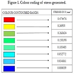
Figure 1. Colour coding of stress generated.
Results
In the present study, deflection of beam (DMX), maximum stress
(SMX), and minimum stress (SMN) along each of three dimensions
i.e. X, Y and Z were calculated using FEM.
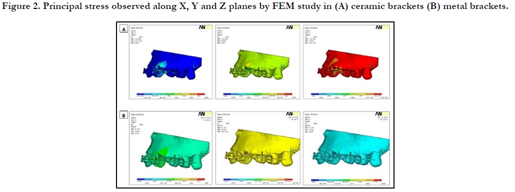
Figure 2. Principal stress observed along X, Y and Z planes by FEM study in (A) ceramic brackets (B) metal brackets.
The rate of canine retraction with the ceramic bracket was 0.40mm along with lingual tipping and intrusion. Evaluation of the resultant stress from ceramic brackets showed maximizationalong the X axis i.e. labio-lingual orientation, reporting a von Mises stress of 2749MPa.
The rate of canine retraction with the metallic bracket was 1.279 mm with von Mises stress of 3019 MPa (Figure 4). Finite element method revealed that metal brackets impose maximum stress onthe Y axis i.e. mesio-distal direction (Figures 2 & 3). Metal brackets also showed a space closure along with lingual tipping and intrusion of canine. Molar displacement was more in the X axis (Figure 5).

Figure 3. Stress levels observed with respect to molars in X, Y and Z planes by FEM study in (a) ceramic brackets (b) metal brackets.

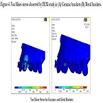
Figure 4. Von Mises stress observed by FEM study in (A) Ceramic brackets (B) Metal brackets.

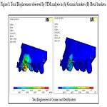
Figure 5. Total Displacement observed by FEM analysis in (A) Ceramic brackets (B) Metal brackets.
Anchor loss measured with ceramic bracket was greater along the X axis (Figure 6).
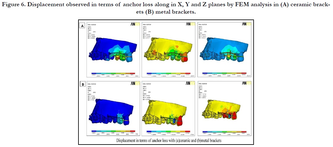
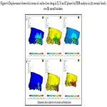
Figure 6. Displacement observed in terms of anchor loss along in X, Y and Z planes by FEM analysis in (A) ceramic brackets (B) metal brackets.
Discussion
Fixed-appliance structures constitute an integral part of modern
day orthodontics and hence have elicited panoply of orthodontic
studies. However, most studies in this domain canvass the efficacy
and suitability post-treatment, when precious resources have
already been spent. A more predictive and proactive approach is
required which can provide adequate leeway for optimization of
orthodontic bracket design. In this study, the rate of canine retraction,
anchor loss and the stress generated during the canine retraction
have been evaluated by using FEM. The stress generated
was evaluated using von Mises Stress. The magnitude of stress
was compared between the ceramic and metallic brackets andit
was found to be a maximum in metal brackets when used for canine
retraction. The stress intensities for anchoring teeth with the
orthodontic bands were the highest. A generalised distribution of
stresswas seen around the previously extracted premolar socket
area. A higher stress was noted on the converging planes in the
extraction socket.
The application of an orthodontic force results primarily in stress
and strain distributions in the root of the tooth and the supporting
structures, finally resulting in bone re-absorption on the
compression side and bone apposition on the tension side [15].
Importantly when a tooth moves under orthodontic force, two kinds of movement are observedi.e. tipping and bodily movement.
During distal tipping movement, there is a greater stress
distribution in the cervical part of root than that of the apical part
of the root. While in the distal translation, there is quite an even
distribution of stress volume along the root surface [16]. Since the
displacement pattern and speed of teeth are determined by the
orthodontic force generated, the outcome of orthodontic procedures
are directly influenced by the biomechanics of orthodontic
structures. Therefore, the measurement of orthodontic force
generated by appliance loading is imperative for proper bracket
design and optimization, planning, as well as predicting efficacy
of the treatment chosen [17].
In vivo monitoring of orthodontic force is riddled with procedural
complexities and operational constraints [17]. Numerous
in vitro force estimation techniques have been demonstrated previously.
However, finite element method (FEM) uses functions
based on theoretical calculations to facilitate a more meticulous
and multi-variate analysis. Controllable experimental criteria, affordability,
and short experimental cycle are the hallmarks of this
method [18-20].
A 3-D quantitative analysis like FEM requires using a geometric
model accurate both in anatomical and physical characteristics,
along with a computer application. This involves the subdivision
or discretization of the structure under consideration into a number of finite sections or elements. These elements are connected
at intersections called nodes [6]. A discretized complex structure
or continuum usually contains many elements, which can be arranged
in two or three dimensions in layers. In our study, FEM
was instrumental in the precise quantification and location of
stress. General distribution of stresses was apparent around the
previously extracted premolar socket area, with higher stress on
the converging planes. Stress intensities for anchoring teeth with
the orthodontic bands were highest.
A study by Kojima et al. studied maxillary canine retraction using
the FEM model, wherein on application of a constant force to
the brackets the canine tipped initially but then showed steady
movement [21]. This tipping of the canine was found to reduce
on decreasing either the wire size or force applied. Similarly, Ammaret
al. noted that as the hook length on the orthodontic bracket
increases, a corresponding decline in canine PDL stress from 80
kPa to 22 kPa was observed [22].
Li et al. presented a study on the three-dimensional finite element
analysis of the mechanical stress on the root from orthodontic
tooth movement by sliding mechanics and concluded that when
orthodontic forces were applied to the tooth, the stress was mainly
concentrated at the neck of the tooth decreasing uniformly to
the apex and crown [23]. The highest stress on the root was on
the cervical margin of the canine, followed by the apical region
of the canine [23]. These results are congruent with the results of
the present study. The stress was more at the apical region than
the coronal part of tooth in both the metal as well as the ceramic
brackets.It was reported in a previous study thatthe sliding force
of 2N was ideal to ensure the bodily orthodontic tooth movement
[24]. Hence, a force higher than 50 grams was used in the present
study, which would lead to bodily movement of the canine. FEM
study on stress generation during rapid canine distraction carried
out by Kalili et al., showed results congruent with the present
study. They also mentioned the need for anchorage preservation
as the stress generated was high in the molar region [25].
The stainless-steel wire (0.019×0.025inch SS) used in the present
studyacted as a stabilizer to maintain the obtained position
of the canine during the earlier levelling and aligning stages of
the treatment. The present study results are in concordance with
the study done by Tanneet al.which reported that the amount of
tooth movement produced by ceramic bracket was significantly
less than the movement produced by a metal bracket [26]. The
wire surfaces were scratched more obviously by ceramic brackets
than by metal bracket. AlSayagh et al. concluded that the canine
retraction with the standard ceramic bracket with elastomeric
chain could be regarded as the best combination variable that
produced less tipping [24]. In comparison,the results in present
study showed that the total displacement was more with the pre
adjusted edgewisemetallic brackets than the pre adjusted edgewise
ceramic brackets. Even the stress on all the three-principal axes
i.e. X, Y and Z was more in the metallic bracket than the ceramic
bracket.The friction at wire and bracket interface was more in the
ceramic brackets than the metallic bracket.
The efficiency of the tooth movement was slightly lost in the ceramic
bracket as compared to the metallic bracket. The loss of
efficiency seems to be due to the friction at the wire and bracket
interface in ceramic bracket while the friction in the metallic
bracket at the wire and bracket interface was comparatively less,
with more tooth displacement as indicated by the microscopic
findings of the wire surfaces [28].
Anchor loss was also studied and it was observed that a very minimal
stress was generated at the molar region with both metallic
and ceramic brackets with almost negligible displacement. Hence,
the results cannot be stated significant. But in comparison the
stress generated was more at the apical region with the ceramic
bracket than the metal bracket. It can be stated that the anchor
loss would be greater with the ceramic bracket than the metal
bracket.
Even though the results of the study concluded that the space
closure was more effective with the metallic brackets, there were
a few limitationsof the present study. The frictional parameters
were not incorporated in the study or model design. Also predictive
analysis of FEM could have been compared to outcomes observed
on clinical assessment. So, these parameters can be included
and evaluated in future studies. The performance of different
orthodontic brackets in relation to the varying bone morphology
or in patients with concomitant dental pathologies can also be
investigated. The novelty of this study lies in its use of FEM, to
conduct a detailed analysis of the components constituting stress
and displacement in ceramic and stainless steel MBT Preadjusted
Edgewise Brackets. Multidimensional observations of these two
types of routinely used brackets have been brought to fore their
benefits and deficiencies, that must be considered while establishing
suitability, and creating novel designs.
Conclusion
The rate of canine retraction between preadjusted edgewise ceramic
and conventional metallic brackets highlighted a clinical difference.
It was observed that the retraction was maximum with
metal bracket in all the three planes compared to the ceramic
bracket. Metallic brackets proved to be more efficient in the closure
of space. Frictional values are high for the ceramic bracket
when compared to the metallic bracket. The use of FEM demonstrated
that the amount of orthodontic force required to move
a tooth depends upon the amount of friction created. Selection
of material with a low coefficient of friction is required to optimize
the treatment. Hence, it is recommended that orthodontic
appliances must first be evaluated by FEM to adjudge its suitability
following which clinical trials are recommended. As FEM is a
building foundation to any innovation, so as not to compromise
the treatment progress and provide a more fructuous outcome.
References
- Kulshrestha RS, Tandon R, Chandra P. Canine retraction: A systematic review of different methods used. J Orthod Sci. 2015; 4(1):1-8. PMID: 25657985.
- Abu-Shahba R, Alassiry A. Comparative evaluation of the maxillary canine retraction rate and anchorage loss between two types of self-ligating brackets using sliding mechanics. J Orthodont Sci. 2019; 8:3. PMID: 31001495.
- Creekmore TD, Eklund MK. The possibility of skeletal anchorage. J ClinOrthod. 1983; 17(4): 266-9. PMID: 6474142.
- Gemmi C. A Brief History of Orthodontics [Internet]. Orthodontics Limited. 2018 [cited 25 August 2020]. Available from: https://www.orthodonticslimited. com/orthodontics/orthodontics-history/#:~:text=By%20the%20 1960s%20gold%20was,to%20be%20used%20a%20lot.
- Talapaneni AK, Supraja G, Prasad M, Kommi PB. Comparison of sagittal and vertical dental changes during first phase of orthodontic treatment with MBT vs ROTH prescription. Indian J Dent Res. 2012; 23: 182-6. PMID:22945707.
- Padmaraj AV, Kapila S, Duncausan MG, Nanda RS. Evaluation of friction between ceramic brackets and orthodontic wires of four alloys. Am J Orthod and Dentofac Orthop. 1990; 98: 499-506. PMID: 2248227.
- Karamouzos A, Athanasiou AE, Papadopoulos MA. Clinical characteristics and properties of ceramic brackets: A comprehensive review. Am J. Orthod Dentofacial Orthop. 1997; 112: 34-40. PMID: 9228839.
- Halimia A, Doukkalib HBA, Azeroualc MF, Zaouia F. A systematic review of force decay in orthodontic elastomeric power chains. International Orthodontics. 2012; 10(3): 223–240. PMID: 22906378.
- Arango Santiago, Ossa Claudia. Stainless Steel: Material Facts for the Orthodontic Practitioner. RevistaNacional de Odontología. 2015; 11(20): 73-82.
- Oh KT, Choo SU, Kim KM, Kim KN. A stainless steel bracket for orthodontic application. Eur J Orthod. 2005; 27(3): 237-244. PMID: 15947222.
- Singh JR, Kambalyal P, Jain M, Khandelwal P. Revolution in Orthodontics: Finite element analysis. J Int Soc Prev Community Dent. 2016; 6(2):110- 114. PMID: 27114948.
- Penedo ND, Elias CN, Pacheco MCT, de Gouvea JP. 3D simulation of orthodontic tooth movement. Dental Press J Orthod. 2010; 15(5): 98-108.
- Dixit US. Finite Element Method: An introduction [Internet]. Department of Mechanical Engineering, Indian Institute of Technology Guwahati- 781039, India. [Cited on 21st April 2020] Available from: http://www. iitg.ac.in/engfac/rtiwari/resume/usdixit.pdf
- Burrstone CJ, Grooves MH. Threshold and optimum force values for maxillary anterior tooth movement. J Dent Res. 1960; 39: 694.
- Liou EJ, Huang CS. Rapid canine retraction through distraction of the periodontal ligament. Am J Orthod Dentofacial Orthop. 1998; 114(4): 372-82. PMID: 9790320.
- Jing Y, Han XL, Cheng BH, Bai D. Three-dimensional FEM analysis of stress distribution in dynamic maxillary canine movement. Chinese Sci Bulletin. 2013; 58: 2454–9.
- Zhou X, Gan Y, Zhao Q, Xiong J, Xia Z. Simulation of orthodontic force of archwire applied to full dentition using virtual bracket displacement method. Int J Numer Method Biomed Eng. 2019; 35(5): e3189.
- Zhang Z, Chen J, Li E, Li W, Swain M, Li Q. Topological design of all‐ceramic dental bridges for enhancing fracture resistance. Int J Numer Methods Biomed Eng. 2016; 32(6): e02749. PMID: 26444905.
- Zhang D, Han X, Zhang Z, Jie Liu, Chao Jiang, Nobuhiro Yoda, et al. Identification of dynamic load for prosthetic structures. Int J Numer Methods Biomed Eng. 2017; 33(12): 12. PMID: 28425209.
- Cheng Y, Lin D, Jiang C, Lin Y. Dental implant customization using numerical optimization design and 3‐dimensional printing fabrication of zirconia ceramic. Int J Numer Methods Biomed Eng. 2017; 33(5): 1-12. PMID: 27539228.
- Kojima Y, Fukui H. Numerical simulation of canine retraction by sliding mechanics. Am J Orthod Dentofacial Orthop. 2005; 127(5): 542-551. PMID: 15877034.
- Ammar HH, Ngan P, Crout RJ, Mucino VH, Mukdadi OM. Three-dimensional modeling and finite element analysis in treatment planning for orthodontic tooth movement. Am J Orthod Dentofacial Orthop. 2011; 139(1): e59-e71. PMID: 21195258.
- Li P, Mao J, Peng Z. Three-dimensional finite element analysis of the mechanical stress on root from orthodontic tooth movement by sliding mechanics. J HuazhongUniv Sci Technolog Med Sci. 2007; 27(6): 745-7. PMID: 18231760.
- Reitan K. Clinical and histological observations on tooth movement during and after orthodontic treatment. Am J Orthod Dentofacial Orthop. 1967; 53: 721-45. PMID: 5233926.
- Kalili T, Caputo A, Lai E, Nishimura, Ichiro, Gordillo P. Stress generated by laminated aligners for class iii mandibular distraction. Paper presented at: COAST: Conferences on Orthodontic Advances in Science and Technology. 2008.
- Tanne K, Matsubara S, Shibaguchi T, Sakuda M. Wire friction from ceramic brackets during simulated canine retraction. Angle Orthod. 1991; 61(4): 285-90. PMID: 1763839.
- Al-Sayagh NM, Ismael AJ. Evaluation of Canine Tipping During its Retraction with Sliding Mechanics. (An in Vitro Study) Al – Rafidain Dent J. 2012; 12(1): 43-51.
- Guerrero AP, Filho OG, Tanaka O, Camargo ES, Vieira S. Evaluation of frictional forces between ceramic brackets and archwires of different alloys compared with metal brackets. Braz Oral Res. 2010; 24(1): 40-5. PMID: 20339712.


