Diagnostic Potential of Mandibular Second Premolar and its Relation to Physiological and Skeletal Maturity in Girls
MSV Kishore1, Sravya R.L2*, Dasari.AK3, Varalakshmi C2, Vishal G2
1 Professor and Head, Department of Orthodontics, SVS Institute of Dental Sciences, Mahabubnagar, Telangana, India.
2 Post Graduate Student, Department of Orthodontics, SVS Institute of Dental Sciences, Mahabubnagar, Telangana, India.
3 Senior Lecturer, Department of Orthodontics, SVS Institute of Dental Sciences, Mahabubnagar, Telangana, India.
*Corresponding Author
Dr. Rajulapudi Lakshmi Sravya,
Post Graduate Student, Department of Orthodontics,
SVS Institute of Dental Sciences, Mahabubnagar, Telangana, India.
Tel: 7799130050
E-mail: lakshmirajulapudi@gmail.com
Received: April 27, 2016; Accepted: July 23, 2016; Published: July 25, 2016
Citation: MSV Kishore, Sravya R.L, Dasari AK, Varalakshmi C, Vishal G (2016) Diagnostic Potential of Mandibular Second Premolar and its Relation to Physiological and Skeletal Maturity in Girls. Int J Dentistry Oral Sci. 3(7), 291-295.DOI : dx.doi.org/10.19070/2377-8075-1600059
Copyright: Sravya RL© 2016. This is an open-access article distributed under the terms of the Creative Commons Attribution License, which permits unrestricted use, distribution and reproduction in any medium, provided the original author and source are credited.
Abstract
Aims and objectives: 1). To estimate the association between mandibular second premolar calcification stages and cervical vertebrae maturation 2). To estimate relationship of dental maturation and sexual maturation in female subjects 3). To analyse discriminatory ability of premolar calcification in assessing subjects having potential growth.
Materials and Methods: Panaromic radiographs and lateral cephalograms of 90 girls subjects (before menarche) were selected for the study. Mandibular second premolar calcification stage was estimated according to Demirjian index-DI and cervical vertebrae maturation index (CVMI) was estimated using Hassel and Farman index.
Results: Highly significant statistical correlation(C= 0.603) was found between DI and CVMI stages in girls.CVMI-2 correlated with stage DI-E, CVMI-3 correlated with stage DI-F, CVMI-4 and 5 correlated with stage DI-G. Dental maturation preceeded sexual maturation and 7.8% of the sample population showed completion before attaining sexual or skeletal maturity.
Conclusion: Mandibular second premolar can be used as a preliminary method of estimating skeletal maturity. Calcification stage DI–F of premolar corresponds to peak growth in females.However, completion of calcification may be misleading, therefore other diagnostic methods should be considered for confirming the growth status of an individual.
2.Introduction
3.Materials and Methods
3.1.Evaluation of Skeletal Maturity According to Hassel And Farman Method
3.2.Evaluation of Dental Maturity of Mandibualr Second Premolar According to Demirjian Index
4.Statistics
5.Results
6.Discussion
7.Conclusions
8.References
Keywords
Skeletal Maturation; Dental Maturation; Demirjian Index; Tooth Calcification; Cervical Vertebrae Maturation; Mandibular Second Premolar; Menarche.
Introduction
Physiological maturity shows considerable variation among different persons. Orthodontic treatment planning involves anticipation of future growth potential of facial skeleton [1]. Few clinical considerations such as use of functional appliances, extra-oral traction etc.are strongly related to rates of craniofacial growth. Therefore, it is essential to establish whether pubertal growth spurt of the patient has been reached or completed.
Rate of progress towards maturity can be estimated by various indicators such as somatic, sexual, skeletal and dental, which are explained below.
Somatic: Annual growth increments in height to estimate growth velocity are valid means determine spurt. Maximum growth in height coincides with maximum facial growth in majority of subjects [2]. However , this method is of little value to orthodontists as identification of spurt is retrospective.
Sexual: Development of secondary sexual characters was related to growth spurt in several studies. It was estimated that peak height velocity precedes menarche by approximately 2 year in female subjects [3], while voice change may be related to growth spurt. Beginning of voice change marks the most intense phase of spurt and transformation to adult relates to deceleration of growth [4].
Skeletal: Hand wrist radiographs [5-7] and cervical vertebrae maturation [8, 9] were proposed as best indicators to assess growth by many scientists. Ossification time of hook of the hamate, adductor sesamoid [10] and capping of epiphysis of middle phalynx of third finger [11, 12] were strongly correlated with peak height velocity in many previous studies [13].
Dental: Dental maturity in subjects can be estimated by using either dental emergence stages or dental calcification stages. Estimation of these dental indicators is relatively simple, requiring a routine diagnostic panaromic or a periapical radiograph [14]. Dental emergence stages (DES) were previously identified as not so useful in indicating pubertal growth [15]. Dental calcification stages of various teeth has been widely studied to estimate its correlation with skeletal maturity. Few studies reported no correlation between development of lower premolars, second molars and the maturational state of the patient [16]. Green et al suggested that skeletal age, height, weight are possibly controlled to some degree by the same forces of growth and development, which is unrelated to dental development [13]. However, several other studies recognized dental indicators to reliably estimate skeletal maturity. Mandibular canine calcification stage G was found to coincide with various skeletal indicators of the pubertal growth spurt [6-8]. A recent report on relation between calcification stages of various teeth to skeletal maturation reported that mandibular second premolar showed highest correlation among all the teeth in Thai subjects [17], another report suggested second mandibular molar showed high correlation with CVMI [18]. Some studies show that dental maturity may vary with gender and racial factors [1, 19, 20], suggesting a caution for applying these results universally. The present study attempts to establish a correlation between mandibular second premolar calcification stages and other indicators of growth among Indian females to check its validity in estimating growth.
The aims of this study were:
- To estimate the association between mandibular second premolar calcification stages and cervical vertebrae maturation
- To estimate relationship of dental maturation and sexual maturation in female subjects
- To analyse discriminatory ability of premolar calcification in assessing subjects having potential growth.
Materials and Methods
The study followed a cross sectional design. 90 pre pubertal girls with age ranging from 10-13 years were selected from out patients reporting to SVS institute of dental sciences Mahabubnagar. Informed consent was obtained from their guardians, dental checkup was supplemented with radiographic analyses for diagnosing overall dental condition of the patient. This radiation dosage was within the levels permitted for routine diagnostic purpose. High quality digital images and adequate use of radiation shields (thyroid collar and lead apron) was used for the exposing the subjects. Radiographic exposure for lateral cephalogram and orthopantomogram was done on the same day.
Inclusion criteria :
- Chronological age between 10-13 years.
- Moderately built girls who haven’t reached menarche.
- Good general health with absence of any nutritional problems.
- No use of any anti-inflammatory agents or antibiotics.
- No active orthodontic treatment
Cervical vertebral maturation was analysed based of index proposed by Hassel and Farman [21] ranging from CVM I TO CVM VI as shown in Table 1.
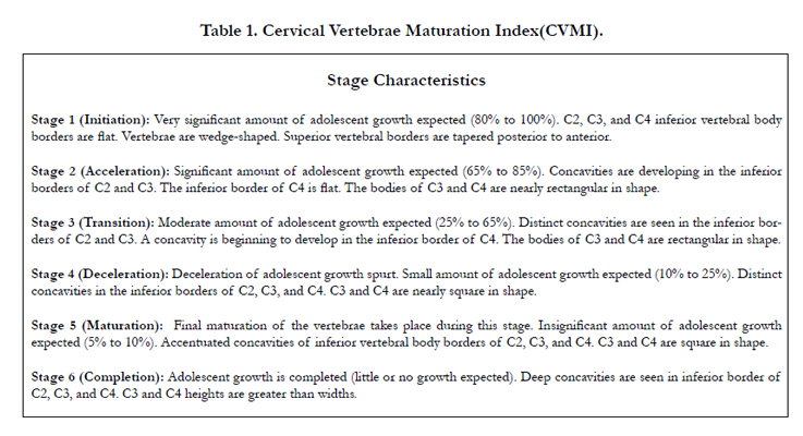

Table 1. Cervical Vertebrae Maturation Index(CVMI).
Mandibular left second premolar calcification stage was assessed according to Demirjian index [22] ranging from DI-A TO DI-H as shown in Table 2.
Comparison between skeletal maturity and dental maturity was carried out to estimate the potential of second premolar in assessing skeletal growth.
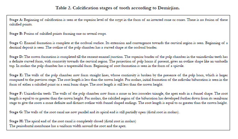
Table 2. Calcification stages of tooth according to Demirjian.
Statistics
We performed statistical analysis using SPSS software version 17.0. Two observers were blinded to asses all the radiographs according to the indices. Inter observer agreement and variability was estimated using Cronbach’s alpha value. To estimate relationship between DI and CVMI Spearman’s rho correlation coefficient and Pearson contingency coefficient were estimated.
Results
Table 3 shows highly significant inter observer agreement (p=0.001) in estimating calcification stages and cervical vertebrae maturation stages. Cronbachs alpha value was 0.872 showing relatively high internal consistency. Table 4 shows distribution of pre pubertal sample population among various stages of premolar calcification and cervical vertebrae stages. Complete calcification of the tooth with DI-H was noticed in 8 girls of the pre pubertal sample population. While, CVMI-VI was not noticed in the sample and CVMI-V was evident in 5 subjects. Correlation between calcification stages and cervical vertebrae maturation stages was positive. Spearman’s rho Correlation Coefficient was 0.465 with high significance (p < 0.001). Pearson contingency coefficient was 0.603 (p < 0.001; Highly Significant). CVMI-2 correlated with stage DI-E, CVMI-3 correlated with DI-stage F, CVMI-4 and 5 correlated with stage G.
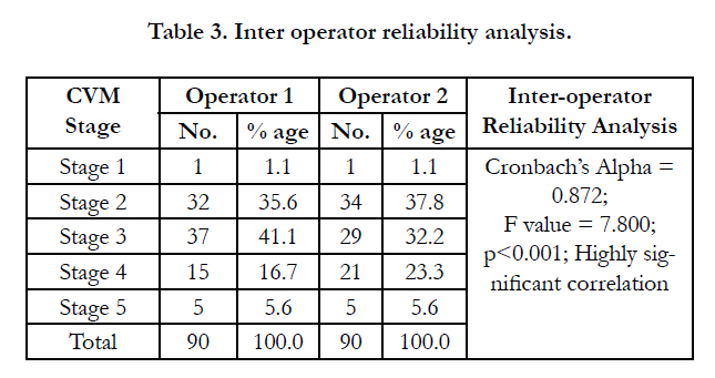
Table 3. Inter operator reliability analysis.
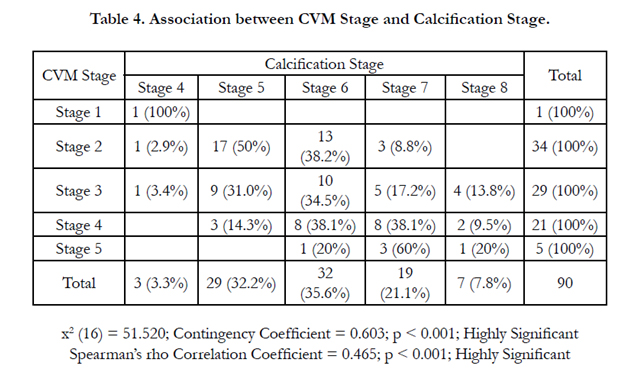
Table 4. Association between CVM Stage and Calcification Stage.
Discussion
Cervical vertebrae maturation was suggested as a reliable indicator for estimating skeletal maturity [13, 23]. CVMI as given by Hassel and Farman was used to analyse the lateral cephalograms obtained from sample population. The variability and repeatability of this method was previously questioned in a study by Gabriel et al., [24]. However, our study showed good interobserver agreement (Cronbachs alpha=0.872) which was statistically significant (p<0.001), Table 3 also shows good inter-observer agreement in estimating Demirjian index.
Age of second premolar calcification and eruption coincides with the age of circumpubertal growth spurt in females, which make its correlation to skeletal maturity skeptical. Few studies suggested tooth calcification showed correlation with chronological age and was independent of skeletal maturity [13] while, few other studies reported good correlation between calcification stages and skeletal maturity. Table 4 reported correlation with high statistical significance between stages of CVMI and stages of 2nd premolar calcification. Spearman correlation coefficient of value 0.465 and Pearson contingency coefficient of value 0.603 was recorded in this study, suggesting second mandibular premolar calcification stages can be reliably used to asses skeletal maturity [25]. Stage E of second premolar calcification corresponded to pre pubertal growth phase as reported in previous study [26]. Stage F of premolar calcification correlated with stage III (transition) phase of CVMI and therefore can be used to estimate peak growth spurt in female subjects.
Sexual maturity was taken as an additional guide to ensure that the sample population had significant growth potential. of second premolar calcification stages in assessing growth. In girls the menarche provides a stage of maturation recordable around puberty, menarche did not occur before peak pubertal skeletal growth and is recorded up till few years later [27]. All girls had the menarche before the end of the spurt [28]. Association between sexual maturity, dental maturity and peak height velocity has been previously reported in study by Demirjian, according to which 90% dental maturity occurred approximately 2 years before sexual maturity. Subjects chosen for the study haven’t attained sexual maturity(menarche).Assuming active growth is remaining in all the subjects, distribution of CVM stages and DI stages in the sample population was determined to check the ability of these indices in discriminating individuals who had growth remaining and who have minimal growth potential. CVMI reported to have good discriminatory ability [21] as it was observed that of the total 90 sample population CVM –stage –VI was not identified, CVM–V was noticed in 5 subjects. This confirms the reliability of CVM index in assessing skeletal maturity as suggested by previous studies [29]. Calcification stages of second premolar showed distribution among all stages ranging from D-H. Stage H was noticed in 7 subjects. Completion of tooth calcification when skeletal growth is still active indicates premolar calcification stages cannot discriminate individuals having active growth from those having minimal growth.
Conclusions
- Mandibular second premolar calcification stages correlates positively to cervical vertebrae maturation stages
- Mandibular second premolar calcification stages can be used as indicator of skeletal maturity
- Peak pubertal growth may be noticeable during stage F of premolar calcification stages.
- Initial and peak phases of pubertal spurt can be estimated by second premolar calcification stages. However, growth completion cannot be estimated using this method.
References
- Chertkow S (1980) Tooth mineralization as an indicator of the pubertal growth spurt. Am J Orthod 77(1): 79-91.
- Hunter CJ (1966) The Correlation Of Facial Growth With Body Height And Skeletal Maturation At Adolescence*. Angle Orthod 36(1): 44-54.
- Demirjian A, Buschang PH, Tanguay R, Patterson DK (1985) Interrelationships among measures of somatic, skeletal, dental, and sexual maturity. Am J Orthod 88(5): 433-438.
- Hägg U, Taranger J (1980) Menarche and voice change as indicators of the pubertal growth spurt. Acta Odontol Scand 38(3): 179-86.
- Greulich WW, Pyle SI (1959) Radiographic atlas of skeletal development of the hand and wrist. Am J Med Sci 238(3): 393.
- Fishman LS (1982) Radiographic evaluation of skeletal maturation: a clinically oriented method based on hand-wrist films. Angle Orthod 52(2): 88- 112.
- Singer J (1980) Physiologic timing of orthodontic treatment. Angle Orthod 50(4): 322-33.
- Baccetti T, Franchi L, McNamara Jr JA (2002) An improved version of the cervical vertebral aturation (CVM) method for the assessment of mandibular growth. Angle Orthod 72(4): 316-323.
- San Román P, Palma JC, Oteo MD, Nevado E (2002) Skeletal maturation determined by cervical vertebrae development. Eur J Orthod 24(3): 303-311.
- Chapman SM (1972) Ossification of the adductor sesamoid and the adolescent growth spurt. Angle Orthod 42(3): 236-244.
- Helm S, Siersbaek-Nielsen S, Skieller V, Björk A (1971) Skeletal maturation of the hand in relation to maximum puberal growth in body height. Tandlaegebladet 75(12): 1223-1234.
- Hägg U, Taranger J (1980) Skeletal stages of the hand and wrist as indicators of the pubertal growth spurt. Acta Odontol Scand 38(3): 187-200.
- Green LJ (1961) The Interrelationships Among Height, Weight And Chronological, Dental And Skeletal Ages*. Angle Orthod 31(3): 189-193
- Coutinho S, Buschang PH, Miranda F (1993) Relationships between mandibular canine calcification stages and skeletal maturity Am J Orthod Dentofacial Orthop 104(3): 262-268.
- Hagg U, Taranger J (1982) Maturation indicators and the pubertal growth spurt. Am J Orthod 82(4): 299-309.
- Chertkow S, Fatti P (1979) The relationship between tooth mineralization and early radiographic evidence of the ulnar sesamoid. Angle Orthod 49(4) : 282-288.
- Krailassiri S, Anuwongnukroh N, Dechkunakorn S (2002) Relationships between dental calcification stages and skeletal maturity indicators in Thai individuals. Angle Orthod 72(2): 155-166.
- Kumar S, Singla A, Sharma R, Virdi MS, Anupam A, et al., (2011) Skeletal maturation evaluation using mandibular second molar calcification stages. Angle Orthod 82(3): 501-506.
- Mappes MS, Harris EF, Behrents RG (1992) An example of regional variation in the tempos of tooth mineralization and hand-wrist ossification. Am J Orthod Dentofacial Orthop 101(2): 145-151.
- Soegiharto BM, Cunningham SJ, Moles DR (2008) Skeletal maturation in Indonesian and white children assessed with hand-wrist and cervical vertebrae methods. Am J Orthod Dentofacial Orthop 134(2): 217-226.
- Hassel B, Farman AG (1995) Skeletal maturation evaluation using cervical vertebrae. Am J Orthod Dentofacial Orthop 107(1): 58-66.
- Demirjian A, Goldstein H, Tanner JM (1973) A new system of dental age assessment. Hum Biol 45(2): 211-227.
- Soegiharto BM, Moles DR, Cunningham SJ (2008) Discriminatory ability of the skeletal maturation index and the cervical vertebrae maturation index in detecting peak pubertal growth in Indonesian and white subjects with receiver operating characteristics analysis. Am J Orthod Dentofacial Orthop134(2): 227-237.
- Gabriel DB, Southard KA, Qian F, Marshall SD, Franciscus RG, et al., (2009) Cervical vertebrae maturation method: poor reproducibility. Am J Orthod Dentofacial Orthop 136(4): 478-e1
- Lewis AB, Garn SM (1960) The relationship between tooth formation and other maturational factors. Angle Orthod 30(2): 70-77.
- Surendran S, Thomas E (2014) Tooth mineralization stages as a diagnostic tool for assessment of skeletal maturity. Am J Orthod Dentofacial Orthop 145(1): 7-14.
- Björk A, Helm S (1967) Prediction of the age of maximum puberal growth in body height. Angle Orthod 37(2): 134-143.
- Uysal T, Sari Z, Ramoglu SI, Basciftci FA (2004) Relationships between dental and skeletal maturity in Turkish subjects. Angle Orthod 74(5): 657- 664.
- Gandini P, Mancini M, Andreani F (2006) A comparison of hand-wrist bone and cervical vertebral analyses in measuring skeletal maturation. Angle Orthod 76(6): 984-989.


