Incidentally Detected Spindle Cell Tumor on F-18 FDG PET/CT for Staging of Lung Cancer
Kang S
Department of Nuclear Medicine, Catholic University of Daegu School of Medicine, Korea.
*Corresponding Author
Sungmin Kang M.D,
Department of Nuclear Medicine,
Catholic University of Daegu School of Medicine 3056-6,
Daemyung 4-Dong, Nam-Gu, Daegu 705-718,
Korea.
Tel: +82-53-650-4954
E-mail: kufa77@hanmail.net
Article Type : Case Report
Received: August 29, 2015; Accepted: October 09, 2015; Published: October 12, 2015
Citation: Kang S (2015) Incidentally Detected Spindle Cell Tumor on F-18 FDG PET/CT for Staging of Lung Cancer. Int J Cancer Stud Res. 4(6), 91-92. doi: dx.doi.org/10.19070/2167-9118-1500015.
Copyright: Kang S© 2015. This is an open-access article distributed under the terms of the Creative Commons Attribution License, whichpermits unrestricted use, distribution and reproduction in any medium, provided the original author and source are credited.
2.Introduction
3.Ethical Standard
4.References
Keywords
Spindle Cell Tumor; Mimicking Carcinoma; F-18 FDG PET/CT; Lung Cancer.
Introduction
A 77-year-old male visited the hospital due to dyspnea, cough and sputum production, and a lung mass on chest radiography.
F-18 fluorodeoxyglucose (FDG) positron emission tomography/ computed tomography (PET/CT) demonstrated a mass with intense hypermetabolism (maximum standardized uptake value (SUVmax) 9.8) at the upper lobe of the right lung, and the presence of a soft tissue mass with hypermetabolism (SUVmax 4.5) at the left thigh.
A subsequent biopsy and histological study confirmed squamous cell carcinoma in the lung mass and a spindle cell tumor in the thigh mass. In this case, the spindle cell tumor was incidentally detected by F-18 FDG PET/CT that was performed for staging of the lung cancer.
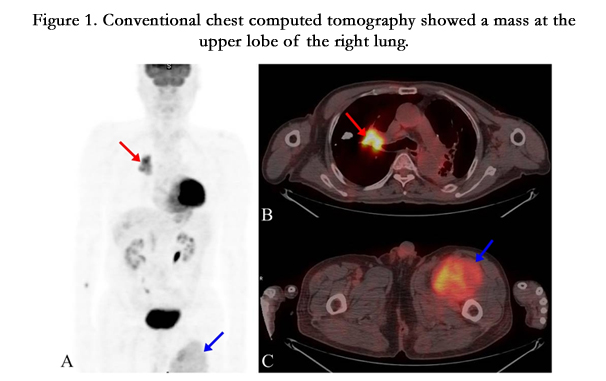
Figure 1. Conventional chest computed tomography showed a mass at the upper lobe of the right lung.
Figure 1. A 77-year-old male visited the hospital due to dyspnea, cough and sputum production. Conventional chest computed tomography showed a mass at the upper lobe of the right lung. F-18 fluorodeoxyglucose (FDG) positron emission tomography/computed tomography (PET/CT) was performed for staging of the lung cancer. F-18 FDG PET/CT showed a mass with intense hypermetabolism (maximum standardized uptake value (SUVmax) 9.8) at the upper lobe of the right lung (A and B, red arrow) and a soft tissue mass with hypermetabolism (SUVmax 4.5) at the left thigh (A and C, blue arrow).
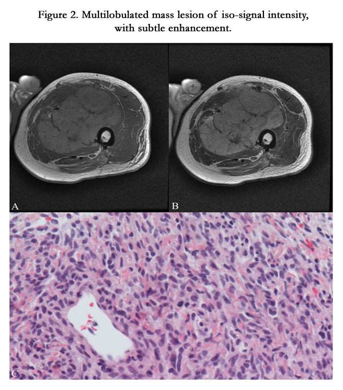
Figure 2. Multilobulated mass lesion of iso-signal intensity, with subtle enhancement.
Figure 2. Axial T1-weighted (A) and T2-weighted (B) magnetic resonance image (MRI) shows a multilobulated mass lesion of iso-signal intensity, with subtle enhancement. The histological examination (magnification × 400, C) shows high cellular proliferation of spindle and plump cells with moderate cellular atypia. Frequent mitoses are present. These findings indicate a spindle cell tumor.
Spindle cell tumor is a rare malignant tumor that typically begins on the skin or in the soft tissues surrounding an organ, although they can also be found in the bone. They are made up of cells which have a spindle shape.
Occasionally, F-18 FDG PET/CT reveals unexpected malignant or premalignant conditions that are unrelated to the disease for which the test has been ordered. In larger series, up to 50 % of these incidental findings are either malignant or premalignant [1]. The use of F-18 FDG PET/CT for the detection and monitoring of patients with musculoskeletal lesions has also been reported [2].
Autopsy series have detected soft tissue (ST) metastases in 0.75~9% of patients who have died from metastatic lung carcinoma [3]. Pain and the presence of a palpable mass are the most frequent clinical features. Biopsy is recommended after the MRI for diagnosis. Due to the rarity of ST metastases, the differential diagnosis poses a problem, especially with primary ST sarcomas. The type of treatment depends on the patient’s clinical status and prognosis, and includes observation, radiotherapy, chemotherapy and surgery. But in our case, the patient did not have any discomfort in the left thigh and the mass was incidentally detected by F-18 FDG PET/CT imaging which was done for the staging of lung cancer, and it mimicked a soft tissue metastasis.
Ethical Standard
A waiver of institutional review board approval was obtained for this case report.
References
- Agress H Jr, Cooper BZ (2004) Detection of clinically unexpected malignant and premalignant tumors with whole-body FDG PET: histopathologic comparison. Radiology 230(2): 417-422.
- Kern KA, Brunetti A, Norton JA, Chang AE, Malawer M, et al. (1988) Metabolic imaging of human extremity musculoskeletal tumors by PET. J Nucl Med 29(2): 181-186.
- Perisano C, Spinelli MS, Graci C, Scaramuzzo L, Marzetti E, et al. (2012) Soft tissue metastases in lung cancer: a review of the literature. Eur Rev Med Pharmacol Sci 16(14): 1908-1914.


