Iron Chelating and Antioxidant Activities and Cytotoxicity Effect of the Pistachio (Pistachio vera L.) hull and kernel Extracts in the A549, HT29 and MCF-7 Cancerous Cell lines
Dahooee F1*, Fatemi SJ1, Mandegary A2,3, Sharififar F4
1 Department of Chemistry, Shahid Bahonar University of Kerman, 22 Bahman Blvd, Kerman, Iran.
2 Gastroenterology and Hepatology Research Center, Institute of Basic and Clinical Physiology, Afzalipour’s Hospital, Imam Highway, Kerman, Iran.
3 Department of Toxicology & Pharmacology, Neuropharmacology Institute, School of Pharmacy, Haft-Bagh Blvd., Kerman, Iran.
4 Department of Pharmacognosy and Herbal and Traditional Medicines Research Center, School of Pharmacy, Haft-Bagh Blvd., Kerman, Iran.
*Corresponding Author
Faezeh Dahooee,
Department of Chemistry, Shahid Bahonar University of Kerman,
22 Bahman Blvd, Kerman, P.O. Box 76169-133, Iran.
Tel/Fax: +98 03433257433
E-mail: fatemijam@yahoo.com
Received: November 18, 2015; Accepted: January 09, 2016; Published: January 13, 2016
Citation: Dahooee F, Fatemi SJ, Mandegary A, Sharififar F (2016) Iron Chelating and Antioxidant Activities and Cytotoxicity Effect of the Pistachio (Pistachio vera L.) hull and kernel Extracts in the A549, HT29 and MCF-7 Cancerous Cell lines. Int J Clin Pharmacol Toxicol. 5(1), 195-201. DOI : dx.doi.org/10.19070/2167-910X-1600033
Copyright: Dahooee F© 2016. This is an open-access article distributed under the terms of the Creative Commons Attribution License, which permits unrestricted use, distribution and reproduction in any medium, provided the original author and source are credited.
Abstract
Synthetic iron-chelators have been considered for treatment of some clinical situations such as iron overloading and cancer. However, their side effects and incomplete effectiveness limit their usages. Natural products could be a source of safe and novel metal chelating agents. The aim of this study was to investigate the cytotoxicity effect, iron chelatory and antioxidant activities of different parts of pistachio (Pistachio vera L.). The iron chelatory effects and Total phenolic content of hull and kernel of pistachio extracts were determined by Ferrozine reagent and Folin Ciocalteu method, respectively. Antioxidant activities were measured by DPPH and FRAP assays. The cytotoxicity of the extracts and deferiprone (synthetic Fe-chelator) were evaluated in the A-549 lung and HT-29 colon and MCF-7 breast cancerous cell lines using MTT cytotoxicity assay. The extracts show potential of iron chelation and antioxidant activity. The cytotoxicity potency of extracts and deferiprone are rated as follows: MCF-7 > A549 > HT-29. The MCF-7 cell lines have the most sensitivity to the cytotoxicity of samples. Our results show that Pistachio hull and kernel are containing cytotoxic compounds for lung (A549 cancer cell line), colon (HT-29 cancer cell line) and breast (MCF-7 cancer cell line). Also, pistachio may be a great natural source for iron chelating, anti-cancer and antioxidant agents in some applications including food and medicinal.
2.Introduction
3.Materials and Methods
3.1.Chemicals
3.2.Samples
3.3.Preparation of the extracts
3.4.Preliminary phytochemical tests
3.5.Determination of total phenolic contents (TPC)
3.6.DPPH radical-scavenging activity
3.7.Ferric Reducing Antioxidant Power (FRAP) assay
3.8.Iron chelating activity
3.9.Cell culture and cytotoxicity assay
3.10.Statistical methods
4.Results
4.1.Preliminary Phytochemical tests
4.2.Total phenolic contents (TPC)
4.3.DPPH radical-scavenging antioxidant assay
4.4.Total antioxidant capacity (TAC)
4.5.Iron chelating activity
4.6.Cytotoxicity assay
5.Discussion
6.Conclusion
6.Acknowledgments
7.References
Keywords
Pistachio; Fe-Chelating Activity; Cytotoxicity; Antioxidant Activity; Deferiprone.
Introduction
Iron is an essential element in human nutrition, it plays a crucial role in oxygen transport and storage and is similarly as vital for processes such as ATP generation, DNA synthesis, cell proliferation and cell cycle progression and various enzymatic activities [1-3]. While iron is essential for life, an excess of iron is toxic. There is no active mechanism for the excretion of iron in the human body. However, iron overloading in situations like iron intoxication and in the patients with chronic anemia who may receive excess iron with each blood transfusion, cause gradual accumulation of iron and damage in various tissues including liver [4], cardiovascular system [5] and brain [6]. Production of reactive oxygen species (ROS) is closely connected to the toxic effects of iron. The link between Fe and production of ROS is Fenton reaction which is responsible to formation of hydroxyl radicals [7]. ROS, especially hydroxyl radicals are active oxygen compounds causes oxidative stress which play an important role in the onset of different diseases, including atherosclerosis, rheumatoid arthritis, cancer and neurodegenerative diseases [8]. Iron has also been associated in the development of cancer [9]. Several in vitro, in vivo, animal and epidemiological studies have confirmed the association between excess iron and cancer [10, 11]. Importantly, iron is an essential cofactor for ribonucleotide reductase (RR), which catalyzes conversion of nucleotides to deoxynucleotides which are necessary for DNA synthesis [12]. Not surprisingly, because of considerably high rate of proliferation, cancer cells require more iron than normal cells.
Moreover, disturbance in regulation of iron homeostasis has been considered as one of the mechanisms in cancer development [13, 14]. The anti-cancer effects of Fe chelators have been focused in recent years. Considering this considerable need for iron in cancer cells, iron deprivation using chelating agents provide a promising form of treatment for cancer therapy which has been reviewed in some studies [10, 13, 14]. Currently, three iron chelating agents are in routine clinical use; desferrioxamine (DFO), deferiprone and deferasirox. Despite the well documented clinical efficacy of these Fe chelators, occurrence of adverse effects such as poor oral bioavailability, short plasma half-life, toxicity, cost and effectiveness, restricts their clinical use [15, 16]. Therefore, finding natural herbal with iron-chelating activity could be a good policy because they are more environmental-friendly and their production is economical [17]. Herbal medicines as a source of natural medicinal compounds have been getting acceptance during the last few decades, especially in developed countries [18]. The iron chelatory effect of several herbs and natural herbal ingredients such as tannins and flavonoids has been reported [16, 19, 20].
Pistachia vera L., (Anacardiaceae) is a perennial plant native to Iran that is cultivated in the Mediterranean and Middle East regions, as well as California. The biological activities of different parts of pistachio such as leaves, kernels, hulls and gum have been reported extensively [21, 22]. There are few reports about the iron-chelating activity of P. vera [23] and P. lentiscus [24] gum extract.
In order to finding herbal compounds with iron chelatory, cytotoxicity effect and antioxidant activity, this study was carried out to evaluate these activities of the methanolic extracts of pistachio hull and kernel in in vitro models. Meanwhile, to find the role the compounds involved in these activities, the preliminary phytochemistry and total phenolic contents of the extracts was determined.
Materials and Methods
2,2-Diphenyl-1-picrylhydrazyl (DPPH) radical, 2,4,6-Tri (2-pyridyl)-s-triazine (TPTZ), Ferrozine (Iron Reagent), Folin- Ciocalteu's reagent, 3-(4, 5-dimethylthiazol-2-yl)-2, 5-diphenyl- 2H-tetrazolium bromide (MTT) were supplied by Sigma–Aldrich (St. Louis, MO, USA). Gallic acid, methanol, iron (II) chloride. Iron (III) chloride, ferric sulfate hydrate, sodium carbonate, sodium acetate, hydrogen chloride, Dimethyl sulfoxide (DMSO) were purchased from Merck (Darmstade, Germany). Fetal bovine serum (FBS), Dulbecco's modified Eagle medium (DMEM) and antibiotics were supplied by Gibco (Life Sciences Inc., USA). All chemicals and solvents were analytical grade.
Pistachio (Pistachio Vera L.) was collected from Rafsanjan region in Kerman province, Iran. After authentication of the plants, a voucher specimen of the plant was deposited in Herbarium center of Faculty of Pharmacy, Kerman University of Medical Sciences, Kerman, Iran (KF 1700).
Dried powder of hull (50 g) and the fresh kernels (100 g) were respectively extracted by sonication method (three times, 35 centigrade degree, 15 min.) with 80% aqueous methanol and 100% methanol respectively at room temperature for 72 h in the dark. The extract was separated every 24h and was replaced with fresh solvent. Finally all the extracts were mixed thoroughly and concentrated under vacuum at 40°C by using a rotary evaporator. The residue was re-extracted with the same solvent and the extracts were stored in freezer in dark until use.
The qualitative phytochemical tests of tannins, flavonoids, saponins, and alkaloids were performed according to Harborne [25]. These tests are based on the visual observation of the colorimetric changes or the precipitate formation after an addition of specific reagents.
The total phenolic content of the medicinal plant samples were determined according to the Folin-Ciocalteu method [26] with some modifications. Briefly, 0.1 ml of extract solution (1 mg/ml) was mixed with 0.1 ml of 10-fold-diluted Folin-Ciocalteu reagent in volumetric flask. After 2 min, 0.4 ml of 7.5% sodium carbonate solution was added. Volume was made up to 2 ml with distilled water and mixture was allowed to stand for 2 h by intermittent shaking. Gallic acid (GA) was used as standard (5-500 μg/ml). Absorbance was measured at 765 nm (Jenway 6505 UV/Vis spectrophotometer). The estimations of phenolic compounds inthe extracts were taken in triplicate and the results were expressed as mg Gallic acid equivalents (GAE)/g dried weight of extracts.
The antioxidant activity of extracts determinates in term of hydrogen atoms, electron donation or radical scavenging ability, using the stable radical DPPH. The inhibition potential of extracts on DPPH radical was measured according to Braca et al [27] with some modifications. Fifty micro-litters of different concentrations of the extracts were added to a 150 μl of a 0.004% methanol solution of DPPH. The mixture was shaken and allowed standing for 30 min in darkness; the absorbance of the solution was determined at 517 nm using microplate reader (Bio Tek ELX 800 ELIZA Reader). Methanol and BHT were used as control and as a standard of the assay, respectively. Inhibition activity of DPPH radical was calculated in following way.(Equation 1):
A0= Absorbance of control (containing only DPPH)
A1= Absorbance of extract in DPPH solution
Ab= Absorbance of extract without DPPH
The concentration of sample required to decrease 50% of DPPH radicals (IC50) was computed by Probit analysis program (SPSS software).
The total antioxidant capacity (TAC) of the extracts was determined by FRAP method [28], a simple, speedy and repeatable method, which can be used to the assay of antioxidants in plasma or botanicals [29, 30]. The FRAP assay based on the ability of antioxidant compounds to reduce complex (Fe (III)–TPTZ) to (Fe(II)–TPTZ) which gives a blue color with an absorbance maximum at 593 nm. The FRAP reagent composed of 10mM TPTZ solution in 40mM HCl, 20 mM FeCl3 solution and 300 mM acetate buffer (pH= 3.6) in a ratio of 1:1:10 (v/v). Then, 50 μl of extracts (100 μg/ml) were added to 3 ml of freshly prepared FRAP reagent and reaction mixtures incubated at 37°C for 30 min. Aqueous solutions of ferrous sulfate (0.1-2 mM) were used to construct standard curve. Absorbance was determined at 593 nm. Triplicate measurements were taken and the FRAP values were expressed as mmol of Fe (II)/g dry weight of extract.
The Fe chelating activity was determined by measuring the formation of the Fe2+-ferrozine complex according to the method of Carter [31] with some modifications. Ten milliliter of various concentrations of extracts were mixed with 12.5 ml sodium acetate buffer (100 mM, PH 4.9) and 1.5 ml FeCl2 (0.01 %, w/v). After incubation for 1h at room temperature, Ferrozine (20μl, 40mM) was added. Binding of Fe (II) ions to ferrozine generates a colored complex that was measured at 562 nm. After 5 min incubation at room temperature, the absorbance at 562 nm was recorded. Distilled water and EDTA were used as control and as a standard metal chelator of the assay, respectively. Iron chelating activity was calculated using the equation 2:
AbsSample= Abs1 – Abs2
Abs Conrol= Absorbance of Control (Fe2+ - Ferrozine Complex)
Abs1= Absorbance of extract and Fe2+ with ferrozine solution
Abs2= Absorbance of extract and Fe2+ without ferrozine solution.
The concentration of sample required to chelate 50% of Fe2+ ions (IC50) was calculated by Probit analysis program (SPSS software).
A549 (human lung epithelial adenocarcinoma), HT-29 (human colorectal adenocarcinoma), and MCF-7 (human breast adenocarcinoma) cell lines were obtained from the National Cell Bank of Iran, Pasteur Institute of Iran (Tehran, Iran). The cells were maintained in DMEM medium supplemented with FBS (10%, v/v) and antibiotics [penicillin (100 units/mL) and streptomycin (100 μg/mL)] at 37°C in a CO2 incubator (5% CO2 and 95% relative humidity). In order to evaluate the cytotoxic effect of extracts, deferiprone (positive Fe chelator control), the cell lines were harvested in the exponential phase of growth, seeded separately into 96-well tissue culture plates (104 per well) and allowed to adhere for 24 h. Thereafter, extracts were added to the desired wells to reach different concentrations. After 24 h of incubation, 20 μL of DMEM medium containing MTT (5 mg/ ml) was added to each well and incubated for 3 h. Consequently, the medium was replaced with 100 μl of DMSO, and optical densities were determined at 595 nm. The MTT assay measures the reduction of a tetrazolium component into an insoluble formazan product by the mitochondria of viable cells. The color intensity generated is directly proportional to the number of viable cells. All experiments were performed in triplicates, standard deviations were calculated.
Results were expressed as mean ± standard deviation (SD) for at least triplicate analyses on the same sample. The probit analysis was used to calculate the IC50s. The potency of compounds in tests showed as IC50 (95% confidence interval). The Excel Pearson function was used to calculate the Pearson correlation coefficient for two sets of values.
Results
The phytochemical screening of the extracts is shown in Table 1. Flavonoids and tannins found in both extracts. Amount of alkaloids was negligible in kernel extracts. None of the extracts contains saponins.
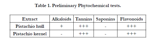

Table 1. Preliminary Phytochemical tests.
The TPC of the methanolic extracts, expressed as mg gallic acid equivalent (GAE)/gr extract is shown in Table 2.
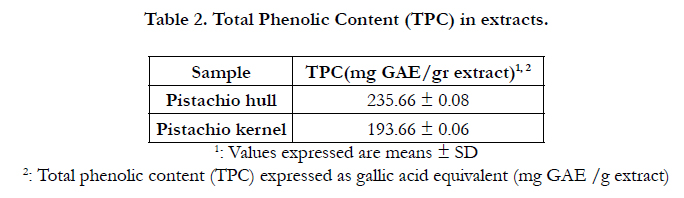
Table 2. Total Phenolic Content (TPC) in extracts.
The IC50 values obtained by the DPPH assay for various extracts are demonstrated in Table 3. The studied extracts displayed antioxidant activity in a concentration-dependent method (Figure 1). Pistachio hull and kernel extracts reached a maximal inhibition activity of % 94 and % 91 at 80 μg/ml respectively. IC50 values of DPPH radical inhibition by pistachio hull and kernel extracts represent % 48 and % 12 of the inhibition activity of BHT (IC50= 4.9 μg/ml), respectively.
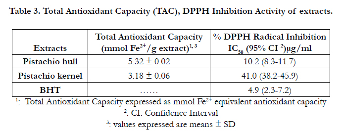
Table 3. Total Antioxidant Capacity (TAC), DPPH Inhibition Activity of extracts.
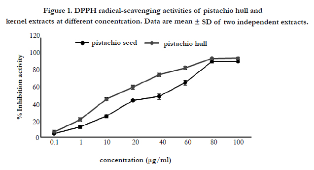
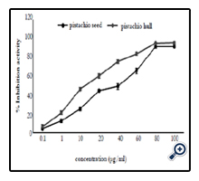
Figure 1. DPPH radical-scavenging activities of pistachio hull and kernel extracts at different concentration. Data are mean ± SD of two independent extracts.
The antioxidant capacity obtained by the FRAP assay for pistachio hull and kernel extracts are shown in Table 3. The extracts have an antioxidant activity in order of pistachio hull > pistachio kernel.
The assay used to determine the chelating activity of Fe2+ was based on the chelation of this metal ion with ferrozine to yield a red colored complex. The potential of Fe2+-chelating was measured by assessing ability of the extracts to compete with ferrozine for the ferrous ions and further ferrous complexes formation, thereby resulting in a decrease in the absorbance at 562 nm. Pistachio hull and kernel extracts and EDTA, as a positive control, and deferiprone (synthetic Fe chelator) could chelate Fe2+ ion in a concentration dependent manner (Figure 2). The maximum value of chelating activity of pistachio hull and kernel extracts were achieved at 2 mg/ml with % 91.1 and % 80.21 Fe2+ chelating activity, respectively. The IC50 value of Fe2+ chelating ability of studied extracts was determined by measuring the iron-ferrozine complex and results compared to synthetic
chelator (deferiprone) are summarized in Table 4.
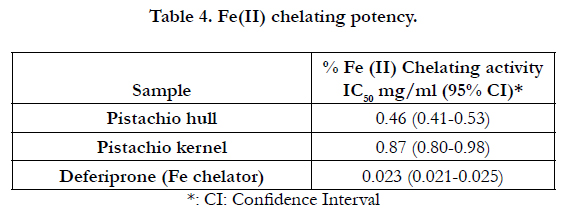
Table 4. Fe(II) chelating potency.
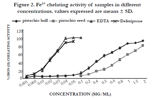
Figure 2. Fe2+ chelating activity of samples in different concentrations. values expressed are means ± SD.
The cytotoxicity of deferiprone, crude extracts on different cell linesare shown in Table 5. The IC50 values, the concentration of material required to achieve 50% reduction in cell viability in comparison to untreated controls, show that pistachio hull extract has more potent effect than pistachio kernel extract in all three cell lines. Meanwhile, the MCF-7 breast cancer cell line showed the highest sensitivity to the tested materials. Although similar for the doxorubicin, the cytotoxicity of extracts and deferiprone was considerably different between the cell lines.
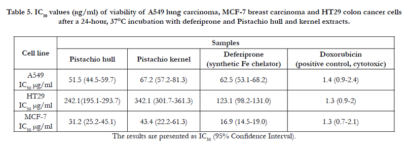
Table 5. IC50 values (μg/ml) of viability of A549 lung carcinoma, MCF-7 breast carcinoma and HT29 colon cancer cells after a 24-hour, 37°C incubation with deferiprone and Pistachio hull and kernel extracts.
Discussion
Although iron is crucial for life, an excess of iron is toxic. Induction of oxidative stress through raising in ROS production is considered as the main mechanism for toxicity of ion metals including iron [32, 33]. Removal of iron which there is no physiologic mechanism for its elimination from the body is the first priority in some instances such as iron poisoning and iron overload. Because of cancer cells rapidly proliferate, these cells have higher requirement for iron than normal cells [10, 34]. The increased requirement of cancer cells to iron has led to sensitivity of these cells to cytotoxic effects of Fe chelators [1, 10, 35]. Considering the vital role of iron in cellular proliferation and its potential to mediate deleterious oxidative damage when in excess, Fe-chelating agents provide a promising form of treatment for both iron overload disease and cancer therapy. Even though several Fe-chelators such as desferrioxamine (DFO), deferiprone and deferasirox has been introduced for using in clinic [16], the obstacles such as poor oral bioavailability, worsening liver fibrosis, hematological abnormalities, short plasma half-life and severe side effects restricted the use of synthetic chelators [36, 37]. Due to the problems with the current synthetic drugs, finding natural products with iron-chelating activity could be a good approach for treating cancer.
Relying on some evidence, the methanolic extracts of pistachio hull and kernel were selected for evaluating their iron chelatorty activity and cytotoxicity effect. There are reports about the ironchelating activity of the plants of genius pistachio including P. vera [23] and P. lentiscus [24] gum extract.
The potency of Fe-chelatory of the extracts of hull and kernel of Pistachio is shown in table 4. In this study we reported for the first time the strong Fe-chelating activity of pistachio hull (IC50= 0.46, 95% CI= 0.41-0.53 mg/ml) and kernel (IC50 = 0.87, 95% CI= 0.80- 0.98 mg/ml). Phytochemistry of the extracts showed presence of tannins and flavonoids in large amounts. It has been proved that Pistachio nuts are a rich source of phenolic compounds and have been considered because of high antioxidant potential [21]. There are reports about Fe chelating activity of tannins [19, 20, 38]. Taking together, the Fe chelating activity of pistachio hull and kernel can be mainly attributed to the higher content of tannins and polyphenols in the extracts.
The cytotoxicity potency of the extracts was rated as follows: MCF7 (IC50 hull=31.2 μg/ml and IC50 kernel=43.4 μg/ml)> A549 (IC50 hull=51.5 μg/ml and IC50 kernel=67.2 μg/ml)> HT29 (IC50 hull=242.1 μg/ml and IC50 kernel=347.5 μg/ml). The ironchelating capacity of extracts followed the same order as did their cytotoxicity effects (Table 5). There are few reports about the anti-cancer activity of the different parts of pistachio [39], there was no studies have been reported the role of iron chelatory on the anti-cancer effect on pistachio.
One of the interesting aspects of our findings that should be emphasized is that the pistachio hull and kernel extracts like deferiprone showed selective cytotoxicity, whilst doxorubicin, a common cytotoxic drug, had nonselective cytotoxicity for the studied cell lines. Therefore, one can conclude that iron chelatory plays a crucial role in the anti-cancer effect of pistachio. Association between iron and breast cancer have been widely reported. It is believed that iron can cause breast cancer not only by it’s the classical role as a critical element for cell proliferation and co-carcinogenic behavior, but it may also synergistically increase the proliferative effect of estrogen on ER+ breast cancer cells [40]. Estrogen and iron activate oxidative stress pathways, via ROS production, induce and maintain the oncogenic phenotype of cancer [40, 41].
The cytotoxicity of the Pistachio hull extract was higher than synthetic chelator (deferiprone) in A549 lung cancer cell lines (Table 5). Although identifying individual active ingredients in extracts would be ideal, it is interesting to note that many studies have observed the extract to be more beneficial compared to the individual or purified ingredient. This suggests the existence of a chemical synergy when using an extract [42].
The extracts of different part of pistachio (hull and kernel) also showed antioxidant activity with the greater activity of the pistachio hull extract. The antioxidant activities of different parts of pistachio such as kernel, gum, hull and leaves have been reported by other authors [21, 23, 43]. Phenolic compounds are a class of antioxidant compounds which act as free radical scavenging [44]. Pistachio is a rich source of phenolic compounds including catechins, gallic acid, quercetin and genistein [43] which their antioxidant activity is established [45, 46]. There was a correlation between the antioxidant properties (DPPH, FRAP) and total phenolic contents of the studied extracts (TPC hull (235.66 ± 0.08mg GAE/gr extract) > TPC kernel (193.66 ± 0.06mg GAE/gr extract)). Antioxidants protect biological systems from oxidative damage produced by reactive oxygen species (ROS) and are therefore considered as health-promoting compounds in nutrition. There is a mutual relation between Fe chelatory and antioxidant properties of the compounds and antioxidant activity of natural antioxidants is partly associated with iron chelation. Chelation of iron and quenching of singlet oxygen are the major characteristics of antioxidant activity [47].
Conclusion
Our results show that Pistachio hull and kernel are containing cytotoxic compounds for A549 lung carcinoma, MCF-7 breast carcinoma and HT29 colon cancer cells. We suggest that anticancer effect can be attributed to the presence of Fe chelatory compounds in the extract such as polyphenols. We conclude that pistachio may be a great natural source for iron chelating, anticancer and antioxidant agents in some applications including food and medicinal. Further in vivo studies and also fractionation of the extracts to find the effective pharmacologic constituents are recommended.
Acknowledgments
We would like to thank Ms. MandanaJafari and Ms. Zahra Mahdavi for their helps in this project.
References
- Kalinowski DS, Richardson DR (2005) The evolution of iron chelators for the treatment of iron overload disease and cancer. Pharmacol Rev 57(4): 547-583.
- Radulescu S, Brookes MJ, Salgueiro P, Ridgway RA, McGhee E, et al. (2012) Luminal iron levels govern intestinal tumorigenesis after Apc loss in vivo. Cell Rep 2(2): 270-282.
- Jomova K, Valko M (2011) Advances in metal-induced oxidative stress and human disease. Toxicology 283(2-3): 65-87.
- Ramm GA, Ruddell RG (2005) Hepatotoxicity of iron overload: mechanisms of iron-induced hepatic fibrogenesis. Semin Liver Dis 25(4): 433-449.
- Kremastinos DT, Farmakis D (2011) Iron overload cardiomyopathy in clinical practice. Circulation 124(20): 2253-2263.
- Ward RJ, Zucca FA, Duyn JH, Crichton RR, Zecca L (2014) The role of iron in brain ageing and neurodegenerative disorders. Lancet Neurol 13(10): 1045-1060.
- Galaris D, Pantopoulos K (2008) Oxidative stress and iron homeostasis: mechanistic and health aspects. Crit Rev Clin Lab Sci 45(1): 1-23.
- Lin MT, Beal MF (2006) Mitochondrial dysfunction and oxidative stress in neurodegenerative diseases. Nature 443(7113): 787-795.
- Huang X (2003) Iron overload and its association with cancer risk in humans: evidence for iron as a carcinogenic metal. Mutat Res 533(1–2): 153-171.
- Bedford MR, Ford SJ, Horniblow RD, Iqbal TH, Tselepis C (2013) Iron chelation in the treatment of cancer: a new role for deferasirox? J Clin Pharmacol 53(9): 885-891.
- Kim JH, Hue JJ, Kang BS, Park H, Nam SY, et al. (2011) Effects of selenium on colon carcinogenesis induced by azoxymethane and dextran sodium sulfate in mouse model with high-iron diet. Lab Anim Res 27(1): 9-18.
- Kus R, J Blinowska K, Kamiński M, Basińska-Starzycka A (2008) Transmission of information during Continuous Attention Test. Acta Neurobiol Exp (Wars) 68(1): 103-112.
- Richardson D, Kalinowski DS, Lau S, Jansson PJ, Lovejoy DB (2009) Cancer cell iron metabolism and the development of potent iron chelators as anti-tumour agents. Biochim Biophys Acta 1790(7): 702-717.
- J BH, M S, T D, R C, W BB, et al. (2008) [Retroperitoneal sarcomas: a single center experience]. Cancer Radiother 12(5): 331-335.
- Maggio A (2007) Light and shadows in the iron chelation treatment of haematological diseases. Br J Haematol 138(4): 407-421.
- Poggiali E, Cassinerio E, Zanaboni L, Cappellini MD (2012) An update on iron chelation therapy. Blood Transfus 10(4): 411-422.
- Sharpe PC, Richardson DR, Kalinowski DS, Bernhardt PV (2011) Synthetic and natural products as iron chelators. Curr Top Med Chem 11(5): 591-607.
- Clement YN, Williams AF, Khan K, Bernard T, Bhola S, et al. (2005) A gap between acceptance and knowledge of herbal remedies by physicians: the need for educational intervention. BMC Complement Altern Med 5: 20.
- Hider RC, Liu ZD, Khodr HH (2001) Metal chelation of polyphenols. Methods Enzymol 335: 190-203.
- Karamac M (2009) Chelation of Cu(II), Zn(II), and Fe(II) by tannin constituents of selected edible nuts. Int J Mol Sci 10(12): 5485-5497.
- Hosseinzadeh H, Sajadi Tabassi SA, Milani Moghadam N, Rashedinia M, Mehri S (2012) Antioxidant Activity of Pistacia vera Fruits, Leaves and Gum Extracts. Iran J Pharm Res 11(3): 879-887.
- Parham M, Heidari S, Khorramirad A, Hozoori M, Hosseinzadeh F, et al. (2014) Effects of pistachio nut supplementation on blood glucose in patients with type 2 diabetes: a randomized crossover trial. Rev Diabet Stud 11(2): 190-196.
- Sehitoglu MH, Han H, Kalin P, Gülçin İ, Ozkan A, et al. (2015) Pistachio (Pistacia vera L.) Gum: a potent inhibitor of reactive oxygen species. J Enzyme Inhib Med Chem 30(2): 264-269.
- Mahmoudi M, Ebrahimzadeh MA, Nabavi SF, Hafezi S, Nabavi SM, et al. (2010) Antiinflammatory and antioxidant activities of gum mastic. Eur Rev Med Pharmacol Sci 14(9): 765-769.
- Harborne JB (1973) Phytochemical methods, a guide to modern techniques of plant analysis. Springer, Netherlands.
- Slinkard K, Singleton VL (1977) Total phenol analysis: automation and comparison with manual methods. American Journal of Enology and Viticulture 28(1): 49-55.
- Braca A, De Tommasi N, Di Bari L, Pizza C, Politi M, et al. (2001) Antioxidant principles from Bauhinia tarapotensis. J Nat Prod 64(7): 892-895.
- Benzie IF, Strain JJ (1996) The ferric reducing ability of plasma (FRAP) as a measure of "antioxidant power": the FRAP assay. Anal Biochem 239(1): 70-76.
- Prior RL, Wu X, Schaich K (2005) Standardized methods for the determination of antioxidant capacity and phenolics in foods and dietary supplements.J Agric Food Chem 53(10): 4290-4302.
- Pulido R, Bravo L, Saura-Calixto F (2000) Antioxidant activity of dietary polyphenols as determined by a modified ferric reducing/antioxidant power assay. J Agric Food Chem 48(8): 3396-3402.
- Carter P (1971) Spectrophotometric determination of serum iron at the submicrogram level with a new reagent (ferrozine). Anal Biochem 40(2): 450-458.
- Britton RS, Leicester KL, Bacon BR (2002) Iron toxicity and chelation therapy. Int J Hematol 76(3): 219-228.
- Kohgo Y, Ikuta K, Ohtake T, Torimoto Y, Kato J (2008) Body iron metabolism and pathophysiology of iron overload. Int J Hematol 88(1): 7-15.
- Andrews NC (2008) Forging a field: the golden age of iron biology. Blood 112(2): 219-230.
- Merlot AM, Kalinowski DS, Richardson DR (2013) Novel chelators for cancer treatment: where are we now? Antioxid Redox Signal 18(8): 973-1006.
- Delea TE, Edelsberg J, Sofrygin O, Thomas SK, Baladi JF (2007) Consequences and costs of noncompliance with iron chelation therapy in patients with transfusion-dependent thalassemia: a literature review. Transfusion 47(10): 1919-1929.
- Jamuar SS, Lai AH (2012) Safety and efficacy of iron chelation therapy with deferiprone in patients with transfusion-dependent thalassemia. Ther Adv Hematol 3(5): 299-307.
- Brewer MS (2011) Natural Antioxidants: Sources, Compounds, Mechanisms of Action, and Potential Applications. Comprehensive Reviews in Food Science and Food Safety 10(4): 221-247.
- Shahraki J, Zareh M, Kamalinejad M, Pourahmad J (2014) Cytoprotective Effects of Hydrophilic and Lipophilic Extracts of Pistacia vera against Oxidative Versus Carbonyl Stress in Rat Hepatocytes. Iran J Pharm Res 13(4): 1263-1277.
- Mittal R, Chaudhry N, Pathania S, Mukherjee TK (2014) Mechanistic insight of drug resistance with special focus on iron in estrogen receptor positive breast cancer. Curr Pharm Biotechnol 15(12): 1141-1157.
- Valko M, Rhodes CJ, Moncol J, Izakovic M, Mazur M (2006) Free radicals, metals and antioxidants in oxidative stress-induced cancer. Chem Biol Interact 160(1): 1-40.
- Adhami VM, Khan N, Mukhtar H (2009) Cancer chemoprevention by pomegranate: laboratory and clinical evidence. Nutr Cancer 61(6): 811-815.
- Tomaino A, Martorana M, Arcoraci T, Monteleone D, Giovinazzo C (2010) Antioxidant activity and phenolic profile of pistachio (Pistacia vera L., variety Bronte) seeds and skins. Biochimie 92(9): 1115-1122.
- Rice-Evans CA, Miller NJ, Paganga G (1996) Structure-antioxidant activity relationships of flavonoids and phenolic acids. Free Radic Biol Med 20(7): 933-956.
- Lu Z, Nie G, Belton PS, Tang H, Zhao B (2006) Structure-activity relationship analysis of antioxidant ability and neuroprotective effect of gallic acid derivatives. Neurochem Int 48(4): 263-274.
- Boots AW, Haenen GR, Bast A (2008) Health effects of quercetin: from antioxidant to nutraceutical. Eur J Pharmacol 585(2-3): 325-337.
- Fibach E, Rachmilewitz EA (2010) The role of antioxidants and iron chelators in the treatment of oxidative stress in thalassemia. Ann N Y Acad Sci 1202: 10-16.


