Photoallergic Reaction to Ficus Pumila
Leibl M1,2*, Al-Ali1,2, Ring J1,2, Eberlein B1,2
Department of Dermatology and Allergy Biederstein, Technische Universität München, Munich, Germany.
2 Christine Kühne Center for Allergy Research and Education (CK-CARE), Munich, Germany.
*Corresponding Author
Dr. med. Maria Leibl,
Klinik und Poliklinik für Dermatologie und Allergologie am Biederstein,
Technische Universität München, Biedersteiner Str. 29, D-80802 München, Germany.
Tel: +49-89-4140-3324
Fax: +49-4140-3526
E-mail: maria.leibl@lrz.tum.de
Received: November 17, 2015; Accepted: August 31, 2016; Published: September 02, 2016
Citation: Leibl M, Al-Ali, Ring J, Eberlein B. Photoallergic Reaction to Ficus Pumila. Int J Clin Med Allergy. 2017;4(2):52-54. doi: dx.doi.org/10.19070/2332-2799-1600013
Copyright: Leibl M© 2016. This is an open-access article distributed under the terms of the Creative Commons Attribution License, which permits unrestricted use, distribution and reproduction in any medium, provided the original author and source are credited.
Abstract
Phyto photoallergic contact dermatitis has rarely been reported and seems to be under reported. We present a 47-year-old patient, who developed erythematous, unsharply marginated skin lesions on the dorsum of the hands and in the face after contact with various plants. Phototesting revealed a reduced MED (minimal erythema dose) as well as a positive photopatch test reaction to Ficus pumila leaves. A photoallergic reaction to this Ficus species has only rarely been described until now.
2.Introduction
3.Case Report
3.1.History
3.2.Skin Examination
3.3.Treatment and Course
3.4.Experimental design
3.5.Allergy Diagnostics
4.Discussion
5.References
Keywords
Ficus Pumila; Photoallergic Reaction; Phytophotodermatitis; Psoralene; Furocoumarine.
Introduction
Photoallergic contact dermatitis is an uncommon reaction, which is induced by cell-mediated hypersensitivity: A photo-activated substance behaves as an hapten and elicits an immunologic response.
The main group of photo contact allergens are the absorbent sunscreen chemicals, but also plants containing furocoumarins are described.
A 47-year old male who initially admitted to our clinic for grouped blisters on the hands and in the face was diagnosed with a photoallergic reaction to a rarely described plant species.
A 47-year-old patient presented in our outpatients department in the middle of April with skin lesions on both hands that existed since one week as well as with skin lesions in the face since 2 days. The patient reported to have collected Humulus lupulus (hop shoots) in the forest on a sunny day and cooked a stew with celery 3 days before. The patient, a trained biologist, also had a terrarium with Ficus pumila among other plants and trimmed plant growth regularly. For 1 day the patient was treated with Aciclovir 400 mg 4x daily because the skin lesions on the hand were diagnosed as herpes simplex infection by colleague.
The patients past medical history were negative for other diseases and medications.
The dermatological examination showed grouped blisters on the left thumb and disseminated lesions on both hands up to the lower arms (Figure 1).
In the nasal and forehead area, strictly left-sided, grouped blisters were visible in the form of stripes on an erythematous base. Yellowish scabs were found in the nasal region (Figure 2).
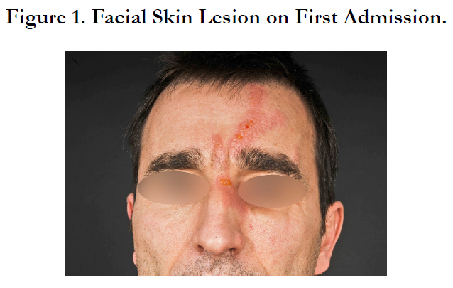
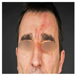
Figure 1. Facial Skin Lesion on First Admission.
Figure 2. Skin Lesion of the Left Hand.
The patient had Fitzpatrick skin type II.
In direct immunofluorescence (sample taken after treatment start with Aciclovir), HSV (herpes simplex virus) 1 and 2 as well as VZV (varicella zoster virus) were negative.
Systemic treatment with Prednisolone 60 mg with gradual step-wise reduction and topical therapy with Prednicarbate was started. Skin lesions improved rapidly, but reddening on hands persisted for approximately 2 weeks.
Increasing UVB doses (25, 50, 100, 150, 200, 250 mJ/cm2) from TL 20W/12 light bulbs (Philips, Hamburg, Germany) with a main emission range of 275 to 365 nm (maximum approximately 315nm) were applied on the lower back of the patient.
The minimal erythema dose (MED) read after 24 h was decreased with 25 mJ/cm2.
Photopatch testing with standard allergens (Almirall Hermal GmbH, Reinbek, Germany) as well as with hop shoots, Apium graveolans (celery), Ficus pumila was performed 3 weeks after first appearance of skin lesions according to the consensus methodology for Europe (1). For the testing the patient brought part of celery bulb, single hop shoots and the ficus plant with stem and leaves. The identification of the plants was done by the patient himself, a biologist. Stem and leaves of Ficus pumila as well as the other plants were cut up into small pieces, put onto Finn Chambers and applied occlusively. The agents were applied in duplicate on the mid upper back skin on either side of the vertebrae. Stems and leaves of the Ficus pumila were mixed up in one Finn Chamber. After 24 h one side of the patch test was removed and the skin was irradiated with 5 J/cm2 UVA (UVA 40W, Philips Electronics, Eindhoven, Netherlands). The other side remained unirradiated and served as control.
24 h after irradiation an unsharply demarcated erythema was visible in the irradiated area where Ficus pumila had been applied.
72 h after irradiation also papulo-vesicles and an infiltrate were visible (Figure 3).
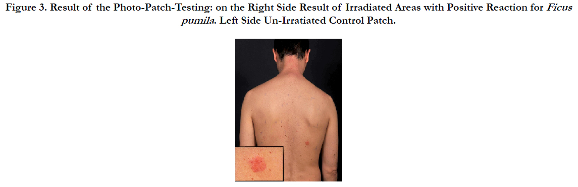
Figure 3. Result of the photo-patch-testing: on the right side result of irradiated areas with positive reaction for Ficus pumila. Left side un-irratiated control patch.
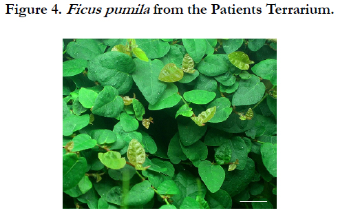
Figure 4. Ficus pumila from the patients terrarium.
The patient reported that burning and itching was strongest in this area 24 h after irradiation. All other tests substances as well as the dark control were negative.
Photopatch testing in controls was not done because of ethical reasons.
Discussion
Ficus pumila, which is native to East Asia (China, Japan, Taiwan) is also known under the name creeping fig and belongs to the mulberry family (moraceae). Its heart-shaped leaves reach a size of approx. 1-2 cm, the plant itself does, in contrast to other species of ficus, not bear fruits. Because it is easy to care for, it is a popular ornamental plant for terrariums and office rooms, but it is often also planted as a creeping plant near house walls [2].
Like umbelliferae and rutaceae, the milky juice of the creeping fig contains furocumarines [3]. These α,β-unsaturated carbonyl compounds are photosensitizing in combination with UV irradiation and can cause burn-like reactions, as e.g. in case of Berloque dermatitis. Moist skin, e.g. during sweating and bathing, increases the penetration of the furocumarines. In addition, furocumarines can be carcinogenic due to UV-induced covalent binding of pyrimidine bases with DNA (cross-linking) [4]).
In our part of the world, furocumarines are found above all in the juice of the giant cow parsnip (giant hogweed heracleum) [5]. The furocumarines bergapten and psoralen are used for the treatment of of vitiligo and psoriasis (systemically and topically). There are reports dating back to 1400 B.C. about the application of psoralen for the treatment of vitiligo.
In the 1930s Stephen Epstein first distinguished between phototoxic and photoallergic reactions: In part of the volunteers painful erythemas occurred after injection of a photosensitizer (sulfonamide) within the first 24 h. With a latency of several days intense itching, highly inflammatory urticarial reactions were observed which were clearly distinguishable from the immediate reaction and were considered to be photoallergic [6].
Phototoxic reactions are obligatory in contact with the sensitizer, and show a dose-dependency. Usually phototoxic reactions are described with blister formation, painful burning and sharply demarcated reddening. The reaction is sharply limited; e.g. the occurrence of Berloque dermatitis with trickle marks was observed after application of perfume and subsequent UV exposure. The reaction usually occurs with some latency and heals with long lasting hyperpigmentation.
In contrast to this, a photoallergic reaction occurs only after prior immunological sensitization to the respective photosensitizer. Clinically it is identical to an allergic contact dermatitis. The reaction is usually dose-independent and the developing erythema unsharply demarcated. This is defined as a so-called spreading reaction. A crescendo reaction in the photopatch test is typical for a photoallergic reaction [7-9]. Photoallergic reactions to 8-methoxypsoralen were described [12].
In the present case, the patient possessed a humid terrarium with Ficus pumila which facilitates skin permeability for furocoumarines. Because of the long-term contact, sensitization may be assumed. The skin changes were not sharply demarcated and occurred also in the face which may had indirect contact with the photosensitizer because the patient reported that he might have touched his forehead with his hand. Phototoxic reactions to figs are often described in the literature, but in contrast photoallergic reactions to this Ficus species are very rare [10-12].
References
- Bruynzeel DP, Ferguson J, Andersen K, Gonçalo M, English J, Goossens A, et al. Photopatch testing: a consensus methodology for Europe. J Eur Acad Dermatol Venerol. 2004 Nov;18(6):679-682. PubMed PMID: 15482294.
- Starr F, Starr K, Loope I. Ficus pumila; Maui. United States Geological Survey, Biological Resources Division; 2003.
- Kitajima J, Kimizuka K, Arai M, Tanaka Y. Constituents of Ficus pumila leaves. Chem Farm Bull. 1998;46(10):1647-1649.
- Song PS, Tapley KJ. Photochemistry and photobiology of psoralens. Photochem Photobiol. 1979 Jun;29(6):1177-1197.
- Pathak MA. Phytophotodermatitis. Clin Dermatol. 1986 Jun;4(2):102-121. PubMed PMID: 2941124.
- Epstein J. Chlorpromazine photosensitivity to sulfonamide. J Invest Dermatol. 1939;2:43-51.
- Hölzle E. Photodermatosen und Lichtreaktionen der Haut. Wissenschaftliche Verlagsgesellschaft. 2003; 225-257.
- Ferguson J, Dover JS. Photodermatology. 2006 Aug;22(4):228.
- Weissmann I, Wagner G, Plewig. Contact allergy to 8-methoxypsoralen. Br J Dermatol. 1980 Jan;102(1):113-5.
- Watemberg N, Urkin Y, Witztum A. Phytophotodermatitis due to figs. Cutis. 1991 Aug;48(2):151-152. PubMed PMID: 1935241.
- Bonamonte D, Foti C, Lionetti N, Rigano I, Angelini G. Photoallergic contact dermatitis to 8-methoxypsoralen in Ficus carica. Contact Dermatitis. 2010 Jun;62(6):343-348. PubMed PMID: 20557340.
- Rademaker M, Derraik J. Photodermatitis caused by Ficus pumila. Contact Dermatitis. 2012 Jul;67(1):53-56. PubMed PMID: 22681467.


