Obligate Bipedal Locomotion in the Modern Human: A Review of Musculoskeletal Modifications
Al-Imam A1,2*
1 Novel Psychoactive Substances Research Unit, School of Life and Medical Sciences, University of Hertfordshire, United Kingdom.
2 Department of Anatomy and Cellular Biology, Faculty of Medicine University of Baghdad, Iraq.
*Corresponding Author
Dr. Ahmed Al-Imam
Novel Psychoactive Substances Research Unit,
Department of Postgraduate Medicine,
University of Hertfordshire, United Kingdom.
E-mail: a.m.al-imam@herts.ac.uk / tesla1452@gmail.com / aalimam@asu.edu
Received: April 17, 2017; Accepted: May 27, 2017; Published: June 03, 2017
Citation: Al-Imam A (2017) Anatomical Adaptations for Obligate Bipedal Locomotion in Humans. Int J Anat Appl Physiol. 3(1), 63-68. doi: dx.doi.org/10.19070/2572-7451-1700010
Copyright:Al-Imam A© 2017. This is an open-access article distributed under the terms of the Creative Commons Attribution License, which permits unrestricted use, distribution and reproduction in any medium, provided the original author and source are credited.
Abstract
Several attributes are only found in primates, some of these are even more unique and noticeable in humans. These may not be limited to; energy-saving bipedal posture and locomotion, stable supine posture, complex manual skills and tool making abilities, hierarchical social structure and cultural organisation, augmented cranial capacity, a more developed frontal lobe, and language proficiency. From a Darwinian point of view, in erect posture and locomotion have evolved to free the hands for the purpose of tool-making. However, more recent theories suggested that bipedal locomotion was related to environmental factors which led to the advent of distinctive and remarkable anatomical features for a form of locomotion that is more energy-economic than quadrupedal locomotion in non-human primates and other members of the animal kingdom. These anatomic features and adaptations include; adjustments to the general body architectural plan, in addition to cranial and postcranial modifications. Postcranial modifications are debated to be the most critical for a stable and fuel-efficient upright walking, other anatomic adaptation were complimentary. The most significant of these anatomical changes took place post-cranially particularly at the level of the pelvis. These changes can be traced back to Australopithecus afarensis dating to at least 3.6 million years ago. The overall level-of-evidence of this article is estimated to be of level-2b, which is well-positioned within the pyramidal hierarchy of level-of-evidence.
2.Background
3.Materials And Methods
4.Discussion
4.1 General Modifications
4.2 Post-Cranial Adaptations
4.3 Cranial Adaptations
5.Conclusion
6.Acknowledgements
7.References
KeyWords
Physical Anthropology; Bipedalism; Locomotion; Hominidae; Hominin; Genus Homo; Australopithecus; Critical Analysis; Anatomy; Comparative Anatomy; Pelvis; Femur; Gluteal musculature..
Background
The modern human is an obligate bipedal creature; several features are distinctive to humans including; an efficient bipedal (upright) walking, stable supine posture, an augmented cranial capacity, an advanced frontal lobe neuroarchitecture, ability to speak and interpret speech, complex tool-making abilities, and an elaborate hierarchical social structure [1, 2]. Cultural anthropologists and physical anthropologists consider that all these features contributed eventually to the birth of the first real culture, rather than a proto-culture as seen in non-human primates [3-6]. It is estimated that the earliest of these traits to be established was the upright posture. However, these anatomical characteristics were much more advancedin the modern human compared to Lucy's species (Australopithecus afarensis) [7-10].
Numerous anatomic adaptations materialised independently across time; these changes exist both cranially and post-cranially, principally serving the purpose of superior energy efficiency. Compared to apes and quadrupeds, humans spend much less energy during bipedal locomotion [11]. Humans have also acquired longer lower limbs to serve the propulsive function during walking. Besides, humans have less body weight (BW) above the waist (68% of the total BW in genus Homo versus 82% in Apes) [12]. Additionally, the line of gravity passes behind the ears and slightly anterior to the spine, and anterior to the knees [11, 12]. Bipedalism appears to be more advanced in the Neanderthals and the modern “wise” humans (Homo sapiens) than in other members of the genus Homo.
It is important to bring the attention of those who will read this manuscript to appreciate that the purpose of this study is not to take sides neither with the theory of Darwinian’s evolution nor the theory of intelligent design; this manuscript will focus on discussing the anatomic adaptations for bipedal locomotions in humans. The renowned Darwinian evolution theory by Charles Darwin and Alfred Russel did theorise that the organic lifeforms are in a constant and a slowly progressive evolution across vast aeons of time, and all species had common ancestry [13-15]. On the other hand, some other scholars have claimed the theory of an intelligent design of life and humans to be specific, and that randomness is not destined to create an intelligent and a fully-aware creature as in the modern humans [16-18].
Materials And Methods
A review of the literature was carried out using a specific set of keywords. Several medical and paramedical databases were explored including; PubMed/Medline, the Cochrane Library, CINAHL Plus, Scopus, Embase, Web of Science, and Google Scholar. Gray literature databases were also explored including; Open Grey, news fora and blogs, and specific courses on the renowned Coursera and edX online learning environment. The implemented keywords included; bipedal, bipedalism, upright walking, two-footed, Genus Homo, hominid, humanoid, hominoid, locomotion, physical Anthropology; Hominidae; Hominin; Genus Homo; Australopithecus; critical Analysis; Anatomy; Comparative Anatomy, Musculoskeletal System, Pelvis, Femur; Gluteal musculature. These keywords were used either alone or in combination with Boolean operators (AND, OR, NOT) to either expand or narrow down the number of hits displayed in a particular search engine.
The literature review was systematic and took place in the period from the 26th of February 2017 to the 28th of March 2017. The number of hits was not filtered by the date of publication. However, and emphasis was given to manuscripts published in the past five years. The total number of bibliographic materials was 53 in total. Each reference was critically appraised using critical appraisal tools including CASP appraisal tools, the aim of this appraisal was to evaluate the level-of-evidence for each manuscript and to assess whether it was suitable to be included in this study or not [19]. Accordingly, one manuscript was not found appropriate and had been excluded.
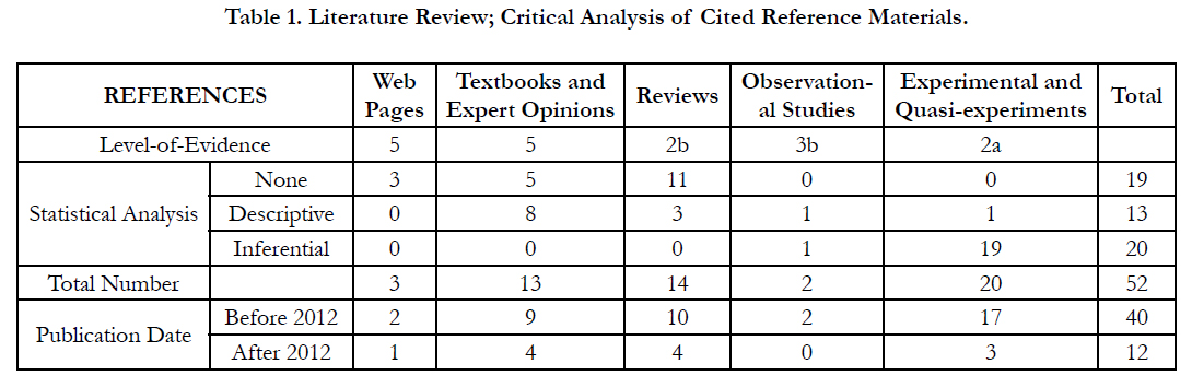
The critical appraisal was successful to categorise the level-of evidence (Table 1) for each manuscript in compliance with the classification system imposed by the Oxford Center for Evidence- Based Medicine (CEBM) for the year 2009 [20]. Correspondingly, the included (cited) manuscripts fell into five main categories; textbooks and Expert Opinions (1), reviews (2), observational studies (3), experimental and Quasi-experiments (4), and web pages (5). The vast majority of papers were either experimental or Quasi-experimental (38.8%) or review articles (29.2%); most of these studies were carried out prior to 2012 (77.1%). The level-of-evidence could be as low as level-5 to as high as level-2a; papers of low evidence were a minority (8.3%). The overall levelof- evidence of this study is estimated to be of level-2b, which is well-positioned within the pyramidal hierarchy of levelof- evidence.

Discussion
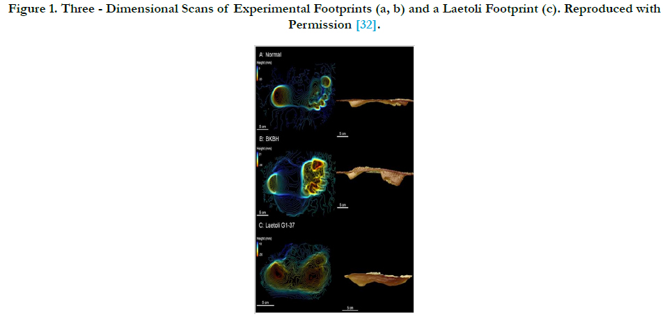
Musculoskeletal and anatomical adaptations can be specifically categorised into; general (1), post-cranial (2), and cranial (3). These changes started to appear in Australopithecus afarensis at approximately 3.6 million years ago (Mya); it has been extensively studied via the Laetoli footprints (Figure 1) discovered by Mary Leaky in 1978 near Olduvai Gorge in Tanzania [21-23]. On the other that hominids adaptations, particularly pelvic re-designing, were far different for example that of Oreopithecus bambolii [24].
Bipedal walking in humans is estimated to be at least 75% less demanding for energy than both quadrupedal and bipedal locomotion in chimpanzees [8, 25, 26]. There was a change in lower limb length and the leg length specifically, the purpose is to provide an efficient lever mechanism in pushing (propelling) the body forward, and to reduce the need for the muscular efforts during the swinging phase of upright walking [27-29].
On the other hand, the upper limbs were excluded from locomotions in humans, except during infancy. Hence, the upper limbs, particularly the hands, became optimised to carry and manipulate objects with a high degree of manual precisions. The implications of this change in motor function had led to a reduction in the ratio of humerus-to-femur length. Humans have a different distribution of weight above and below the level of the waist. It is estimated that 68% (versus 82% in apes) of the total body weight in humans is located above the waist [12]. Furthermore, the line of gravity is located slightly anterior to the vertebral columns and the knees [11, 12, 30].
Post-cranial Adaptations were of paramount importance for a coherent and a cost-effective bipedal form of locomotion. The foot has evolved to function as the propulsive organ and to have no grasping functionality; the heal became enlarged and positioned beneath the centre of gravity, the toes were shorter and more straight (not for grasping), the hallux became fully adducted and non-apposable [31, 32]. It can be deduced from the Laetoli footprints (Figure 1) of Australopithecus afarensis that the body weight was primarily transmitted down via the line of gravity to; the heel, the ball of the foot, the lateral foot border, and the big toe (hallux). Though the foot became arched, with two longitudinal arches and one transverse arch, it became more rigid when compared to monkeys, apes, and non-human primates [31-33].
The knee became bigger, with a larger articular surface area (SA) of femoral-tibial condyles. Compared to apes, the condyles are more flattened and of longer anteroposterior (AP) diameter [11, 33]. Furthermore, the lateral femoral condyle possessed a unique anterior lip to prevent lateral displacement (dislocation) of the patella from the patellofemoral unit; this lateral displacement occurs due to the pulling effect quadriceps femoris muscle in the presence of the valgus angle of the knee. The knees were allowed to be fully extended during the swinging phase of walking which potentiates propulsion from the ground. The knees developed a valgus angle (Q angle), thus positioning the knees right underneath the centre of gravity, which also enabled the knee to be locked in full extension for an extended period of standing upright while requiring a minimal muscular effort [34].
At the hip, the surface area of the coxo-femoral (hip) joint became increased, while the femur developed a conspicuous angle of torsion-inclination to accommodate the tensile, compressive, and torsional forces across the femoral neck and head [33, 35]. The cortex of the femoral neck became more thickened inferiorly; its trabecular patterns were rearranged to prevent and reduce the incidence of catastrophic fractures of the femoral neck [36]. The iliopsoas, the most powerful flexor muscle of the hip, became more developed and an imprint was created in front the coxal bone in relation to the iliopubic eminence [11, 33].
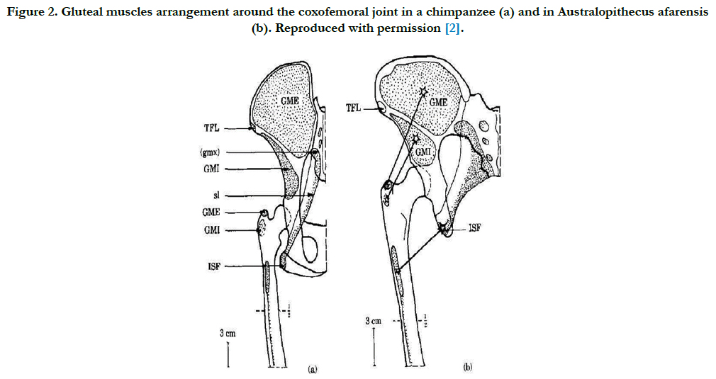
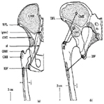
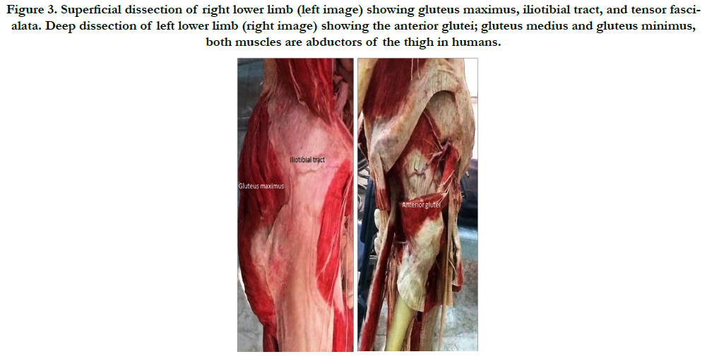
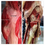
At the pelvis, there was an enlargement of the sacroiliac joints connecting the axial to the peripheral skeleton, both joints (bilaterally) became in a more precise alignment with; the acetabulum of the innominate bone [37], and the line of gravity [2]. The Ilia changed in shape from being long and narrow to short and broad. Additionally, there was broadening of the pelvis, the sacrum became broader with the sacral ala expanded more laterally, the sacrum also became more inclined backwards in continuity with lumbar lordosis, thus the volume of the pelvic cavity became significantly larger, these changes are more evidently noticed in females in an aim to accommodate the presenting part of the newborn baby at the time of delivery [38]. The ischial spines became more prominent medially providing robust attachments for the sacrospinous and sacrotuberous ligaments, which led to the formation of a basinlike support for abdominal viscera; this basin-like support relies on the thoracic cage in quadrupeds [37, 38]. One of the most significant changes in the pelvis took place at the iliac blades (Figure 2 and 3); the blades have rotated along the sagittal axis for each, thus repositioning the anterior glutei muscles (gluteus medius and gluteus minimus) more anteriorly, while the gluteus maximus (the most powerful hip extensor muscle) was fixed posteriorly to persist as the most powerful extensor muscle of the coxo-femoral joint, thus preventing the body from pitching forward in an upright posture and during walking. The new post of anterior gluteal muscles (Figure 3) enabled them to function as the main abductor muscles of the hip joint, which led to an efficient and an accurate tilting of the pelvis during walking. Finally, the anterior iliac spines became robust, for anchorage of sartorius and rectus femoris, which enabled these two muscles to function as stronger flexors of the thigh [7]. Other muscles that played an importance in relation to adaptation for obligate bipedal locomotions are the muscles of the calf. These muscles include three major bulky muscles; gastrocnemius, soleus, and plantaris [39]. There has been changes in relation several morphometric parameters of the muscles including; the dimension and volume of the muscles [37], proximal and distal bony attachments [7], the metabolic status and energy consumption [36], and the number of proprioceptive nerve endings [9, 16, 32, 40]. Additionally, the plantaris muscle is widely considered as a proprioceptive organ of the lower limb due to its higher density of muscles spindles, intrafusal muscle fibres and Golgi tendon organs. In fact, plantaris and other muscle of the upper limb, palmaris longus, are well-known to have an evolutionary significance and inter-ethnic variations [41, 42].
The vertebral column developed four curvatures in the sagittal plane; cervical, thoracic, lumbar, and sacral. It was Leonardo da Vinci who was the first to study the double-Sigmoid (double-S) curvature of the spinal column [43-45]. The aim of this double-S curvature of the spine is to withstand compressive forces more efficiently and to function as a shock absorber. The combined lumbar lordosis and thoracic kyphosis have positioned the centre of gravity directly above feet, preventing the body from toppling forward during walking. Moreover, the size of vertebrae, specifically the vertebral bodies and their facet joints (zygapophysial joints), significantly increased as we go down the spinal column till reaching the first piece of the sacrum at the pelvic inlet [46-48].
These adaptations were significantly less diverse and less vital when compared with post-cranial adaptation. The head has become well positioned and accurately balanced on top of the cervical segment of the vertebral column; this has occurred due to the central positioning of the foramen magnum in relation to the AP diameter of the cranium. Furthermore, the face has become orthognathic rather than prognathic (as in non-human primates and Australpethicines). There was also a reduction in the amount of musculature mass of neck extensors. Similarly, there was a decrement in the mass of the supra-orbital ridges and its functioning Occipitofrontalis muscle [11, 33]. The endocranial capacity has significantly increased up to 1500 cubic centimeters over the period from 2.4 to 0.5 Mya, this has also led to a change in the weight distribution within the cranium and around the pivotal Atlanto-occipital joint [49-52].
Conclusion
The upright walking, also known as bipedalism, is a not strict feature for humans. However, a stable bipedal stance and an energy-economic bipedal form of locomotion are unique in humans compared with the great apes and non-human primates. Bipedalism is a shared biomechanical trait between several different species of human and non-human primates. However, bipedal upright walking seems to be obligatory and far more evolved in genus Homo, particularly in modern humans. The erect bipedal posture and locomotion were not easily achieved by humans; the evolutionary timeline and natural selection have struggled to intelligently design a bipedal creature, and create an energy-efficient bipedalism. Nevertheless, the economic characteristic of this form of locomotion came at a price; several pathologies are affecting multiple elements of the musculoskeletal system including; the vertebral column, the hip joint, and the knee joints. Arthritis is one of the most devastating conditions affecting the musculoskeletal system, which may simply occur as a consequence of the ageing process and excessive use of the joints.
In this study, the anatomical and biomechanical properties of human adaptations for bipedal walking were explored; these included cranial and postcranial adaptations and some other miscellaneous adaptation. It is apparent that anatomic modifications at the level of the spine, pelvis, hip, and knee joint were the most critical for a successful bipedal locomotion. Each of these adaptations possibly occurred independently from the other and in a non-simultaneous fashion across aeons of the evolutionary timeline. Perhaps, bipedalism is the oldest of the unique traits in humans; other traits include; advanced tool making abilities, progressively enlarged frontal lobe capacity, culture, verbal and comprehensive linguistic abilities, and written language.
Acknowledgments
The authors would like to acknowledge with gratitude the supportive efforts by Professor Dr Ashok Sahai. Professor Sahai is an eminent anatomist from India; he is also a fellow and the vice president of the Anatomical Society of India (ASI). Our gratitude goes to Raichlen DA, Berge C, and colleague for granting their permission to reproduce figures (Figure 1 and 2) from their own original studies.
References
- Matsuzawa T (2009) Symbolic representation of number in chimpanzees. Current opinion in neurobiology. 19(1): 92-8.
- Matsuzawa T (2012) What Is Uniquely Human? A View from Comparative Cognitive Development in Humans and Chimpanzees In the:The Primate Mind: Built to Connect with Other Minds by Frans B. M. de Waal, Pier Francesco Ferrari.
- Daly GB (2015) Why Japanese primatology ? A perspective from sociocultural anthropology. In Primate Research Supplement. The 31th Congress Primate Society of Japan. 106-106.
- Gamkrelidze TV, Ivanov VV (1995) Indo-European and the Indo-Europeans: A Reconstruction and Historical Analysis of a Proto-Language and Proto-Culture. Part I: The Text. Part II: Bibliography, Indexes. Walter de Gruyter. Germany.
- McGrew WC (2015) The cultured chimpanzee: nonsense or breakthrough. Hum Ethol Bull. 30(1): 40-51.
- Williams N (2008) Japan wildlife boost. Curr Biol. 18(9): R358-9.
- Berge C (1994) How did the australopithecines walk? A biomechanicalstudy of the hip and thigh of Australopithecus afarensis. J Human Evolution. 26(4): 259-73.
- Sellers WI, Cain GM, Wang W, Crompton RH (2005) Stride lengths, speedand energy costs in walking of Australopithecus afarensis: using evolutionaryrobotics to predict locomotion of early human ancestors. J Royal SocietyInterface. 2(5): 431-41.
- Stern Jr JT, Susman RL (1983) The locomotor anatomy of Australopithecusafarensis. Am J Phys Anthropol. 60(3): 279-317.
- Ward CV (2013) Postural and locomotor adaptations of Australopithecus species. The paleobiology of Australopithecus. Springer Netherlands. 235- 245.
- Morbeck ME (1986) "Primate morphophysiology, locomotor analyses andhuman bipedalism." 423-425.
- Rodman PS, McHenry HM (1980) Bioenergetics and the origin of hominid bipedalism. Am J Phys Anthropol. 52(1): 103-6.
- Gould SJ (1987) Is a new and general theory of evolution emerging?. Self-Organizing Systems . Springer US, 113-130.
- Huxley TH (1968) On the origin of species. University of Michigan.
- Smith JM (1993) The theory of evolution. Cambridge University Press.
- Dembski WA, McDowell S (2008) Understanding intelligent design. Harvest House Publishers: US.
- Dembski WA (2002) Intelligent design: The bridge between science & theology. InterVarsity Press: US.
- Pennock RT (2001) Intelligent design creationism and its critics: Philosophical, theological, and scientific perspectives. MIT Press, UK.
- Norman GR, Shannon SI. (1998) Effectiveness of instruction in critical appraisal (evidence-based medicine) skills: a critical appraisal. Canadian Medical Association Journal. 1998. 158(2):177-81.
- Howick J, Chalmers I, Glasziou P, Greenhalgh T, Heneghan C, et al., (2011) Explanation of the 2011 Oxford Centre for Evidence-Based Medicine (OCEBM) levels of evidence (background document). Oxford Center for Evidence-Based Medicine.
- Hatala KG, Demes B, Richmond BG (2016) Laetoli footprints reveal bipedal gait biomechanics different from those of modern humans and chimpanzees. Proc R Soc B. The Royal Society. 283: 1836 (20160235).
- Masao FT, Ichumbaki EB, Cherin M, Barili A, Boschian G, et al., (2016) New footprints from Laetoli (Tanzania) provide evidence for marked body size variation in early hominins. eLife. 5: e19568.
- Raichlen DA, Gordon AD, Harcourt-Smith WE, Foster AD, Haas Jr WR (2010) Laetoli footprints preserve earliest direct evidence of human-like bipedal biomechanics. PLoS One. 5(3): e9769.
- Kohler M, Moya-Sola S (1997) Ape-like or hominid-like? The positional behavior of Oreopithecus bambolii reconsidered. Proc Natl Acad Sci U S A. 94(21): 11747-50.
- Sellers WI, Dennis LA, Crompton RH (2003) Predicting the metabolic energy costs of bipedalism using evolutionary robotics. J Exp Biol. 206(7): 1127-36.
- Wheeler PE (1991) The influence of bipedalism on the energy and water budgets of early hominids. J Hum Evol. 21(2): 117-36.
- Gatesy SM, Biewener AA (1991) Bipedal locomotion: effects of speed, sizeand limb posture in birds and humans. J Zoology. 224(1): 127-47.
- Steudel-Numbers KL, Tilkens MJ (2004) The effect of lower limb length on the energetic cost of locomotion: implications for fossil hominins. J Hum Evol. 47(1): 95-109.
- Steudel-Numbers KL (2006) Energetics in Homo erectus and other early hominins: the consequences of increased lower-limb length. J Hum Evol. 51(5): 445-53.
- Le Huec JC, Saddiki R, Franke J, Rigal J, Aunoble S (2011) Equilibrium of the human body and the gravity line: the basics. Eur Spine J. 20(5): 558-563.
- Harcourt‐Smith WE, Aiello LC (2004) Fossils, feet and the evolution of human bipedal locomotion. J Anatomy. 204(5): 403-16.
- Latimer B, Lovejoy CO (1989) The calcaneus of Australopithecus afarensis and its implications for the evolution of bipedality. Am J Phys Anthropol. 78(3): 369-86.
- Crompton RH, Sellers WI, Thorpe SK (2010) Arboreality, terrestriality and bipedalism. Philosophical Transactions of the Royal Society B: Biological Sciences. 365(1556): 3301-14.
- Mizuno Y, Kumagai M, Mattessich SM, Elias JJ, Ramrattan N, et al., (2001) Q‐angle influences tibiofemoral and patellofemoral kinematics. J Orthop Res. 19(5): 834-40.
- Musculoskeletal Key. Structure and Function of the Hip.
- Brown TD, Ferguson AB (1980) Mechanical property distributions in thecancellous bone of the human proximal femur. Acta Orthop Scand. 51(1-6): 429-37.
- Berge C (1998) Heterochronic processes in human evolution: an ontogenetic analysis of the hominid pelvis. Am J Phys Anthropol. 105(4): 441-59.
- Snell CA, Donhuysen HW.(1968) The pelvis in the bipedalism of primates. American journal of physical anthropology. 1968 May 1;28(3):239-46.
- Kardong KV (2006) Vertebrates: comparative anatomy, function, evolution. Boston: McGraw-Hill.
- Bruner E, Grimaud-Herve D, Wu X, de la Cuetara JM, Holloway R (2015) A paleoneurological survey of Homo erectus endocranial metrics. Quaternary Intl. 368: 80-7.
- Roohi SA, Choon-Sian L, Tan GH, Naicker AS, Med Rehab M (2007) Astudy on the absence of palmaris longus in a multiracial population. Malaysian Orthopaedic J. 1(1): 26-8.
- Sebastin SJ, Puhaindran ME, Lim AY, Lim IJ, Bee WH (2005) The prevalence of absence of the palmaris longus–a study in a Chinese population and a review of the literature. J Hand Surg Br. 30(5): 525-7.
- Crisco Iii JJ, Panjabi MM (1991) The intersegmental and multisegmental muscles of the lumbar spine: a biomechanical model comparing lateral stabilizing potential. Spine. 16(7): 793-9.
- Jose AM (2001) Anatomy and Leonardo da Vinci. Yale j biol med. 74(3): 185.
- Sanan A, Rengachary SS (1996) The history of spinal biomechanics. Neurosurgery. 39(4): 657-69.
- Nakatsukasa M, Hayama S, Preuschoft H (1995) Postcranial skeleton of a macaque trained for bipedal standing and walking and implications for functional adaptation. Folia Primatologica. 64(1-2): 1-29.
- Rosenberg K, Trevathan W (1995) Bipedalism and human birth: The obstetrical dilemma revisited. Evolutionary Anthropology: Issues, News, and Reviews. 4(5): 161-8.
- Whitcome KK, Shapiro LJ, Lieberman DE (2007) Fetal load and the evolution of lumbar lordosis in bipedal hominins. Nature. 450(7172): 1075-8.
- Bruner E, Grimaud-Herve D, Wu X, de la Cuetara JM, Holloway R (2015) A paleoneurological survey of Homo erectus endocranial metrics. Quaternary Intl. 368: 80-7.
- Bruner E, Iriki A (2016) Extending mind, visuospatial integration, and the evolution of the parietal lobes in the human genus. Quaternary Intl. 405: 98-110.
- Stringer C (2016) The origin and evolution of Homo sapiens. Phil Trans R Soc B. 371(1698): 20150237.
- Zhang Y, Wu X, Schepartz LA (2015) Comparing methods for estimating cranial capacity in incomplete human fossils using the Jingchuan 1 partial cranium as an example. Quaternary Intl. 434: 57-64.


